-
Články
Top novinky
Reklama- Vzdělávání
- Časopisy
Top články
Nové číslo
- Témata
Top novinky
Reklama- Videa
- Podcasty
Nové podcasty
Reklama- Kariéra
Doporučené pozice
Reklama- Praxe
Top novinky
ReklamaAutoregulation of the Noncoding RNA Gene
Most genes along the male single X chromosome in Drosophila are hypertranscribed about two-fold relative to each of the two female X chromosomes. This is accomplished by the MSL (male-specific lethal) complex that acetylates histone H4 at lysine 16. The MSL complex contains two large noncoding RNAs, roX1 (RNA on X) and roX2, that help target chromatin modifying enzymes to the X. The roX RNAs are functionally redundant but differ in size, sequence, and transcriptional control. We wanted to find out how roX1 production is regulated. Ectopic DC can be induced in wild-type (roX1+ roX2+) females if we provide a heterologous source of MSL2. However, in the absence of roX2, we found that roX1 expression failed to come on reliably. Using an in situ hybridization probe that is specific only to endogenous roX1, we found that expression was restored if we introduced either roX2 or a truncated but functional version of roX1. This shows that pre-existing roX RNA is required to positively autoregulate roX1 expression. We also observed massive cis spreading of the MSL complex from the site of roX1 transcription at its endogenous location on the X chromosome. We propose that retention of newly assembled MSL complex around the roX gene is needed to drive sustained transcription and that spreading into flanking chromatin contributes to the X chromosome targeting specificity. Finally, we found that the gene encoding the key male-limited protein subunit, msl2, is transcribed predominantly during DNA replication. This suggests that new MSL complex is made as the chromatin template doubles. We offer a model describing how the production of roX1 and msl2, two key components of the MSL complex, are coordinated to meet the dosage compensation demands of the male cell.
Published in the journal: . PLoS Genet 8(3): e32767. doi:10.1371/journal.pgen.1002564
Category: Research Article
doi: https://doi.org/10.1371/journal.pgen.1002564Summary
Most genes along the male single X chromosome in Drosophila are hypertranscribed about two-fold relative to each of the two female X chromosomes. This is accomplished by the MSL (male-specific lethal) complex that acetylates histone H4 at lysine 16. The MSL complex contains two large noncoding RNAs, roX1 (RNA on X) and roX2, that help target chromatin modifying enzymes to the X. The roX RNAs are functionally redundant but differ in size, sequence, and transcriptional control. We wanted to find out how roX1 production is regulated. Ectopic DC can be induced in wild-type (roX1+ roX2+) females if we provide a heterologous source of MSL2. However, in the absence of roX2, we found that roX1 expression failed to come on reliably. Using an in situ hybridization probe that is specific only to endogenous roX1, we found that expression was restored if we introduced either roX2 or a truncated but functional version of roX1. This shows that pre-existing roX RNA is required to positively autoregulate roX1 expression. We also observed massive cis spreading of the MSL complex from the site of roX1 transcription at its endogenous location on the X chromosome. We propose that retention of newly assembled MSL complex around the roX gene is needed to drive sustained transcription and that spreading into flanking chromatin contributes to the X chromosome targeting specificity. Finally, we found that the gene encoding the key male-limited protein subunit, msl2, is transcribed predominantly during DNA replication. This suggests that new MSL complex is made as the chromatin template doubles. We offer a model describing how the production of roX1 and msl2, two key components of the MSL complex, are coordinated to meet the dosage compensation demands of the male cell.
Introduction
Some long noncoding RNAs have the ability to recruit chromatin modifying enzymes to specific genes thereby controlling their expression [1]. Other noncoding RNAs behave as transcriptional enhancers to flanking protein coding genes [2]. The two roX RNAs that participate in dosage compensation of the single male X chromosome in Drosophila are some of the best characterized examples of noncoding RNAs that target chromatin remodeling enzymes to large domains [3]. The roX RNAs assemble into a complex containing at least five MSL protein subunits that bind actively transcribed genes along the male X chromosome, but not autosomes or the two X chromosomes in females [4]. This has been termed the dosage compensation complex or the MSL complex. One function of the complex is acetylation of histone H4 at lysine 16, carried out by the MOF (males absent on first) histone acetyltransferase resulting in an essential ∼two-fold increase in transcription [5]–[8]. Another modification is ubiquitylation of histone H2B at K34 by the MSL2 RING finger protein [9].
Flies carry two roX genes that differ greatly in size and sequence [10]. The roX1 gene is located on the X chromosome at polytene band 3F and produces a 3.7 kb RNA. The roX2 gene is located at 10C on the X and makes a ∼600 nt RNA. Both RNAs ‘paint’ the length of the male X in a banded pattern [11]. The only obvious sequence similarity between them is limited to short repeated elements near the 3′ end of each gene [12]. These repeats are essential for function and predicted to fold into conserved secondary structures [13], [14]. Neither roX RNA is maternally deposited in eggs. Zygotic transcription of roX1 RNA occurs in both male and female embryos beginning at blastoderm [15]. Females lose roX1 RNA midway through embryogenesis, but males maintain expression through adulthood. By contrast, roX2 RNA first appears a few hours after roX1 but only in male embryos [16]. Despite the vast differences in size, sequence, and regulation, the two roX RNAs are functionally redundant [17].
Little is known about how production of the roX RNAs and MSL protein subunits are coordinated. Unusual cis spreading behavior of MSL complex from sites of autosomal roX transgene has been attributed to cotranscriptional assembly of free MSL subunits onto growing nascent roX transcripts [13], [18], [19], although direct biochemical evidence is lacking. Most MSL protein subunits are made in both males and females, except for MSL2 which is translationally repressed in females by the action of SXL [20], [21]. MSL2 is a RING finger protein that binds DNA in a sequence independent manner through a second cysteine-rich motif [22]. The H83M2 transgene removes the 5′ and 3′ UTRs containing SXL binding sites from msl2 mRNA and has been used extensively to drive ectopic MSL2 expression in females [5], [11], [15], [17], [23]–[29]. Forcing females to make MSL2 using the H83M2 transgene induces production of roX RNAs, resulting in ectopic dosage compensation that is toxic to females [15], [30]. These observations led to the idea that MSL2 protein alone, or acting with the other MSL proteins drives transcription of roX1 RNA [31]–[33].
We reexamined the question of how MSL proteins regulate transcription of the roX1 gene using flies missing the roX2 locus. Deleting roX2 allowed us to study expression of the wild type endogenous roX1 gene without the confounding effects of a second functionally redundant RNA species. In this way we found an RNA-dependent autoregulatory loop controlling roX1 expression. We propose that the early burst of roX1 transcription at blastoderm initiates this cycle. Furthermore, production of the key male-limited MSL2 protein subunit is not only regulated at the translational level as has been extensively documented, but we find msl2 transcription is associated with DNA replication. We propose a model where pre-existing roX RNA, assembled in mature MSL complexes, drives bursts of roX1 transcription during S phase when its chromatin target is doubling.
Results
roX1 transcription is dependent on roX2 RNA
Male embryos normally establish dosage compensation by the onset of gastrulation [16], [34]. Ectopic expression of MSL2 in females leads to roX1 transcription and dosage compensation [15]. We asked whether dosage compensation can only be initiated during early embryogenesis, or could it be artificially started later during larval development. To achieve that, we used an inducible Flp-out system [35] to create clones of GAL4 expressing cells on day 4 AEL (after egg laying) that in turn drove expression of UAS-GFP and UAS-MSL2 in female larvae (Figure 1A and Methods). The purpose of this was to assay females in which the early burst of roX1 RNA had decayed away leaving cells devoid of any roX RNA. When no clones were induced, we never observed GFP or MSL painting in any cell showing that expression was tightly blocked prior to induction (data not shown). Third instar larvae in which late GAL4+ clones were induced displayed overlapping expression of GFP and MSL2 (Figure 1B–1B″). More importantly, MSL2 appeared as subnuclear punctate staining in imaginal disc cells suggesting that it was concentrated on the X chromosome (Figure 1B″). MSL2 immunostaining of polytene squashes confirmed binding along the X (Figure 1C). Unfortunately, we were unable to perform GFP immunostaining and reliable roX1 FISH (Fluorescence in situ hybridization) in the same glands as the proteinase K treatment necessary to expose roX RNA often destroyed protein epitopes. We took comparable salivary glands from GFP positive larvae (Figure 1D) and processed them for roX1 FISH. The results show that roX1 transcription was successfully induced (Figure 1E–1F), consistent with previous reports that MSL2 drives roX1 transcription [15], and confirms an earlier report that dosage compensation can be initiated long after it normally occurs [13]. Figure 1G shows a wild type male X for comparison.
Fig. 1. MSL proteins alone cannot drive roX1 expression late in development. 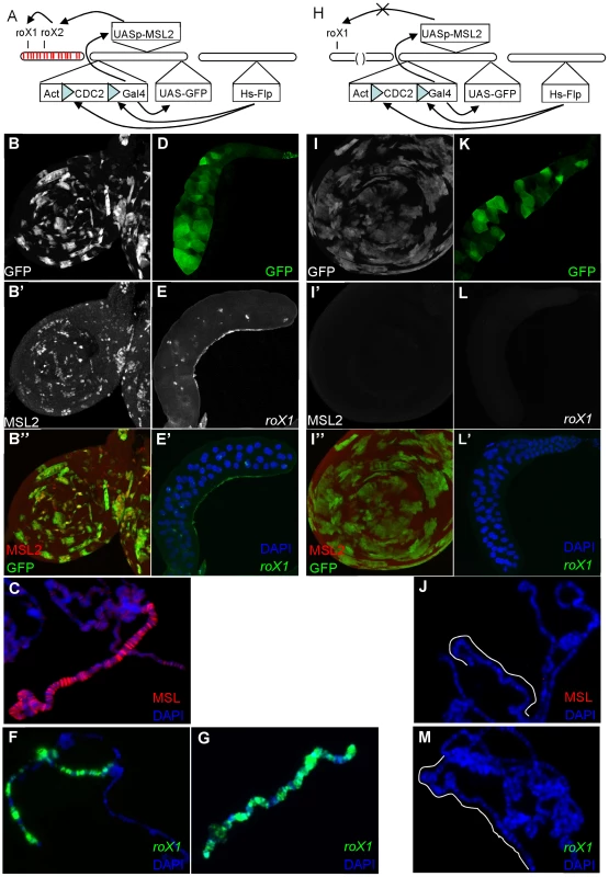
A) 4 day old larvae were heatshocked to induce expression of Flp, resulting in the removal of the blocking sequence from GAL4 and subsequent expression of both MSL2 and GFP. MSL2 is expected to initiate roX transcription and MSL complex assembly. (B) GFP+ clones mark imaginal disc cells that have successfully removed the blocking sequences from GAL4 (B′–B″). Induction of MSL2 results in punctate subnuclear foci in imaginal disc cells. (C) MSL2 immunostaining of polytene chromosome shows late MSL2 paints the entire X chromosome. (D) Whole salivary gland showing GFP induced in some cells. (E–E′) roX1 FISH of whole mount of similar GFP+ salivary glands or (F) polytene squashes shows successful induction of roX1 expression in a subset of cells. (G) roX1 FISH of wildtype males (H) The same experiment was repeated in roX1+roX2− larvae. However, in the absence of roX2, MSL2 fails to drive roX1 expression. (I) Despite the presence of GFP+ (late MSL2 expressing) cells, MSL2 is not detectable over the X in (I′–I″) imaginal disc cells or (J) polytene chromosomes. (K) Whole salivary gland showing successful GFP expression in roX1+roX2− larvae. (L–L′) Expression of roX1 is never observed painting the X or as nascent transcripts at band 3F in separately processed GFP+ glands or on (M) polytene squashes. We repeated the experiment in roX1+ roX2− females (Figure 1H). While GFP+ clones were recovered at similar frequencies indicating successful MSL2 induction (Figure 1I, 1K), no MSL2 accumulation was observed in imaginal discs (Figure 1I′) or on polytene chromosomes (Figure 1J). We will later demonstrate that the failure to detect MSL2 is due to reduced protein stability in the absence of roX RNA. More importantly, we also could not detect roX1 expression in any cell. Absence of roX1 RNA might be attributed to poor RNA stability or transcription failure. We favor the latter since even minute amounts of roX1 transcription can be readily detected when MSL complex accumulates over the roX1 gene [13]. Moreover, we could not detect nascent transcripts from the roX1 locus in these animals although such nascent roX1 transcripts were easily detected in other genotypes (Figure 1L–1L′, 1M). This argues that although late MSL2 readily switches on roX2, it is not sufficient alone or with the other MSL subunits to drive expression of the roX1 gene when roX2 is absent. Without any roX RNA, cells rapidly destroy the ectopic MSL2 as well.
To further test our hypothesis that late expression of the endogenous roX1 locus depends on roX2 RNA, we returned to wildtype (roX1+ roX2+) females to perform RNA in situ hybridization for both RNAs in the same nuclei. Late induction of MSL2 results in roX2 RNA painting the length of X chromosomes in many cells (Figure 2A′). By contrast, roX1 painting over the full X chromosome was seen in only a small minority of nuclei (Figure 2A). A much more common pattern was roX2 over the entire X while roX1 expression confined to either several Mbp (Figure 2B–2B′) around or just at the endogenous roX1 locus at polytene band 3F (Figure 2C–2C′). This suggests that roX2 expression reliably follows MSL2 induction, but roX1 expression lags. Delayed roX1 expression might be explained if fully functional MSL complex containing roX2 RNA must first assemble before transcription of roX1 can occur. We conclude that roX2 RNA, presumably packaged in mature MSL complexes, is necessary to initiate transcription from the endogenous roX1 gene late in development.
Fig. 2. Late induction of roX1 expression requires roX2 RNA. 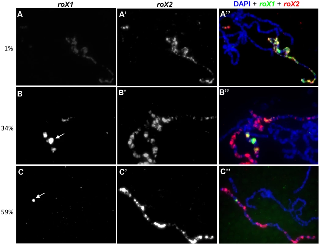
In nuclei where dosage compensation was successfully turned on after late msl2 induction, extensive roX2 was observed painting the entire X chromosome. (A) However, only 1% of the chromosomes showed extensive roX1 painting. 34% and 59% of chromosomes showed roX1 expression confined to several Mbp around (B) or just at the endogenous roX1 locus (C), respectively. The remaining chromosomes (6%) had no roX1 expression despite the presence of roX2 (data not shown). roX1 and roX2 were detected by biotin (green, A–C) and digoxigenin (red, A′–C′) labeled antisense riboprobes, respectively. The merged figure is shown in A″–C″. White arrows denote the endogenous roX1 locus at band 3F. Autoregulation of roX1 expression
Finding unusually late activation of the roX1 gene required preexisting roX2 RNA, we wondered if roX1 RNA could also perform the same role leading to a positive autoregulatory loop. To answer this question, we used a fly stock displaying an unusual mosaic pattern of dosage compensation.
The H83M2 transgene makes MSL2 constitutively using the hsp83 promoter [30]. It lacks the regulatory 5′ and 3′ msl2 UTRs and drives ectopic dosage compensation in 100% of female cells. Females carrying a roX1 deletion also showed MSL X chromosome painting utilizing roX2 RNA in all nuclei (data not shown). However, an entirely different result was obtained in H83M2 females missing only roX2. Roughly half the nuclei adopted a fully male-like pattern of dosage compensation utilizing roX1 RNA, while the other half lacked dosage compensation (Figure 3A and personal communication Art Alekseyenko). The X chromosomes of these negative cells showed very weak staining for MSL2, similar to the situation found in roX1 roX2 double mutant animals [17]. Males also exhibited a similar mosaic phenotype when their only source of MSL2 was the H83M2 transgene demonstrating that failed dosage compensation cannot be due to SXL or some other female factor (Figure 3B). It is unclear how two adjacent cells that are genetically identical containing a full set of MSL subunits adopt opposite dosage compensation fates. However, this fortuitous observation allowed us to test whether failure to activate the endogenous roX1 gene might explain lack of dosage compensation in some cells.
Fig. 3. roX1 RNA is needed to sustain endogenous roX1 transcription in males. 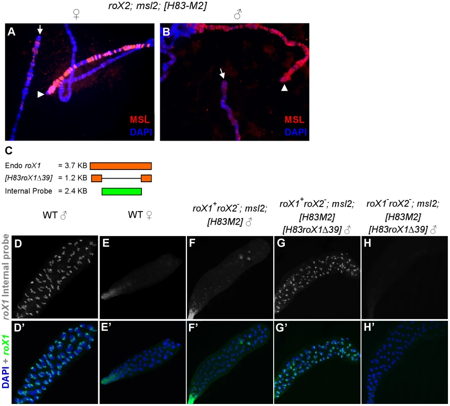
X chromosomes from neighboring cells display a mosaic pattern in which the MSL complex either succeeded (arrowhead) or failed (arrow) to paint the X from roX2; msl2; H83M2 female (A) and male (B) salivary glands. (C) Endogenous roX1 and H83roX1Δ39 transcripts (Orange) and antisense riboprobe recognizing only full length roX1 (green). Whole mount roX1 FISH using the internal probe on salivary glands from (D) wild type male, (E) wild type female, (F) roX1+ roX2−/Y; msl2; H83M2 mosaic male, (G) roX1+roX2−/Y; msl2; H83M2 H83-roX1Δ39/+ male, (H) roX1− roX2 −/Y; msl2; H83M2 H83-roX1Δ39/+ male. The X chromosomes in G are fully painted in all cells with MSL complex relying upon roX1-Δ39 RNA (Figure S1D), but the truncated roX1 RNA is not recognized by the internal probe. In order to test whether pre-existing roX1 RNA is necessary to drive continued transcription of the roX1 gene, we used a 1.2 kb roX1 minigene, H83roX1-Δ39, that is able to form partially active MSL complexes (Figure 3C) [13]. We designed a probe that recognizes only the internal sequence of the endogenous roX1 RNA missing from H83roX1-Δ39. In this way, we could selectively visualize the expression of only the endogenous roX1 RNA in animals also making the shorter transgenic H83roX1-Δ39 RNA (Figure 3D, 3H and Figure S1C). The roX2; H83M2 males (roX1+ roX2−/Y; msl2−; H83M2/+) displayed the same mosaic roX1 pattern (Figure 3F) typically seen in polytene squashes (Figure 3A and 3B). When we introduced H83roX1-Δ39 into these roX2 H83M2 males, the expression of full-length roX1 RNA made from the endogenous locus was restored to all cells (Figure 3G and Figure S1B, lane 3). In addition, while the distribution of roX1 RNA was often limited to a single band or several Mbp around 3F in nuclei from the mosaic H83M2 males (Figure S1E–S1F), the endogenous, full-length roX1 RNA coated the entire length of the X chromosome in nearly all cells when the constitutively expressed H83roX1-Δ39 transgene was also present, (Figure S1G). The H83roX1-Δ39 transgene was ineffective in H83M2 females (Figure S1B, lane 4), perhaps because females have two X chromosomes, depressed MSL1 expression [36] and so require more stimulatory activity than H83roX1-Δ39 can supply.
We conclude that the unexpected mosaic pattern found in roX2 H83M2 animals results from a failure of the wild type roX1 gene to respond to MSL proteins. Providing a constitutive source of roX1 RNA is sufficient to reliably drive transcription of the endogenous wild type roX1 gene. Taken together, these results support an autoregulatory model where new transcription of the wild type roX1 gene requires pre-existing RNA, either roX1 or roX2, in addition to MSL proteins. We postulate that the early MSL-independent burst of roX1 RNA made at blastoderm normally assembles the first MSL complexes needed to set up the future maintenance of roX1 transcription in adult males.
The mosaic expression of roX1 is not limited to the polytene chromosomes
The mosaic pattern of dosage compensation described above has been reported before. Certain hypomorphic alleles of Sxl unreliably initiate sex determination and so contain a mixture of XXAA cells that either correctly adopt a female fate repressing dosage compensation, or mistakenly choose a male fate and paint their X chromosomes with MSL complex [37]. The mechanism underlying the mosaic pattern of dosage compensation found in roX2 H83M2 animals studied here must be different, and we set out to understand its basis.
We first tested if the mosaic pattern was a peculiarity of polytene cells or a general feature in most tissues of these animals. When we performed MSL2 immunostaining on diploid imaginal discs, MSL2 protein decorated a subnuclear domain presumed to be the X chromosome in all the imaginal disc cells when both roX RNAs were present in female cells carrying the H83M2 transgene (Figure 4A). Again, roX1− roX2+ animals also had no trouble establishing dosage compensation in all the cells (data not shown). However, when roX2 was deleted, only a fraction of the cells displayed dosage compensation utilizing roX1 RNA (Figure 4B–4B″), similar to the spotty pattern observed in salivary glands (Figure 3A, 3B and 3F). When both roX genes were deleted, no MSL2 staining was detected (Figure 4C–4C″). We had expected that removing roX RNA would produce a diffuse nuclear cloud of MSL2 protein unable to bind to the X chromosome. Either diffuse MSL2 protein stains too weakly to detect with our antibodies, or MSL2 protein is unstable without roX RNA. The latter is likely to be the case since MSL2 was previously shown to be unstable when not packaged into MSL complexes [29]. To directly test this interpretation, we incubated the imaginal discs and salivary glands with MG132, a proteasome inhibitor, before performing MSL2 immunostaining. Strong nuclear MSL2 staining reappeared (Figure 4D–4D″) in the imaginal disc, showing that MSL2 is synthesized in the absence of roX RNA, but fails to accumulate due to rapid turnover. MSL2 protein stabilized by MG132 showed a dramatically different staining pattern covering all polytene chromosomes, rather than being restricted to the X (Figure S2). This shows that free MSL2 subunits have a general affinity for all chromatin in agreement with earlier biochemical work [22]. Apparently flies have evolved a mechanism to efficiently destroy any MSL2 subunits that fail to assemble into complexes with roX RNA. A somewhat similar situation was reported for the MSL1 subunit. Massively over expressed MSL1 transiently paints all chromosome arms but is quickly lost [36].
Fig. 4. Dosage compensation fails in many diploid cells relying exclusively on roX1 and H83M2. 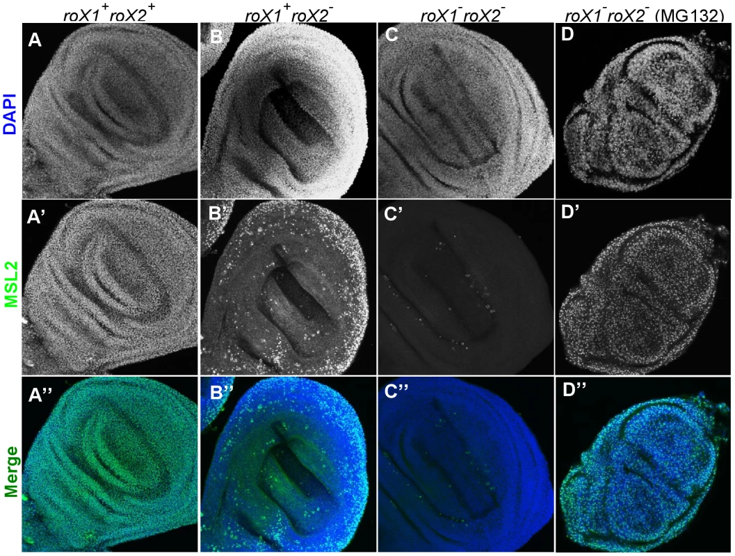
(A–A″) Males and females relying entirely on H83M2 showed subnuclear MSL2 staining in all wing imaginal disc cells when both roX1 and roX2 RNAs were present. (B–B″) Many cells lacked dosage compensation if roX1+ was the only source of roX RNA. (C–C″) MSL2 did not accumulate over the X when both roX1 and roX2 were absent. (D–D″) Nuclear staining of MSL2 was easily detected in the absence of roX RNA after treatment with MG132, a proteasome inhibitor. Our data suggest that many cells lacking roX2 RNA were unable to carry out dosage compensation because the remaining wild type roX1 gene failed to switch on. If true, this should affect the viability of whole animals. As previously reported, ectopic MSL2 produced by H83M2 is toxic to females (Figure 5A) [30] and toxicity requires roX RNA (Figure 5B) [17]. We found that roX2 alone was almost equally toxic to females as when both RNAs were present (compare Figure 5A–5C). The surviving adults produced very few eggs and were sterile. However, roX1 alone was much less toxic to females (Figure 5D) as would be expected if many cells lacked dosage compensation and thus escaped the toxic effects of MSL2 production. These surviving females were fertile. We conclude that the mosaic dosage compensation pattern is not limited to polytene tissues, but instead reflects a widespread failure of MSL2 protein made by the constitutive H83M2 transgene to activate the endogenous roX1 gene.
Fig. 5. roX2 mutant females escape the toxic effects of H83M2. 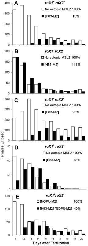
Adult females eclosing each day were counted from a population of eggs laid on day one. White bars show non-transgenic females without ectopic MSL2 (except E), and black bars show females carrying the H83M2 transgene. Each experiment varied the roX genotype. (A) roX1+ roX2+, (B) no roX, (C) roX2+ only, (D) roX1+ only, (E) roX1+ only. Cumulative viability of H83M2 females is shown as a percentage of their non-transgenic sisters. H83M2 does not turn on the expression of roX1 reliably
Males that lack roX2, but are in all other ways wild type, have normal dosage compensation in all cells utilizing the remaining wild type roX1 gene [17]. Finding spotty roX1 activation only in animals relying on H83M2 prompted us to examine why the transgene did not behave like the endogenous msl2 locus in only this unusual genetic background. MSL2 made from the endogenous locus must activate roX1 RNA production more effectively than transgenic MSL2. This was shown by introducing single wild type allele of endogenous msl2 to roX2; H83M2 males and observing dosage compensation in all cells (Figure S3G and S3I lane 5, Figure S4E and S4G lane 5). Next, we turned to the NOPU-M2 transgene that also escapes SXL repression in females, but differs from H83M2 by using the endogenous msl2 promoter [21]. NOPU-M2 is not toxic to females because it makes less MSL2 compared to H83M2. However, addition of the NOPU-M2 transgene to roX2 mutant females makes them sensitive to the toxic effects of H83M2 (Figure 5E). Females carrying both msl2 transgenes also drive activation of roX1 expression in all cells (Figure S3H and S3I lane 6, Figure S4F and G lane 6). This is unlikely to be a simple additive effect of two pools of MSL2 protein since the H83M2 transgene itself was fully capable of driving roX1 expression in 100% of the cells when roX2 was present (Figure S3C–S3D, and S3I lane 1–2 and Figure S4A–S4B and S4G lane 1–2). We propose that H83M2 differs from the endogenous msl2 gene in some way needed to reliably drive the roX1 autoregulatory loop.
The mosaic pattern might arise from the H83M2 transgene suffering from positional effect so MSL2 protein was not made in some cells. This is not the case since MSL2 expression can be clearly observed in all cells when both roX RNAs are present (Figure 4A). A second possibility is that H83M2 initiates expression too late in embryogenesis to capture the early MSL-independent roX1 transcripts we postulate are needed to begin the autoregulatory loop. We performed both MSL2 immunostaining and roX1 FISH on roX2; msl2; H83M2 embryos from 2 h to 20 h AEL. These embryos showed H83M2 expression and subnuclear punctate roX1 accumulation in essentially all cells at early gastrulation and remained on through at least until 20 hrs AEL when cuticle formation prevents reliable staining of internal tissues (Figure S5 and Figure S6). These results suggest that H83M2 does come on during early embryogenesis and is effective in driving roX1 transcription during the rapid cell divisions of embryonic development. However, many cells subsequently lose roX1 transcription, and thus dosage compensation, later during larval stages.
Cell cycle regulation of msl2 expression
The MSL complex associates with many hundreds of actively transcribed genes along the male X. We considered the possibility that like core histone proteins, the MSL complex might need to abruptly increase its abundance following DNA replication. We set out to test if the transcription of msl2 might be coupled to S phase.
We first used in situ hybridization to directly visualize actively growing msl2 transcripts on polytene chromosomes. Most salivary gland cells have completed their endoreplication cycles at the wandering larval stage used in all our experiments, but a few cells are still actively replicating. When males carrying both the wild type endogenous msl2 gene and the H83M2 transgene were treated with antisense msl2 riboprobes, all nuclei showed strong hybridization to a cloud of msl2 RNA over the H83M2 transgene inserted at 87A (Figure 6A). By contrast, a weaker hybridization signal was observed over the endogenous msl2 gene at 23F in only about 20% of the nuclei. Most nuclei lacked detectable msl2 transcripts at the endogenous locus. We tested whether the H83M2 transgene might somehow inhibit expression of endogenous msl2 gene by testing nontransgenic, wild type males. We found that again, only about 20% of male cells actively transcribed msl2 (data not shown). This shows that while wild type males have abundant MSL2 protein painting the X chromosome in all cells, few cells are making new msl2 mRNA.
Fig. 6. Transcription of msl2 is correlated with cell cycle. 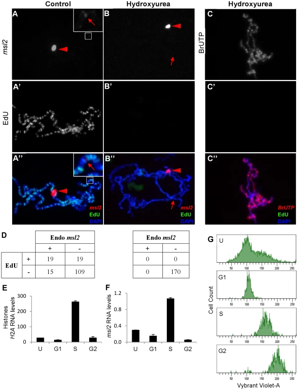
Nascent msl2 transcripts were detected with antisense FISH riboprobe in salivary glands. (A) Transgenic H83M2 expression (bright signal indicated by red triangle) was observed at the 87A insertion site in all nuclei. Hybridization to the endogenous msl2 locus at 23F (faint band indicated by red arrow in inset) was seen in only a minority of nuclei. Because of the difference in signal intensities between the two msl2 loci, the inset is enhanced to show the weaker signal. (A′) EdU incorporation shows that this is one of the few nuclei undergoing endoreplication. (B) After treatment with 1 mM of HU (hydroxyurea), no cell transcribed the endogenous msl2 gene (red arrow) but the transcription of H83M2 continued (red triangle). (B′) HU blocked EdU incorporation from any cell. (C) Simultaneous treatment of salivary glands with HU, BrUTP, and EdU showed that blocking replication did not inhibit bulk transcription in these cells. (D) Many nuclei without (left table, N = 162) or with (right table, N = 170) HU treatment were scored for expression of endogenous msl2 and EdU incorporation. Nascent transcripts were detected at the H83M2 transgene in 100% of the nuclei (data not represented in the table). After sorting growing S2 cells into their respective phase of cell cycle via FACS, qPCR was done to quantify H2A (E) and msl2 (F) transcripts levels normalized to PKA. (G) The FACS profile of unsorted (U) and sorted S2 cells (G1, S and G2 cell cycle). The sorted cells have a slightly higher content of Vybrant Violet-A dye because the cells are collected into tubes containing Vybrant Violet-A. The Y-axes are drawn on different scales. To test if the sporadic transcription of endogenous msl2 might coincide with the replication phase of the cell cycle, we briefly incubated dissected salivary glands in EdU to label newly replicated DNA, fixed the glands, and processed them for msl2 FISH. We found a strong bias (Fisher's exact test p<0.0001) for replicating cells to also be transcribing msl2, but the overlap was not perfect (Figure 6A). For comparison we also examined histone H2A transcription, but even outside S phase every nucleus contained an intense hybridization signal over the endogenous histone gene cluster at polytene band 39DE (Figure S7). Only a portion of histone mRNA regulation occurs at transcription initiation. Most regulation is posttranscriptional [38], [39].
To directly test whether the sporadic msl2 transcription pattern is associated with S phase, we treated our salivary glands with 1 mM of hydroxyurea (HU) to inhibit replication. EdU incorporation was completely blocked (Figure 6B′) but more significantly, transcripts were specifically lost only from the endogenous msl2 locus at 23F while msl2 transcripts made from the H83M2 transgene at 87A remained strong (Figure 6B, 6B″). Likewise, HU treatment had no effect on bulk nascent transcripts as shown by strong incorporation of BrUTP on all chromosome arms (Figure 6C). These results show that msl2 transcription normally occurs around S phase and is blocked by replication inhibitors. By contrast, H83M2 does not show any cell cycle regulation. This difference might impact roX1 expression.
To test whether the replication associated msl2 transcription might be limited to only polytene cells with unusual cell cycles, we examined mitotically dividing diploid S2 cell in culture. Actively growing S2 cells were sorted into G1, S, and G2 (Figure 6G) via FACS (Fluorescence Activated Cell Sorting). RNA from these populations was assayed for msl2, histone H2A, and PKA transcripts by qPCR. As previously reported [38], histone RNA accumulates during S phase but is low in G1 and G2 (Figure 6E). Importantly, msl2 transcripts are most abundant during S while lowest during G2 phase (Figure 6F). These results show that msl2 RNA accumulation and synthesis normally occur predominantly in S phase, but the H83M2 transgene lacks this coordination with replication.
We attempted to determine whether roX1 transcription was also linked to S phase as might be expected, but were unsuccessful. We used both BrUTP and EU (Ethynyl Uridine) to label all new transcripts followed by a cold chase. We anticipated that the bulk of mRNA would leave the nucleus while newly labeled roX1 RNA remained behind on the X. However, we were unable to detect any labeled RNA localized on the male X after chase (data not shown). We do not know if the signal was too weak to detect, or if the substitutions at the 5 position of uridine interferes with the function of RNA [40], leading to problems in roX1 RNA folding, stability, MSL protein assembly, and/or targeting to the X.
Discussion
Previous studies of roX1 transcriptional control argued that either MSL2 alone or with a full set of MSL proteins was sufficient to drive male-specific expression [31]–[33]. Here, we present evidence that the expression of roX1 gene is instead controlled through an autoregulatory loop. Pre-existing roX RNA, presumably in mature MSL complex, is required to drive new transcription. The reason we reached a different conclusion is largely attributed to removing the functionally redundant roX2 in most of our experiments and assaying transcription only from the wild type roX1 locus at its normal location on the X. The pathway we describe shares some elements with the negative regulatory loop between TFIIIA binding 5S rDNA to drive transcription or 5S rRNA for storage during Xenopus oogenesis [41].
A model for autoregulation of roX1
Figure 7 shows a model for how such an autoregulatory loop might operate. Because roX RNA is not maternally deposited into embryos, one problem is how male embryos could build their first MSL complex needed to initiate the cycle. Meller has shown that roX1 transcription switches on in both sexes around blastoderm, just as general zygotic transcription begins [15], [16]. This suggests that an embryonic roX1 promoter is active without MSL complex and could supply the first roX1 RNA molecules to males, but these RNAs are eventually degraded in females. Early RNA assembles with MSL proteins and then drive roX1 transcription from the known male-specific MSL-dependent promoters, setting up a positive autoregulatory loop necessary for the future maintenance of roX1 expression in males. In our experiments, we showed that roX2 RNA or truncated roX1 RNA can also initiate endogenous roX1 expression late in development after the early roX1 transcripts are gone.
Fig. 7. Autoregulation model. 
The earliest roX1 transcripts (red) made at blastoderm originate from an uncharacterized MSL-independent promoter. This RNA may assemble with MSL protein subunits to produce the first functional MSL complexes needed to bind the internal DHS enhancer that drives sustained transcription (blue) from the male-specific promoters. When present, roX2 RNA can also drive roX1 transcription. Components of the replication pre-initiation complex also bind the DHS sequence in male cells (Figure S8A). The msl2 transcripts are made predominantly during replication and new MSL2 protein is needed to assemble and stabilize newly made roX1 RNA. This model requires that male embryos preferentially sequester newly assembled MSL complex at the roX1 gene to drive sustained transcription instead of allowing it to diffuse away to the vastly larger pool of ordinary X linked genes that must be dosage compensated. Only after roX1 transcription is successfully upregulated can MSL complexes be released to the target genes along the X chromosome. Such behavior has been previously documented in cells containing abundant free MSL subunits and low levels of roX1 transcription, exactly the conditions we believe occur as young male embryos initiate dosage compensation [13]. Examining cells shortly after MSL2 first turned on roX1 transcription showed the earliest roX1 transcripts remained near the site of synthesis (Figure 2B and 2C) consistent with the idea that newly formed MSL complexes preferentially act on the roX1 gene. At later times, such as seen in H83roX1-Δ39 animals that have had five days to drive endogenous roX1 expression, every cell painted the entire X with roX1 RNA (Figure 3G). While massive local cis spreading of the MSL complex has been reported for roX1 and roX2 transgenes inserted into autosomes [13], [18], [42], the physiological relevance of this is not widely accepted [25], [26], [43], [44]. Here we instead report striking local cis spreading of newly made MSL complex from the wild type roX1 gene in its normal X chromosome environment (Figure S1E–S1F). Our data agree well with previous reports of local MSL spreading along the X [19] and support a role for cis spreading in the normal process of dosage compensation.
We have not directly determined what region of the roX1 gene is necessary for autoregulation. However, a strong candidate is the ∼200 bp male-specific DNase I hypersensitive site (DHS) found about 1.5 kb downstream of the adult roX1 promoters. The DHS is sufficient to recruit the MSL complex to ectopic sites when moved to the autosomes in a sequence specific manner [45], [46]. While partial complexes lacking MOF or MSL3 remain bound to the DHS, incomplete complexes lacking either roX RNA or the MLE RNA helicase postulated to fold roX RNA bind the DHS poorly [45], [46]. This element stimulates roX1 transcription when MSL complex is bound and represses basal transcription when MSL complex is absent. Also, deleting the DHS greatly reduces transcription of roX1 transgenes [33]. Together, these findings support a model where transcription from the roX1 gene requires pre-existing roX RNA within MSL complexes bound to the internal DHS enhancer. However, this view is likely an oversimplification because very large internal deletions such as roX1ex7B, comparable to our H83roX1-Δ39 transgene (Figure 3A), remain transcriptionally active despite the loss of the DHS enhancer [32], [47].
Cell cycle regulation
Translation of msl2 mRNA is normally subject to elaborate controls acting through the 5′ and 3′ UTRs [48]–[52]. Little attention has been given to its transcription control, although recently anti-MSL2 antibodies were found to precipitate msl2 mRNA [53]. We found that msl2 transcription is associated with replication, and this is likely important for its normal control. Without MSL2 protein, naked roX1 RNA is rapidly destroyed [11]. Here we found the converse; cells rapidly clear any MSL2 protein not bound to roX RNA. When free MSL2 subunits are artificially stabilized with proteosome inhibitors, they coat all the chromosomes indiscriminately. This implies that the synthesis of MSL2 and roX RNAs are closely coordinated so each component stabilizes the other ensuring that only correctly targeted molecules survive. Although MSL2 lacks known RNA binding motifs, previous work of others is consistent with an intimate interaction between MSL2 and roX RNA [31]. We suspect the loss of replication-coupled transcription may contribute to the failure of dosage compensation in some cells relying exclusively on H83M2 for MSL2 protein and roX1 for roX RNA. This defect is corrected when both roX1 and MSL2 are coordinately made from the same hsp83 promoter (Figure 3G). Cells in mosaic animals lose dosage compensation sometime between the end of embryogenesis and third instar larvae. Many tissues undergo significant changes in cell cycle near the end of embryogenesis, particularly the introduction of G1 [54], and we suspect this shift contributes to loss of dosage compensation in our mosaic animals. If MSL complex should ever drop below the level needed to sustain the autoregulatory roX1 loop, it could never recover regardless of later MSL2 production. The details remain unclear because H83M2 is vigorously transcribed at the developmental times we examined, including S phase. We do not know whether the regulatory msl2 UTR sequences removed from H83M2 disrupt additional posttranscriptional controls that might promote efficient translation during replication.
A second issue is that the X is painted with MSL complex throughout the cell cycle. This might drive continuous rather than cyclic roX1 synthesis. While we were unable to directly determine if roX1 transcription is cell cycle regulated, we note that the replication machinery components ORC2 and MCM are bound specifically to the roX1 DHS enhancer only in male cells (Figure S8A). The significance of this is not known, but it is tempting to speculate that components of the pre-initiation complex bound to DHS compete with MSL complex thus inhibiting roX1 transcription in G1. Firing of the replication origins removes ORC2 and MCM possibly allowing MSL complex access to the DHS and so switch on transcription shortly after the onset of S phase. The replication machinery is commonly found near the promoters of many genes [55], including msl2 (Figure S8B), so further experiments will be needed to determine if such binding actually plays any regulatory role here.
Regulation of roX2
While we did not specifically address transcriptional control of roX2, it must differ from roX1 in several important ways. Meller has previously shown that roX2 transcription lags roX1 by a few hours during early embryonic development and is always limited to males [16]. Here we found that roX2 transcription differs by not requiring pre-existing roX RNA and can be switched on several days later during larval development simply by ectopically expressing MSL2. MSL2 protein made by the constitutive H83M2 transgene only sporadically activates roX1, but robustly drives roX2. The roX2 gene also carries a DHS enhancer similar to that found in roX1 [45], but if it plays a comparable role in roX2 regulation, it presumably would not require complete MSL complex. The region near the proline rich domain towards the C-terminus of MSL2 is essential for this regulation [31].
Parallels between roX1 and Sxl
The roX1 autoregulation loop described above shares parallels to the SXL autoregulatory loop controlling all aspects of sex determination and dosage compensation in Drosophila. X∶A counting elements act upon an early establishment Sxl promoter to make SXL in early female embryos. These first SXL proteins stimulate productive splicing of Sxl mRNAs transcribed from a distinct maintenance promoter ensuring further SXL production [56]. MSL1, MSL3, MOF, and MLE are unable to package early roX1 RNA made from the embryonic promoter in female embryos to form a fully functional mature MSL complex. To do that, MSL2 protein made only in males is required. We suspect these earliest MSL complexes sustain roX1 transcription as embryos switch to the MSL-dependent promoters. Recently a new role for MSL2 in females has been described during the brief window when Sxl autoregulation is established [57]. Perhaps females also fleetingly utilize the early burst of roX1 before abundant SXL represses msl2 translation.
Thousands of large noncoding RNAs have recently been discovered in vertebrates [2], [58], many of which are associated with chromatin remodeling enzymes [1], [59]–[61]. It is likely that some of these will face similar regulatory and functional demands as the roX RNAs and may have evolved comparable strategies to control their production. For instance, the short RepA sequence at the 5′ end of mammalian Xist RNA may influence production of full length transcripts [59].
Materials and Methods
Drosophila stocks
Larvae and flies were raised on standard cornmeal-yeast-agar-molasses medium containing propanoic acid at 25°C. In all experiments the roX1 mutation is roX1ex6 and the roX2 allele is Df (1) roX252 [17]. The [w+ 4Δ4.3] transgene supplies essential adjacent genes lost in the roX2 deletion.
The transgenic flies used have been previously described as follows: [w+H83M2-6I] [30], [w+ NOPU-M2] [21], and [w+ H83roX1-Δ39] [13]. The H83M2 transgene was recombined with [P{SUPor-P}KG02776] which contains y+ as a marker so larvae of the correct genotype could be recognized by mouth hook color (Flybase). The P{GAL4-Act5C(FRT.CD2).P}, P{UAS-GFP.S65T} and MKRS-hsFLP transgenic fly stocks were provided by Graeme Mardon.
The UASp-MSL2 transgene was made by digesting the H83M2 transgene with EcoRI and subcloning the MSL2 ORF into the pUAS-P2 plamid vector (a gift from Pernille Rørth).
Heatshock
The female larvae for the flp-out experiment were kept at 25°C and heatshocked at 37°C for 1 hr each at day 4 and 5 AEL. Their salivary glands were then dissected on day 6 AEL, fixed and immunostained as described below.
The full genotypes of the larvae heatshocked in Figure 1 are as follows:
B-F) y w; [w+ y+Act-FRT-CDC2-FRT-Gal4] [w+UAS-GFP] +/+ + [w+UASp-MSL2]; MRKS [Hs-Flp]/+
H-L) y w roX252 [w+4Δ4.3]; [w+ y+Act-FRT-CDC2-FRT-Gal4] [w+UAS-GFP] +/+ + [w+UASp-MSL2]; MRKS [Hs-flp]/+
The full genotypes of the larvae in Figure 2 are identical to Figure 1H–1L.
The full genotypes of the larvae in Figure 3 are as follows:
A) y w roX252 [w+4Δ4.3]; msl2; [w+H83-M2] [w+y+P{SUPor-P}KG02776]/+ +
B) y w roX252 [w+4Δ4.3]/Y; msl2; [w+H83-M2] [w+y+P{SUPor-P}KG02776]/+ +
D) y w/Y
E) y w
F) y w roX252 [w+4Δ4.3]/Y; msl2; [w+H83-M2] [w+y+ P{SUPor-P}KG02776]/+ +
G) y w roX252 [w+4Δ4.3]/Y; msl2; [w+H83-M2] [w+H83-roX1Δ39] [w+y+ P{SUPor-P}KG02776]/+ + +
H) y w roX1ex6roX252 [w+4Δ4.3]/Y; msl2; [w+H83-M2] [w+H83-roX1Δ39] [w+y+ P{SUPor-P}KG02776]/+ + +
The full genotypes of the larvae in Figure 4 are as follows:
A-A″) y w; msl2; [w+H83-M2] [w+y+ P{SUPor-P}KG02776]
B-B″) y w roX252 [w+4Δ4.3]/Y; msl2; [w+H83-M2] [w+y+P{SUPor-P}KG02776]/+ +
C-C″) y w roX1ex6 roX252 [w+4Δ4.3]/Y; msl2/+; [w+H83-M2] [w+y+P{SUPor-P}KG02776]/+ +
D-D″) Same as C
The full genotypes of the parents in the crosses for Figure 5A–5E are as follows:
A) y w/Y; msl2; [w+H83-M2] [w+y+ P{SUPor-P}KG02776]/+ + X y w; msl2
B) y w roX1ex6/Y; msl2; [w+H83-M2] [w+y+ P{SUPor-P}KG02776]/+ + X y w roX1ex6; msl2
C) y w roX252 [w+4Δ4.3]/Y; msl2; [w+H83-M2] [w+y+ P{SUPor-P}KG02776]/+ + X y w roX252 [w+4Δ4.3]; msl2
D) y w roX1ex6 roX252 [w+4Δ4.3]/Y; msl2/+; [w+H83-M2] [w+y+ P{SUPor-P}KG02776] +/+ + [w+GMroX1+] X y w roX1ex6 roX252 [w+4Δ4.3]
E) y w roX252 [w+4Δ4.3]/Y; msl2; [w+H83-M2] [w+y+ P{SUPor-P}KG02776]/+ + X y w roX252 [w+4Δ4.3]; [w+NOPU-M2]
Immunohistochemistry and in situ hybridization
Immunostaining and in situ hybridization on third instar salivary glands was performed as described in [11] except for the following modification. The slides were treated with proteinase K (20 µg/ml in PBT) for 3 min. Each slide was hybridized with 5 ng of roX1, msl2 or H2A biotin-labeled single-stranded antisense riboprobes using the T7 high yield Transcription kit #K0441 (Fermentas). Fluorescent development was done as instructed by the Tyramide Signal Amplification (TSA) kit (NEL700A) (PerkinElmer) with SA-FITC (NEL720) (PerkinElmer) or SA-Texas Red (NEL 721) (PerkinElmer). For double ISH, we labeled the second antisense riboprobes with digoxygenin. Before adding the anti-digoxygenin-HRP conjugate, the first horseradish peroxidase conjugate was quenched in 3% H2O2 for 30 mins. The second color is then developed using Tyramide-Alexa488 (T20922) (Invitrogen). To inhibit MSL2 degradation, we incubate imaginal discs and salivary glands with 10 µM of MG132 (474791) (Calbiochem) dissolved in Schneider Culture Media for 3 hours before proceeding with immunostaining.
EdU labeling
To block replication, salivary glands from 3rd instar larvae were first incubated in 1 mM HU for 1 h. If not, they were simply incubated in S2 cell culture media (see below). After which, the glands were transferred and incubated in EdU for 15 minutes followed immediately by fixation. The detection was then performed as instructed by the Click-iT EdU imaging kit #C10337 (Invitrogen). Both HU and EdU were dissolved in S2 cell culture media before use.
Co-labeling replicating DNA and nascent transcripts via EdU and BrUTP respectively
Salivary glands were initially treated the same way as described above. However, after HU treatment, the glands were incubated in a BrUTP/DOTAP/EdU mixture instead for 15 mins. The BrUTP/DOTAP mixture was previously described to allow efficient nucleotide triphosphate uptake by the cell to label nascent transcripts [62]. EdU detection was performed as described above. Rat Anti-BrdU monoclonal antibodies #NB500-169 (Novus Biological) and Goat anti-rat Alexa 594 #A11007 (Invitrogen) were then used as primary and secondary antibodies respectively, to detect BrUTP.
Cell culture and FACS
Schneider 2 (S2) cells were cultured in 15-cm plates at a density of 1×10⁶ cells/ml in Schneider's Media #11720034 (Invitrogen) supplemented with 10% FCS and penicillin/streptomycin. After reaching a density of 5×10⁶ cells/ml, the cells were re-suspended in new culture media containing 5 µM of Vybrant Dye Cycle Violet #V35003 (Invitrogen) to stain the DNA. The cells were then sorted into their respective phase of cell cycle via FACSAria II (BD Biosciences) at the Cytometry and Cell Sorting Facility located in Baylor College of Medicine.
Real-time PCR
Total RNA was extracted from cells using Trizol® reagent 15596-018 (Invitrogen) as per the manufacturer's protocol. cDNA was synthesized from 0.5–1 µg of total RNA using random hexamers and MMLV Reverse transcriptase #M1701 (Promega) as per manufacturer's protocol. The cDNA was purified using MinElute PCR kit #28004 (Qiagen).
Real-time PCR was performed using an Applied Biosystems 7300 Sequence Detection system. The 25 µl PCR included 1 µl cDNA, 1× SYBR® Green PCR Master Mix #4309155 (Applied Biosystems) and 1 µl of gene specifc primers. The reactions were incubated in a 96-well optical plate at 95°C for 10 min, followed by 40 cycles of 95°C for 15 s and 60° for 10 min. The Ct data was determinate using default threshold settings. The threshold cycle (Ct) is defined as the fractional cycle number at which the fluorescence passes the fixed threshold. PKA is used as an endogenous standard for normalization of histone H2A and msl2. The primers used for qPCR are: H2A forward TGGACGTGGAAAAGGTGGCA; H2A reverse ACGGCAGCTAGGTAAACTGGAG; MSL2 forward GGCGAGTACCAGGGCTTCAATATC; MSL2 Reverse TGCTGCAGCTGGACACGAATAG; PKA forward – AGCCGCACTCGCGCTTCTAC and PKA reverse - AGCCGGAGAATCTGCTGATTG.
Supporting Information
Zdroje
1. KhalilAMGuttmanMHuarteMGarberMRajA 2009 Many human large intergenic noncoding RNAs associate with chromatin-modifying complexes and affect gene expression. Proc Natl Acad Sci U S A 106 11667 11672
2. OromUADerrienTBeringerMGumireddyKGardiniA 2010 Long noncoding RNAs with enhancer-like function in human cells. Cell 143 46 58
3. DengXMellerVH 2006 Non-coding RNA in fly dosage compensation. Trends Biochem Sci 31 526 532
4. GelbartMKurodaM 2009 Drosophila dosage compensation: a complex voyage to the X chromosome. Development 136 1399 1410
5. HilfikerAHilfiker-KleinerDPannutiALucchesiJC 1997 mof, a putative acetyl transferase gene related to the Tip60 and MOZ human genes and to the SAS genes of yeast, is required for dosage compensation in Drosophila. EMBO J 16 2054 2060
6. AkhtarABeckerPB 2000 Activation of transcription through histone H4 acetylation by MOF, an acetyltransferase essential for dosage compensation in Drosophila. Mol Cell 5 367 375
7. BoneJRLavenderJRichmanRPalmerMJTurnerBM 1994 Acetylated histone H4 on the male X chromosome is associated with dosage compensation in Drosophila. Genes Dev 8 96 104
8. SmithERAllisCDLucchesiJC 2001 Linking global histone acetylation to the transcription enhancement of X-chromosomal genes in Drosophila males. J Biol Chem 276 31483 31486
9. WuLZeeBMWangYGarciaBADouY 2011 The RING Finger Protein MSL2 in the MOF Complex Is an E3 Ubiquitin Ligase for H2B K34 and Is Involved in Crosstalk with H3 K4 and K79 Methylation. Mol Cell 43 132 144
10. AmreinHAxelR 1997 Genes expressed in neurons of adult male Drosophila. Cell 88 459 469
11. MellerVHGordadzePRParkYChuXStuckenholzC 2000 Ordered assembly of roX RNAs into MSL complexes on the dosage-compensated X chromosome in Drosophila. Curr Biol 10 136 143
12. FrankeABakerBS 1999 The roX1 and roX2 RNAs are essential components of the compensasome, which mediates dosage compensation in Drosophila. Mol Cell 4 117 122
13. KelleyRLLeeOKShimYK 2008 Transcription rate of noncoding roX1 RNA controls local spreading of the Drosophila MSL chromatin remodeling complex. Mech Dev 125 1009 1019
14. ParkSWKangYSypulaJGChoiJOhH 2007 An evolutionarily conserved domain of roX2 RNA is sufficient for induction of H4-Lys16 acetylation on the Drosophila X chromosome. Genetics 177 1429 1437
15. MellerVHWuKHRomanGKurodaMIDavisRL 1997 roX1 RNA paints the X chromosome of male Drosophila and is regulated by the dosage compensation system. Cell 88 445 457
16. MellerVH 2003 Initiation of dosage compensation in Drosophila embryos depends on expression of the roX RNAs. Mech Dev 120 759 767
17. MellerVHRattnerBP 2002 The roX genes encode redundant male-specific lethal transcripts required for targeting of the MSL complex. EMBO J 21 1084 1091
18. ParkYKelleyRLOhHKurodaMIMellerVH 2002 Extent of chromatin spreading determined by roX RNA recruitment of MSL proteins. Science 298 1620 1623
19. OhHParkYKurodaMI 2003 Local spreading of MSL complexes from roX genes on the Drosophila X chromosome. Genes Dev 17 1334 1339
20. BashawGJBakerBS 1997 The regulation of the Drosophila msl-2 gene reveals a function for Sex-lethal in translational control. Cell 89 789 798
21. KelleyRLWangJBellLKurodaMI 1997 Sex lethal controls dosage compensation in Drosophila by a non-splicing mechanism. Nature 387 195 199
22. FauthTMuller-PlanitzFKonigCStraubTBeckerPB 2010 The DNA binding CXC domain of MSL2 is required for faithful targeting the Dosage Compensation Complex to the X chromosome. Nucleic Acids Res 38 3209 3221
23. BhadraUPal-BhadraMBirchlerJA 1999 Role of the male specific lethal (msl) genes in modifying the effects of sex chromosomal dosage in Drosophila. Genetics 152 249 268
24. BhadraUPal-BhadraMBirchlerJA 2000 Histone acetylation and gene expression analysis of Sex lethal mutants in Drosophila. Genetics 155 753 763
25. DahlsveenIKGilfillanGDShelestVILammRBeckerP 2006 Targeting Determinants of Dosage Compensation in Drosophila. PLoS Genet 2 e5 doi:10.1371/journal.pgen.0020005
26. GilfillanGDKonigCDahlsveenIKPrakouraNStraubT 2007 Cumulative contributions of weak DNA determinants to targeting the Drosophila dosage compensation complex. Nucleic Acids Res 35 3561 3572
27. GuWSzauterPLucchesiJC 1998 Targeting of MOF, a putative histone acetyl transferase, to the X chromosome of Drosophila melanogaster. Dev Genet 22 56 64
28. KindJAkhtarA 2007 Cotranscriptional recruitment of the dosage compensation complex to X-linked target genes. Genes Dev 21 2030 2040
29. LymanLMCoppsKRastelliLKelleyRLKurodaMI 1997 Drosophila male-specific lethal-2 protein: structure/function analysis and dependence on MSL-1 for chromosome association. Genetics 147 1743 1753
30. KelleyRLSolovyevaILymanLMRichmanRSolovyevV 1995 Expression of msl-2 causes assembly of dosage compensation regulators on the X chromosomes and female lethality in Drosophila. Cell 81 867 877
31. LiFSchiemannAHScottMJ 2008 Incorporation of the noncoding roX RNAs alters the chromatin-binding specificity of the Drosophila MSL1/MSL2 complex. Mol Cell Biol 28 1252 1264
32. RattnerBPMellerVH 2004 Drosophila male-specific lethal 2 protein controls sex-specific expression of the roX genes. Genetics 166 1825 1832
33. BaiXAlekseyenkoAAKurodaMI 2004 Sequence-specific targeting of MSL complex regulates transcription of the roX RNA genes. EMBO J 23 2853 2861
34. FrankeADernburgABashawGJBakerBS 1996 Evidence that MSL-mediated dosage compensation in Drosophila begins at blastoderm. Development 122 2751 2760
35. XuTHarrisonSD 1994 Mosaic analysis using FLP recombinase. Methods Cell Biol 44 655 681
36. ChangKAKurodaMI 1998 Modulation of MSL1 abundance in female Drosophila contributes to the sex specificity of dosage compensation. Genetics 150 699 709
37. PalmerMJRichmanRRichterLKurodaMI 1994 Sex-specific regulation of the male-specific lethal-1 dosage compensation gene in Drosophila. Genes Dev 8 698 706
38. SittmanDBGravesRAMarzluffWF 1983 Histone mRNA concentrations are regulated at the level of transcription and mRNA degradation. Proc Natl Acad Sci U S A 80 1849 1853
39. MarzluffWFWagnerEJDuronioRJ 2008 Metabolism and regulation of canonical histone mRNAs: life without a poly(A) tail. Nat Rev Genet 9 843 854
40. SchmittgenTDDanenbergKDHorikoshiTLenzHJDanenbergPV 1994 Effect of 5-fluoro - and 5-bromouracil substitution on the translation of human thymidylate synthase mRNA. J Biol Chem 269 16269 16275
41. WolffeAPBrownDD 1988 Developmental regulation of two 5S ribosomal RNA genes. Science 241 1626 1632
42. LarschanEAlekseyenkoAAGortchakovAAPengSLiB 2007 MSL Complex Is Attracted to Genes Marked by H3K36 Trimethylation Using a Sequence-Independent Mechanism. Mol Cell 28 121 133
43. GilfillanGDStraubTde WitEGreilFLammR 2006 Chromosome-wide gene-specific targeting of the Drosophila dosage compensation complex. Genes Dev 20 858 870
44. FagegaltierDBakerBS 2004 X Chromosome Sites Autonomously Recruit the Dosage Compensation Complex in Drosophila Males. PLoS Biol 2 e341 doi:10.1371/journal.pbio.0020341
45. ParkYMengusGBaiXKageyamaYMellerVH 2003 Sequence-specific targeting of Drosophila roX genes by the MSL dosage compensation complex. Mol Cell 11 977 986
46. KageyamaYMengusGGilfillanGKennedyHGStuckenholzC 2001 Association and spreading of the Drosophila dosage compensation complex from a discrete roX1 chromatin entry site. EMBO J 20 2236 2245
47. DengXRattnerBPSouterSMellerVH 2005 The severity of roX1 mutations is predicted by MSL localization on the X chromosome. Mech Dev 122 1094 1105
48. MedenbachJSeilerMHentzeMW 2011 Translational Control via Protein-Regulated Upstream Open Reading Frames. Cell 145 902 913
49. DuncanKGrskovicMStreinCBeckmannKNiggewegR 2006 Sex-lethal imparts a sex-specific function to UNR by recruiting it to the msl-2 mRNA 3′ UTR: translational repression for dosage compensation. Genes Dev 20 368 379
50. GrskovicMHentzeMWGebauerF 2003 A co-repressor assembly nucleated by Sex-lethal in the 3′UTR mediates translational control of Drosophila msl-2 mRNA. EMBO J 22 5571 5581
51. GebauerFGrskovicMHentzeMW 2003 Drosophila Sex-lethal inhibits the stable association of the 40S ribosomal subunit with msl-2 mRNA. Mol Cell 11 1397 1404
52. GebauerFCoronaDFPreissTBeckerPBHentzeMW 1999 Translational control of dosage compensation in Drosophila by Sex-lethal: cooperative silencing via the 5′ and 3′ UTRs of msl-2 mRNA is independent of the poly(A) tail. EMBO J 18 6146 6154
53. JohanssonAMAllgardssonAStenbergPLarssonJ 2011 msl2 mRNA is bound by free nuclear MSL complex in Drosophila melanogaster. Nucleic Acids Res 39 6428 6439
54. DuWDysonN 1999 The role of RBF in the introduction of G1 regulation during Drosophila embryogenesis. EMBO J 18 916 925
55. MacAlpineHKGordanRPowellSKHarteminkAJMacAlpineDM 2010 Drosophila ORC localizes to open chromatin and marks sites of cohesin complex loading. Genome Res 20 201 211
56. ClineTWMeyerBJ 1996 Vive la difference: males vs females in flies vs worms. Annu Rev Genet 30 637 702
57. GladsteinNMcKeonMNHorabinJI 2010 Requirement of male-specific dosage compensation in Drosophila females–implications of early X chromosome gene expression. PLoS Genet 6 e1001041 doi:10.1371/journal.pgen.1001041
58. GuttmanMAmitIGarberMFrenchCLinMF 2009 Chromatin signature reveals over a thousand highly conserved large non-coding RNAs in mammals. Nature 458 223 227
59. ZhaoJSunBKErwinJASongJJLeeJT 2008 Polycomb proteins targeted by a short repeat RNA to the mouse X chromosome. Science 322 750 756
60. MohammadFMondalTGusevaNPandeyGKKanduriC 2010 Kcnq1ot1 noncoding RNA mediates transcriptional gene silencing by interacting with Dnmt1. Development 137 2493 2499
61. NaganoTMitchellJASanzLAPaulerFMFerguson-SmithAC 2008 The Air noncoding RNA epigenetically silences transcription by targeting G9a to chromatin. Science 322 1717 1720
62. ChangWYWinegardenNAParaisoJPStevensMLWestwoodJT 2000 Visualization of nascent transcripts on Drosophila polytene chromosomes using BrUTP incorporation. Biotechniques 29 934 936
Štítky
Genetika Reprodukční medicína
Článek Physiological Notch Signaling Maintains Bone Homeostasis via RBPjk and Hey Upstream of NFATc1Článek Intronic -Regulatory Modules Mediate Tissue-Specific and Microbial Control of / TranscriptionČlánek Probing the Informational and Regulatory Plasticity of a Transcription Factor DNA–Binding DomainČlánek Repression of Germline RNAi Pathways in Somatic Cells by Retinoblastoma Pathway Chromatin ComplexesČlánek An Alu Element–Associated Hypermethylation Variant of the Gene Is Associated with Childhood ObesityČlánek Three Essential Ribonucleases—RNase Y, J1, and III—Control the Abundance of a Majority of mRNAsČlánek Genomic Tools for Evolution and Conservation in the Chimpanzee: Is a Genetically Distinct Population
Článek vyšel v časopisePLOS Genetics
Nejčtenější tento týden
2012 Číslo 3
-
Všechny články tohoto čísla
- Comprehensive Research Synopsis and Systematic Meta-Analyses in Parkinson's Disease Genetics: The PDGene Database
- Genomic Analysis of the Hydrocarbon-Producing, Cellulolytic, Endophytic Fungus
- Networks of Neuronal Genes Affected by Common and Rare Variants in Autism Spectrum Disorders
- Akirin Links Twist-Regulated Transcription with the Brahma Chromatin Remodeling Complex during Embryogenesis
- Too Much Cleavage of Cyclin E Promotes Breast Tumorigenesis
- Imprinted Genes … and the Number Is?
- Genetic Architecture of Highly Complex Chemical Resistance Traits across Four Yeast Strains
- Exploring the Complexity of the HIV-1 Fitness Landscape
- MNS1 Is Essential for Spermiogenesis and Motile Ciliary Functions in Mice
- A Fundamental Regulatory Mechanism Operating through OmpR and DNA Topology Controls Expression of Pathogenicity Islands SPI-1 and SPI-2
- Evidence for Positive Selection on a Number of MicroRNA Regulatory Interactions during Recent Human Evolution
- Variation in Modifies Risk of Neonatal Intestinal Obstruction in Cystic Fibrosis
- PIF4–Mediated Activation of Expression Integrates Temperature into the Auxin Pathway in Regulating Hypocotyl Growth
- Critical Evaluation of Imprinted Gene Expression by RNA–Seq: A New Perspective
- A Meta-Analysis and Genome-Wide Association Study of Platelet Count and Mean Platelet Volume in African Americans
- Mouse Genetics Suggests Cell-Context Dependency for Myc-Regulated Metabolic Enzymes during Tumorigenesis
- Transcriptional Control in Cardiac Progenitors: Tbx1 Interacts with the BAF Chromatin Remodeling Complex and Regulates
- Synthetic Lethality of Cohesins with PARPs and Replication Fork Mediators
- APOBEC3G-Induced Hypermutation of Human Immunodeficiency Virus Type-1 Is Typically a Discrete “All or Nothing” Phenomenon
- Interpreting Meta-Analyses of Genome-Wide Association Studies
- Error-Prone ZW Pairing and No Evidence for Meiotic Sex Chromosome Inactivation in the Chicken Germ Line
- -Dependent Chemosensory Functions Contribute to Courtship Behavior in
- Diverse Forms of Splicing Are Part of an Evolving Autoregulatory Circuit
- Phenotypic Plasticity of the Drosophila Transcriptome
- Physiological Notch Signaling Maintains Bone Homeostasis via RBPjk and Hey Upstream of NFATc1
- Precocious Metamorphosis in the Juvenile Hormone–Deficient Mutant of the Silkworm,
- Igf1r Signaling Is Indispensable for Preimplantation Development and Is Activated via a Novel Function of E-cadherin
- Accurate Prediction of Inducible Transcription Factor Binding Intensities In Vivo
- Mitochondrial Oxidative Stress Alters a Pathway in Strongly Resembling That of Bile Acid Biosynthesis and Secretion in Vertebrates
- Mammalian Neurogenesis Requires Treacle-Plk1 for Precise Control of Spindle Orientation, Mitotic Progression, and Maintenance of Neural Progenitor Cells
- Tcf7 Is an Important Regulator of the Switch of Self-Renewal and Differentiation in a Multipotential Hematopoietic Cell Line
- REST–Mediated Recruitment of Polycomb Repressor Complexes in Mammalian Cells
- Intronic -Regulatory Modules Mediate Tissue-Specific and Microbial Control of / Transcription
- Age-Dependent Brain Gene Expression and Copy Number Anomalies in Autism Suggest Distinct Pathological Processes at Young Versus Mature Ages
- A Genome-Wide Association Study Identifies Variants Underlying the Shade Avoidance Response
- -by- Regulatory Divergence Causes the Asymmetric Lethal Effects of an Ancestral Hybrid Incompatibility Gene
- Genome-Wide Association and Functional Follow-Up Reveals New Loci for Kidney Function
- A Natural System of Chromosome Transfer in
- Cell Size and the Initiation of DNA Replication in Bacteria
- Probing the Informational and Regulatory Plasticity of a Transcription Factor DNA–Binding Domain
- Repression of Germline RNAi Pathways in Somatic Cells by Retinoblastoma Pathway Chromatin Complexes
- Temporal Transcriptional Profiling of Somatic and Germ Cells Reveals Biased Lineage Priming of Sexual Fate in the Fetal Mouse Gonad
- Rapid Analysis of Genome Rearrangements by Multiplex Ligation–Dependent Probe Amplification
- Metabolic Profiling of a Mapping Population Exposes New Insights in the Regulation of Seed Metabolism and Seed, Fruit, and Plant Relations
- The Atypical Calpains: Evolutionary Analyses and Roles in Cellular Degeneration
- The Silkworm Coming of Age—Early
- Development of a Panel of Genome-Wide Ancestry Informative Markers to Study Admixture Throughout the Americas
- Balanced Codon Usage Optimizes Eukaryotic Translational Efficiency
- The Min System and Nucleoid Occlusion Are Not Required for Identifying the Division Site in but Ensure Its Efficient Utilization
- Neurobeachin, a Regulator of Synaptic Protein Targeting, Is Associated with Body Fat Mass and Feeding Behavior in Mice and Body-Mass Index in Humans
- Statistical Analysis of Readthrough Levels for Nonsense Mutations in Mammalian Cells Reveals a Major Determinant of Response to Gentamicin
- Gene Reactivation by 5-Aza-2′-Deoxycytidine–Induced Demethylation Requires SRCAP–Mediated H2A.Z Insertion to Establish Nucleosome Depleted Regions
- The miR-35-41 Family of MicroRNAs Regulates RNAi Sensitivity in
- Genetic Basis of Hidden Phenotypic Variation Revealed by Increased Translational Readthrough in Yeast
- An Alu Element–Associated Hypermethylation Variant of the Gene Is Associated with Childhood Obesity
- Modelling Human Regulatory Variation in Mouse: Finding the Function in Genome-Wide Association Studies and Whole-Genome Sequencing
- Novel Loci for Adiponectin Levels and Their Influence on Type 2 Diabetes and Metabolic Traits: A Multi-Ethnic Meta-Analysis of 45,891 Individuals
- Polycomb-Like 3 Promotes Polycomb Repressive Complex 2 Binding to CpG Islands and Embryonic Stem Cell Self-Renewal
- Insulin/IGF-1 and Hypoxia Signaling Act in Concert to Regulate Iron Homeostasis in
- EMF1 and PRC2 Cooperate to Repress Key Regulators of Arabidopsis Development
- Three Essential Ribonucleases—RNase Y, J1, and III—Control the Abundance of a Majority of mRNAs
- Contrasted Patterns of Molecular Evolution in Dominant and Recessive Self-Incompatibility Haplotypes in
- A Machine Learning Approach for Identifying Novel Cell Type–Specific Transcriptional Regulators of Myogenesis
- Genomic Tools for Evolution and Conservation in the Chimpanzee: Is a Genetically Distinct Population
- Nos2 Inactivation Promotes the Development of Medulloblastoma in Mice by Deregulation of Gap43–Dependent Granule Cell Precursor Migration
- Intracranial Aneurysm Risk Locus 5q23.2 Is Associated with Elevated Systolic Blood Pressure
- Heritability and Genetic Correlations Explained by Common SNPs for Metabolic Syndrome Traits
- A Genome-Wide Association Study of Nephrolithiasis in the Japanese Population Identifies Novel Susceptible Loci at 5q35.3, 7p14.3, and 13q14.1
- DNA Damage in Nijmegen Breakage Syndrome Cells Leads to PARP Hyperactivation and Increased Oxidative Stress
- DNA Resection at Chromosome Breaks Promotes Genome Stability by Constraining Non-Allelic Homologous Recombination
- Genetic Analysis of Floral Symmetry in Van Gogh's Sunflowers Reveals Independent Recruitment of Genes in the Asteraceae
- A Splice Site Variant in the Bovine Gene Compromises Growth and Regulation of the Inflammatory Response
- Promoter Nucleosome Organization Shapes the Evolution of Gene Expression
- The Nucleoside Diphosphate Kinase Gene Acts as Quantitative Trait Locus Promoting Non-Mendelian Inheritance
- The Ciliogenic Transcription Factor RFX3 Regulates Early Midline Distribution of Guidepost Neurons Required for Corpus Callosum Development
- Phosphorylation of the RNA–Binding Protein HOW by MAPK/ERK Enhances Its Dimerization and Activity
- A Genome-Wide Scan of Ashkenazi Jewish Crohn's Disease Suggests Novel Susceptibility Loci
- Parkinson's Disease–Associated Kinase PINK1 Regulates Miro Protein Level and Axonal Transport of Mitochondria
- LMW-E/CDK2 Deregulates Acinar Morphogenesis, Induces Tumorigenesis, and Associates with the Activated b-Raf-ERK1/2-mTOR Pathway in Breast Cancer Patients
- Mapping the Hsp90 Genetic Interaction Network in Reveals Environmental Contingency and Rewired Circuitry
- Autoregulation of the Noncoding RNA Gene
- The Human Pancreatic Islet Transcriptome: Expression of Candidate Genes for Type 1 Diabetes and the Impact of Pro-Inflammatory Cytokines
- Spo0A∼P Imposes a Temporal Gate for the Bimodal Expression of Competence in
- Antagonistic Regulation of Apoptosis and Differentiation by the Cut Transcription Factor Represents a Tumor-Suppressing Mechanism in
- A Downstream CpG Island Controls Transcript Initiation and Elongation and the Methylation State of the Imprinted Macro ncRNA Promoter
- PLOS Genetics
- Archiv čísel
- Aktuální číslo
- Informace o časopisu
Nejčtenější v tomto čísle- PIF4–Mediated Activation of Expression Integrates Temperature into the Auxin Pathway in Regulating Hypocotyl Growth
- Metabolic Profiling of a Mapping Population Exposes New Insights in the Regulation of Seed Metabolism and Seed, Fruit, and Plant Relations
- A Splice Site Variant in the Bovine Gene Compromises Growth and Regulation of the Inflammatory Response
- Comprehensive Research Synopsis and Systematic Meta-Analyses in Parkinson's Disease Genetics: The PDGene Database
Kurzy
Zvyšte si kvalifikaci online z pohodlí domova
Současné možnosti léčby obezity
nový kurzAutoři: MUDr. Martin Hrubý
Všechny kurzyPřihlášení#ADS_BOTTOM_SCRIPTS#Zapomenuté hesloZadejte e-mailovou adresu, se kterou jste vytvářel(a) účet, budou Vám na ni zaslány informace k nastavení nového hesla.
- Vzdělávání


