-
Články
Top novinky
Reklama- Vzdělávání
- Časopisy
Top články
Nové číslo
- Témata
Top novinky
Reklama- Videa
- Podcasty
Nové podcasty
Reklama- Kariéra
Doporučené pozice
Reklama- Praxe
Top novinky
ReklamaComprehensive Mapping of the Flagellar Regulatory Network
Flagella are surface-associated appendages that propel bacteria and are involved in diverse functions such as chemotaxis, surface attachment, and host cell invasion. Flagella are incredibly complex macromolecular machines that are energetically costly to produce, assemble, and power. Flagellar production is tightly regulated and flagellar components are only synthesized when flagellar motility is advantageous. Regulation also ensures that flagellar components are produced in roughly the same order in which they are needed, increasing efficiency of the assembly process. The transcriptional regulation of flagellar genes has been studied extensively in the model organism Escherichia coli; however, many questions remain. We have used an unbiased, genome-wide approach to comprehensively identify all of the binding sites and regulatory targets for the two key regulators of flagellar synthesis, FlhDC and FliA. Our results redefine the flagellar regulatory network, and suggest that FliA binds many sites that are not associated with productive transcription. This work is important because it suggests possible new functions for FliA outside of the transcription of canonical mRNAs, and it provides new insight into the temporal orchestration of gene expression that coordinates the flagellar assembly process.
Published in the journal: . PLoS Genet 10(10): e32767. doi:10.1371/journal.pgen.1004649
Category: Research Article
doi: https://doi.org/10.1371/journal.pgen.1004649Summary
Flagella are surface-associated appendages that propel bacteria and are involved in diverse functions such as chemotaxis, surface attachment, and host cell invasion. Flagella are incredibly complex macromolecular machines that are energetically costly to produce, assemble, and power. Flagellar production is tightly regulated and flagellar components are only synthesized when flagellar motility is advantageous. Regulation also ensures that flagellar components are produced in roughly the same order in which they are needed, increasing efficiency of the assembly process. The transcriptional regulation of flagellar genes has been studied extensively in the model organism Escherichia coli; however, many questions remain. We have used an unbiased, genome-wide approach to comprehensively identify all of the binding sites and regulatory targets for the two key regulators of flagellar synthesis, FlhDC and FliA. Our results redefine the flagellar regulatory network, and suggest that FliA binds many sites that are not associated with productive transcription. This work is important because it suggests possible new functions for FliA outside of the transcription of canonical mRNAs, and it provides new insight into the temporal orchestration of gene expression that coordinates the flagellar assembly process.
Introduction
Bacterial flagellar synthesis and motility are highly regulated processes involving positive and negative input at the transcriptional, post-transcriptional, translational, and post-translational levels. This complex regulatory system allows for sequential production of flagellar components in roughly the order they are required for assembly. One of the primary ways this temporality is established is through the underlying hierarchical transcription network (reviewed [1]–[3]). Promoters for flagellar genes are divided into three categories (Classes 1, 2, and 3) based on their timing and mode of expression.
The atypical transcription factor FlhDC (more specifically, FlhD4C2) serves as the master flagellar regulator in Escherichia coli, Salmonella enterica, and other related enteric bacteria [4]. The flhDC operon is considered the sole Class 1 transcription unit. FlhDC expression and activity is regulated at a number of levels, allowing it to serve as an integrator of environmental, nutritional, and growth-phase signals [5]–[11]. In turn, FlhDC is responsible for activating the transcription, either directly or indirectly, of all structural and regulatory components of the flagellar machinery.
Class 2 promoters are directly activated by FlhDC and transcribed by RNA polymerase (RNAP) containing the primary σ-factor, σ70 [2]. Contacts between FlhDC and the carboxy-terminal domain of the α-subunit of RNAP are important for activation, but the precise mechanism is not known [12]. The seven commonly accepted FlhDC-dependent operons (flgAMN, flgBCDEFGHIJ, flhBAE, fliAZY, fliE, fliFGHIJK, and fliLMNOPQR) encode important regulatory factors and the structural components of the membrane-spanning basal body and associated export apparatus [1]. Notably, fliA encodes an alternative σ-factor [13]–[15] and flgM encodes its cognate anti-σ-factor [16]. The interplay between these two factors regulates the transition from early flagellar gene expression to late-stage gene expression.
When both FliA and FlgM are present in the cytoplasm, FlgM binds FliA, preventing interaction with RNAP, and repressing FliA-dependent transcription. Upon assembly of the basal body and secretion apparatus (from Class 2 gene products), FlgM is exported out of the cell, freeing FliA and allowing initiation of FliA-dependent transcription from Class 3 promoters. This coupling of transcription and assembly allows for efficient “just-in-time” expression kinetics, as has been described for some metabolic pathways [17]. FliA drives transcription of six commonly accepted Class 3 operons (flgKL, fliDST, flgMN, fliC, tar-tap-cheRBYZ (meche), and motAB-cheAW (mocha)) and can initiate transcription of its own operon, fliAZY. These Class 3 operons encode products needed in late flagellar assembly such as the subunits of the flagellar filament (flagellin), components of the motor, and chemotaxis-related regulatory factors. In addition to these classical Class 3 operons, FliA has been predicted or shown to drive transcription of genes involved in chemotaxis (trg [18] and tsr [19]), aerotaxis (aer [18], [20]), and cyclic-di-GMP regulation of motility (yhjH and ycgR [21]). FliA can also drive transcription of the Class 2 fliLMNOPQR operon [22], and FliA-dependent transcription of other Class 2 operons has been suggested [1], [2], [23].
In addition to their roles in the flagellar transcription network, FlhDC and FliA have been implicated in the regulation of non-flagellar genes. Studies have reported a host of non-flagellar genes and processes, such as cell division and anaerobic metabolism, regulated by FlhDC, or by FlhD alone [24]–[28]. However, the evidence provided for most of these additional target genes, such as changes in gene expression with no information on DNA binding, is far from conclusive. The reports of FlhD acting independently to regulate cell division have been directly refuted by another study [29] and many putative FlhDC targets fail to be repeatedly detected across multiple studies [20], [24], [25], [27], [28]. Furthermore, most previous studies fail to accurately distinguish between direct and indirect FlhDC-dependent regulation, as exemplified by the identification of the flagellar-related gene aer as an FlhDC target [25] when it is actually transcribed by FliA [18], [20]. There are a few well-characterized FliA-dependent promoters, such as modA [30] and flxA [31], that have no obvious function in flagellar synthesis or motility. Furthermore, there have been various bioinformatic studies that predicted FliA-dependent promoters based on sequence identity, finding hundreds of promoters, many with non-flagellar functions [30], [32], [33]; however, these predictions have not been tested experimentally.
Though the E. coli flagellar network has been studied extensively over the past few decades, many issues remain to be addressed. These include (i) the ability of FlhD to have regulatory effects independent of FlhC, (ii) the extent of co-regulation by FlhDC and FliA and the relative contributions of each at known complex promoters such as fliAZY and fliLMNOPQR, and (iii) the extent and composition of the non-flagellar regulons of FlhDC and FliA. Chromatin immunoprecipitation followed by deep sequencing (ChIP-seq) is a powerful technique to study the genome-wide localization of DNA-binding proteins. ChIP-seq provides high-resolution information about binding location and relative binding affinity in vivo [34]. Factors such as local DNA structure and the binding of nucleoid-associated proteins and/or other transcription factors affect binding and regulatory activity, making direct in vivo measurements incredibly valuable [35]. RNA-seq is a high-resolution method for assessing the transcriptomic differences between strains or conditions and allows for identification of novel transcripts such as non-coding RNAs [36]. By combining ChIP-seq and RNA-seq, one can assess the regulatory effect of each binding site and comprehensively establish which regulatory effects are direct and which are indirect. The combination of ChIP-seq and transcriptome analysis has recently been applied to many bacterial transcription factors [37]–[39] and σ-factors [40], [41]. This powerful combination of techniques has primarily been used to investigate single DNA-binding proteins, but can also be used to build global transcription networks [38]. In this study, we performed ChIP-seq on FlhD, FlhC, and FliA, and RNA-seq on motile wild-type, ΔflhD, ΔflhC, and ΔfliA derivatives of E. coli MG1655. These data reveal new binding sites and regulatory targets for both FlhDC and FliA, including the discovery of two FliA-dependent non-coding RNAs. We have comprehensively determined the direct and indirect regulons of these three proteins and demonstrate that this information can be used to build a detailed map of this hierarchical transcription network.
Results
Construction and validation of epitope-tagged strains
In order to perform ChIP experiments, we constructed strains in which FlhD, FlhC, and FliA were chromosomally epitope-tagged with a 3×FLAG tag. Since both termini of FlhD and FlhC have been implicated in complex formation or DNA-binding [42], [43], we inserted epitope tags into internal, unstructured regions (Figure S1A). FliA was tagged at the N-terminus. Tagged strains were constructed from a poorly motile MG1655 derivative, which contained no IS element upstream of flhDC. Spontaneous highly motile isolates were recovered following incubation on motility agar, as has been described previously [44]–[46]. To confirm that all isolates had gained motility through IS element insertion in the region upstream of flhDC, we sequenced the upstream and coding regions of each isolate (File S1). Each motile isolate had an IS element inserted in the region upstream of flhDC although the identity and location of the IS elements varied between strains (Figure S1B). No additional mutations were observed in flhD, flhC, or the region between flhD and uspC, in any of the tagged strains. To ensure that each of the IS element insertions was responsible for the motile phenotype of the strains, we constructed strains in which a selectable marker gene, thyA, was inserted in each observed insertion location. Insertion of the thyA cassette in each of the observed locations resulted in fully motile strains (Figure S1C). This experiment confirmed that while the tagged strains used in this study are not strictly isogenic, they are likely functionally equivalent. Additionally, it lends insight into IS element-associated motility by showing that motility acquisition is largely sequence - and location - independent. This is consistent with the previous hypothesis that IS element insertion up-regulates flhDC by displacing repressors bound in the upstream region [44], [45]. It is formally possible that the tagged strains contain additional mutations that confer motility, but this is extremely unlikely given the frequency with which motile strains were isolated.
While motile isolates were obtained for each tagged strain, all three tagged strains were less motile than a similarly selected wild-type strain, suggesting that epitope-tagging resulted in moderate functional impairment (Figure S1D). It is possible that the diminished functionality of the tagged proteins could prevent the identification of very weak binding sites; however, as discussed in detail below, the tagged proteins resulted in robust ChIP-seq signal at all extensively characterized binding sites and at many novel sites. Furthermore, the ChIP-seq signals generated for FlhD-FLAG and FlhC-FLAG proteins were nearly identical (Figure 1A) although the tags are inserted in distinct locations within the quaternary structure of the FlhD4C2 complex. This suggests that whatever the functional defect is, it is not influencing the ability of the tagged proteins to bind DNA. Finally, the RNA-seq experiments (performed with an isogenic set of strains) do not support the existence of any directly regulated targets not identified by ChIP-seq.
Fig. 1. Genome-wide binding of FlhD and FlhC. 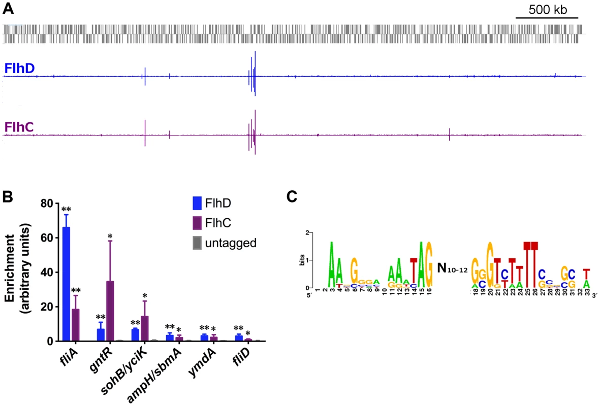
(A) Genome-wide binding of FlhD (blue) and FlhC (purple) determined by ChIP-seq. Gray boxes represent genes in the MG1655 genome. (B) ChIP-qPCR enrichment at one well-known (fliA) and 5 novel FlhDC binding sites (n = 3). ChIP enrichment in tagged strains was compared to untagged control using two-sample T tests: * p<0.05, ** p<0.01 (one-tailed). (C) FlhDC motif derived in this study, using BioProspector [49] (score = 1.49) and visualized by inputing aligned sequenced into WebLogo [76]. Genome-wide binding of FlhD and FlhC
We used ChIP-seq to assess genome-wide binding of FlhD and FlhC in E. coli MG1655 grown to mid-exponential phase (OD600 0.5–0.7) in Lysogeny Broth (LB). The genome-wide binding profiles of the two proteins are shown in Figure 1A. Based on a stringent peak-calling analysis, 10 FlhD binding sites and 8 FlhC binding sites were identified (Table 1). All 8 FlhC binding sites overlapped with FlhD binding sites. The 2 additional FlhD binding sites were also associated with substantial FlhC occupancy, barely missing the threshold in the peak calling analysis. None of the peaks identified in the FlhD or FlhC ChIP-seq experiments overlapped with regions enriched in control ChIP-seq experiments using untagged control strains. The complete colocalization of FlhD and FlhC binding is consistent with the proteins only binding DNA as a heteromeric complex. The 10 sites of FlhDC binding include all 5 binding sites associated with the 7 canonical flagellar Class 2 operons. An FlhDC binding site was identified upstream of yecR, a gene of unknown function, consistent with previous reports [20], [24]. The additional 4 binding sites identified represent novel FlhDC targets. Three of the novel FlhDC binding sites are in intergenic regions, upstream of one or more genes (ampH/sbmA, yciK/sohB, and gntR). The remaining novel binding site is located inside csgC and immediately upstream of a transcription start site for the adjacent gene, ymdA [47].
Tab. 1. FlhDC binding sites and expression of associated genes. 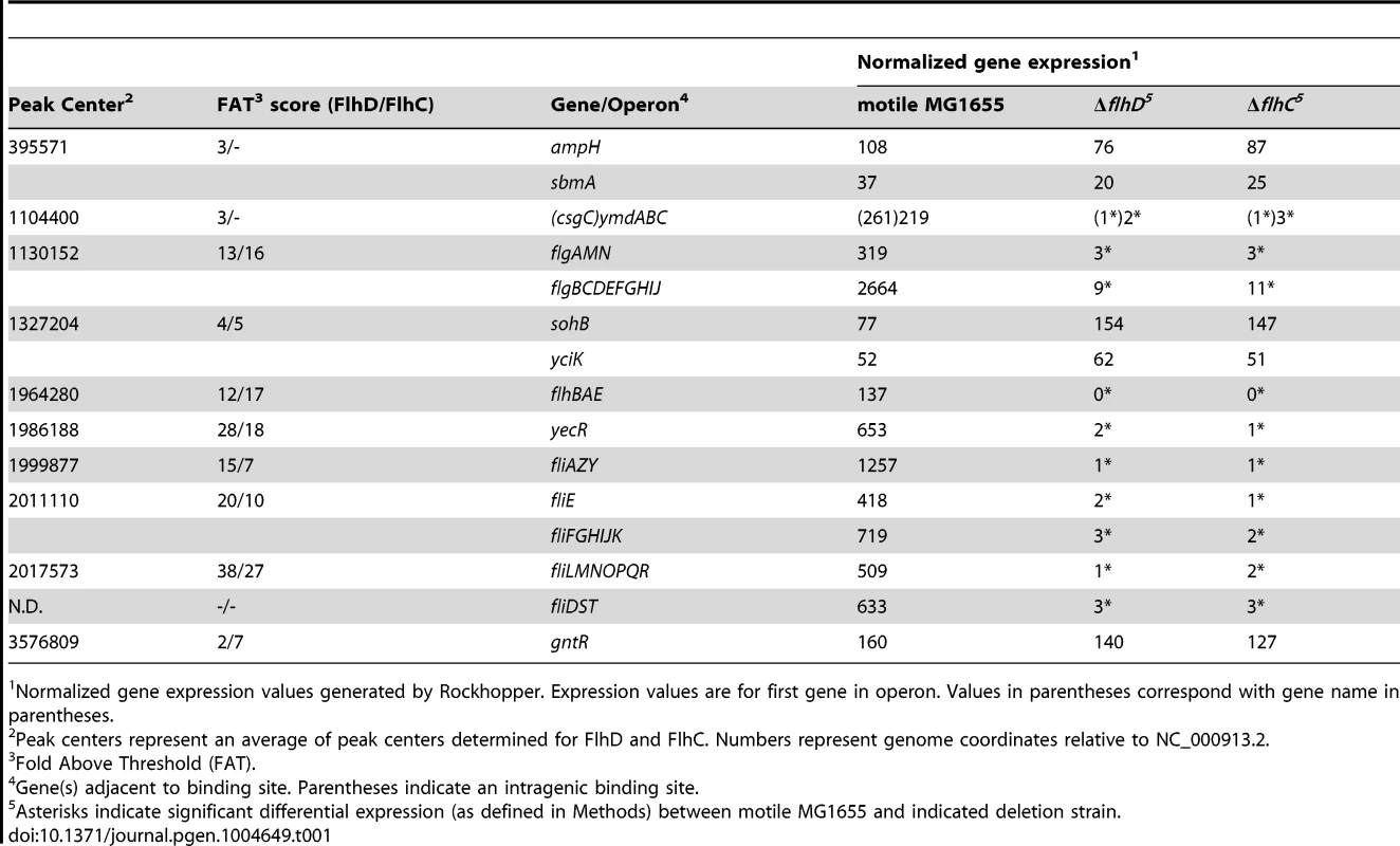
Normalized gene expression values generated by Rockhopper. Expression values are for first gene in operon. Values in parentheses correspond with gene name in parentheses. To confirm FlhDC binding sites identified by ChIP-seq, targeted ChIP-qPCR was performed for all loci (Figure 1B; Table S1). Although ChIP-qPCR is not a completely independent validation method, it allows for analysis of more biological replicates, more straightforward comparison with untagged control strains, and reduces common artifacts enhanced by ChIP-seq library amplification [48]. Consistent with the ChIP-seq findings, FlhD and FlhC occupancy was significantly above that detected in an untagged strain (t-test, p-value≤0.05) for all loci. ChIP-qPCR was also used to evaluate FlhDC occupancy at previously predicted binding sites [24] not identified by ChIP-seq (Figure S2). The fliDST promoter was the only site to show significant occupancy by either protein (Figure 1B, Figure S2). This site was not identified by our ChIP-seq peak-calling analysis, but small, correctly shaped peaks are visible in the raw data for both proteins at this locus (see below). Hence, we consider the fliDST promoter to be a genuine FlhDC binding site and have included it in all downstream analysis, bringing the total to 11 FlhDC-bound sites.
The sequences surrounding the FlhDC binding sites identified with ChIP-seq and/or ChIP-qPCR were extracted and analyzed by BioProspector [49]. The resulting motif, found in 7 out of 11 sites, is low-scoring and degenerate (motif score = 1.49, Figure 1C) but corresponds well with previously reported motifs for FlhDC binding [20], [50].
Direct and indirect regulation by FlhDC
To determine the effect of the identified FlhDC binding sites on the transcriptome, RNA-seq was performed in the motile wild-type MG1655 strain and isogenic single-gene deletions of flhD and flhC. Differential gene expression analysis was performed using Rockhopper, an RNA-seq analysis program optimized for bacterial datasets [36]. Gene expression was almost identical in the ΔflhD and ΔflhC strains (Figure S3), as would be expected since the factors likely regulate the same genes and because the flhD deletion has a minor polar effect on flhC expression. Figure 2 shows a genome-wide comparison of normalized expression values in motile wild-type MG1655 versus the average normalized expression of the ΔflhD and ΔflhC strains. Overall, 228 genes were differentially expressed due to deletion of flhD/flhC (Figure 2). Of those significantly regulated genes, 40 are associated with FlhDC binding sites, indicating direct regulation.
Fig. 2. Genome-wide FlhDC-dependent gene expression. 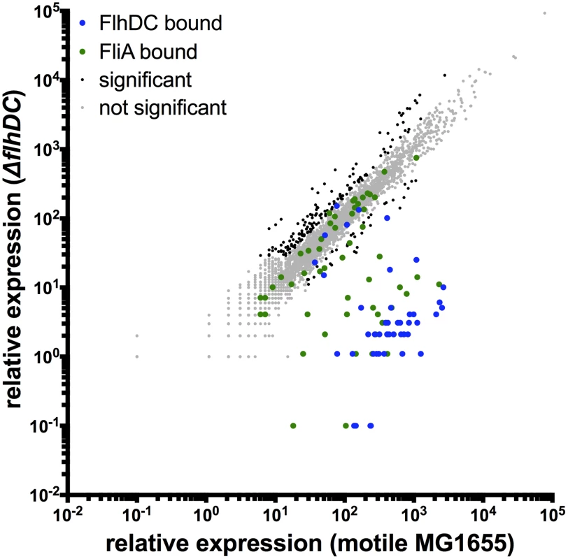
Relative expression of all genes in motile MG1655 versus relative expression in ΔflhDC. Gene expression values represent normalized expression values calculated by Rockhopper. Values on the y-axis represent the average of normalized expression values in the ΔflhD and ΔflhC strains. Color-coding indicates which genes are associated with FlhDC (blue) or FliA (green) binding. Genes not associated with FlhDC of FliA binding are color-coded according to whether they are significantly regulated (Rockhopper, q-value≤0.01) and changed at least 2-fold (black) or not (grey). Of the 11 FlhDC binding sites identified by ChIP, 9 are associated with regulation of one or more adjacent genes (Figure 3A–E, Table 1). We detected strong, positive regulation of all previously reported Class 2 operons by FlhD and FlhC. Additionally, the RNA-seq data demonstrated FlhDC-dependent activation of yecR, fliDST, and the novel target operon ymdABC (Table 1, Figure 3B). The FlhDC binding site upstream of sohB seemed to repress expression by 1.95-fold (Figure 3C, Table 1). The RNA-seq experiments did not provide any evidence for regulation associated with the novel binding sites upstream of ampH/sbmA and gntR (Figure 3D–E, Table 1).
Fig. 3. FhDC binding and regulation at known and novel targets. 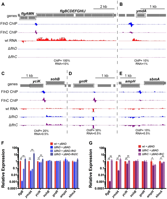
(A–E) Mapped reads from ChIP-seq and RNA-seq experiments. Genes and operons of interest are boxed (dotted black line) and labelled. Within each panel, both lanes of ChIP-seq data are scaled equivalently, and all three lanes of RNA-seq are scaled equivalently. Relative scales are indicated below each panel. Dotted gray lines indicate that mapped reads exceed the scale shown. (F & G) Gene expression, relative to mreB, measured by RT-PCR. Gene names are indicated on the x-axis and strain are indicated in the legend (n = 4–7). Statistical comparisons of were performed using two-sample T tests between the indicated groups: ** p<0.01 (two-tailed). Selected examples of direct FlhDC-dependent regulation, or lack of regulation, were validated using qRT-PCR (Figure 3F–G). The presence or absence of regulation was confirmed for all genes tested, except sohB and the divergently transcribed gene yciK. While the RNA-seq data suggested that sohB was repressed 1.95-fold by FlhDC and yciK was not regulated, the qRT-PCR data clearly indicate the opposite: no regulation of sohB and significant, but slight positive regulation of yciK (1.5-fold for FlhD and 2.29-fold for FlhC; Figure 3F–G). Positive FlhDC-dependent regulation of yciK is also supported by complementation experiments. Small (less than 4-fold) but significant changes in ampH and sbmA levels are detected between wild type and ΔflhC; however, these changes are not detected in the ΔflhD strain or supported by complementation experiments (Figure 3F–G). RNA levels were also determined using qRT-PCR in a variety of complemented strains. As suggested by the motility assays (Figure S1E), flhD overexpression could only partially rescue the flhD deletion, but overexpression of flhDC restored target gene expression to wild-type or higher levels (Figure 3F). Overexpression of flhC alone in the flhD deletion strain did not rescue expression levels at all, demonstrating that the ΔflhD phenotype was not solely due to the polar effect on flhC expression (). The flhC deletion could be completely complemented by flhC overexpression, but target gene expression never exceeded wild-type levels, suggesting that the wild-type levels of flhD expression limit the possible pool of active FlhDC complexes (Figure 3G).
Mechanism of direct regulation of transcription by FlhDC
To provide further insight into the mechanism of regulation at FlhDC-dependent promoters, ChIP-qPCR was used to evaluate σ70 occupancy at FlhDC-dependent promoters in wild-type and flhD deletion strains (Figure 4). For all positively regulated promoters tested, σ70 occupancy was detected in the wild-type strain; however, σ70 occupancy was completely undetectable in the ΔflhD strain, except at the yciK/sohB promoter. At that promoter, deletion of flhD significantly reduced, but did not eliminate, σ70 occupancy, consistent with FlhDC-dependent recruitment to the yciK promoter and FlhDC-independent recruitment to the adjacent sohB promoter. Our data suggest that FlhDC binding is absolutely required for σ70:RNAP recruitment to FlhDC-dependent promoters. This is consistent with reports that these promoters have poor matches to the consensus −10 and −35 hexamers [1].
Fig. 4. FlhDC is required for σ70:RNAP recruitment to FlhDC-dependent promoters. 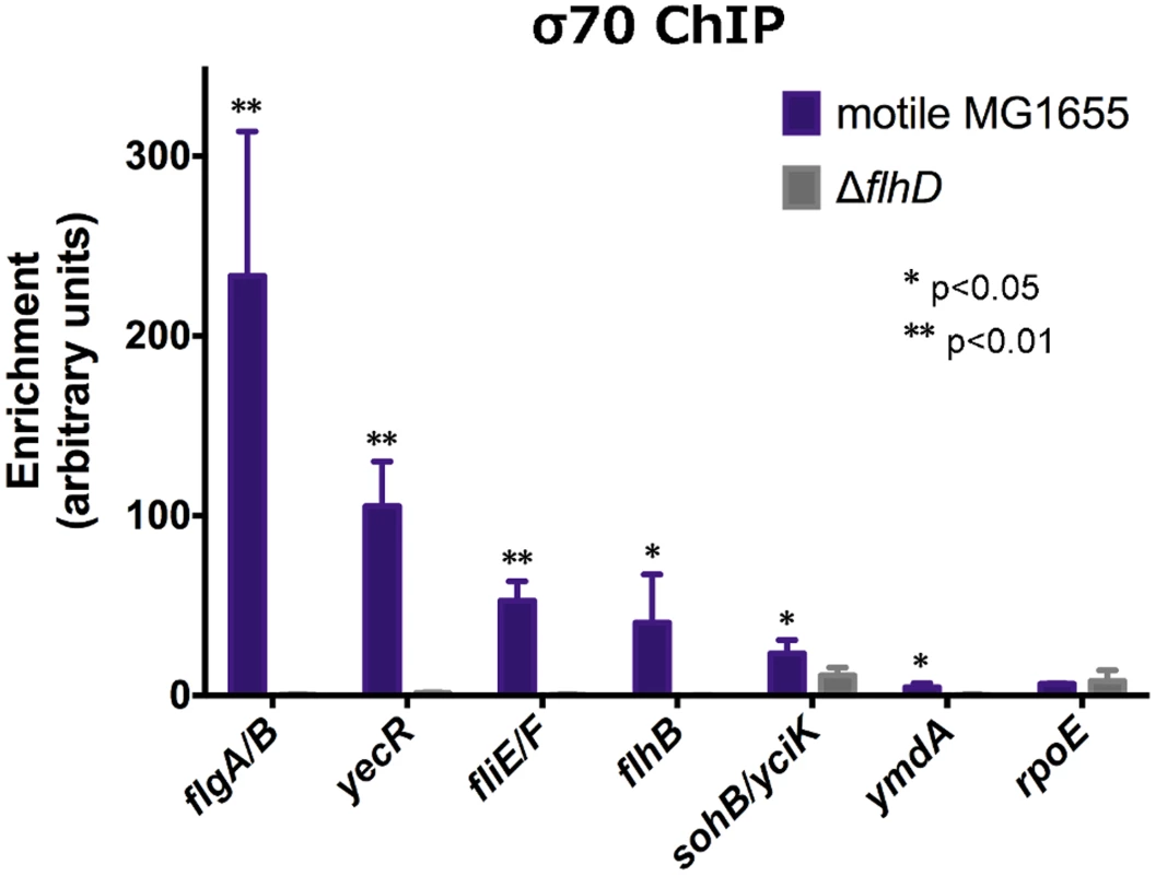
σ70 enrichment, as determined by ChIP-qPCR, at FlhDC-dependent promoters and rpoE (non-FlhDC promoter) in motile MG1655 and ΔflhD (n = 4). σ70 enrichment in motile MG1655 was compared to enrichment in ΔflhD using two-sample T tests: * p<0.05, ** p<0.01 (one-tailed). Genome-wide binding of FliA
To begin assessing the next level of the transcriptional hierarchy, ChIP-seq was used to determine the genome-wide binding of the flagellar σ-factor FliA (Figure 5A). Following a stringent peak-calling analysis, 52 regions were identified that did not overlap with regions enriched in untagged controls (Table 2). These 52 FliA binding sites include 7 canonical flagellar Class 3 promoters and one upstream of the fliLMNOPQR operon. FliA binding sites were also identified upstream of five other flagellar-related genes previously shown or predicted to be FliA-dependent: aer, ycgR, yhjH, trg, and tsr. Additionally, FliA binding sites were identified at 4 other previously reported FliA-dependent promoters of non-flagellar genes: modA [30], ves [20], ynjH [30], and flxA [31]. The remaining 35 FliA binding sites represent novel FliA promoters for which no previous experimental evidence can be found, although a small fraction have been predicted by various bioinformatic approaches [51]. Strikingly, 28 of these novel binding sites are inside genes, with only 4 of these intragenic binding sites <300 bp upstream of a start codon for an appropriately oriented annotated gene.
Fig. 5. Genome-wide binding of FliA. 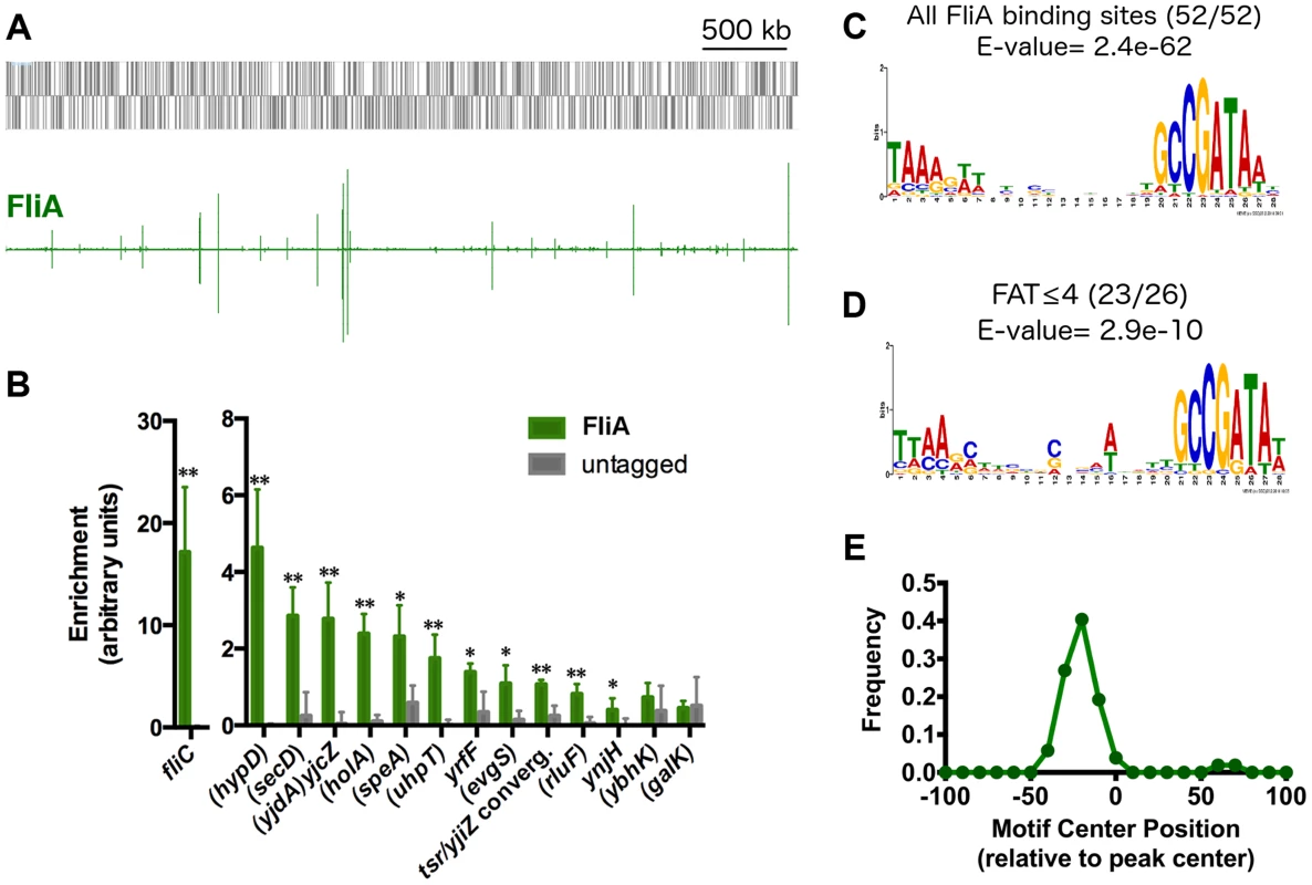
(A) Genome-wide binding of FliA (green) determined by ChIP-seq. Gray boxes represent gene in the MG1655 genome. (B) ChIP-qPCR enrichment at one well-known (fliC) and 13 novel FliA binding sites (n = 3, * p<0.05, ** p<0.01). Gene names in parentheses indicate that the binding site occurs within that gene. (C) FliA motif derived in this study (all binding sites). (D) Motif derived from binding sites with FAT scores ≤4. Motifs in C and D were generated using MEME [52]. (E) Distribution of motifs relative to ChIP-seq peak centers. Motifs cluster ∼25 nt upstream of the peak center, relative to the orientation of the motif. Tab. 2. FliA promoters and expression of associated genes. 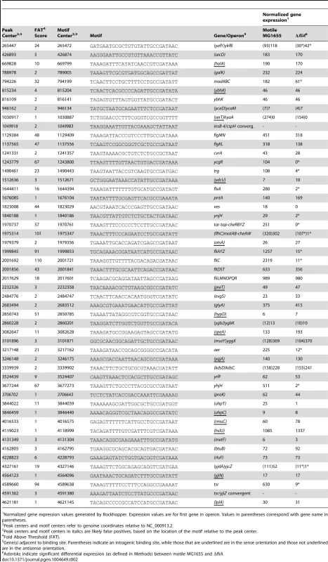
Normalized gene expression values generated by Rockhopper. Expression values are for first gene in operon. Values in parentheses correspond with gene name in parentheses. A subset of the putative FliA binding sites identified by ChIP-seq was also tested using ChIP-qPCR. Of the 13 novel FliA binding sites tested, 11 showed significant enrichment in these targeted experiments (Figure 5B). The 2 novel sites that could not be validated had ChIP-seq “fold above threshold” (FAT) scores of 4 or lower. Twenty-two other peaks had similarly low peak scores (≤4) but were not tested by ChIP-qPCR. Sequences surrounding each of the 52 FliA binding sites were extracted and examined for a motif using MEME [52]. A highly significant motif (E-value = 2.4e−62) was identified in all 52 binding regions and matches previously reported FliA promoter motifs well (Figure 5C, Table 2). This MEME analysis identified motifs associated with all low-scoring sites, including the two that could not be validated by ChIP-qPCR. To further assess whether low-scoring sites, as a group, show evidence of sequence-specific FliA binding, motifs were identified separately for high-scoring (FAT>4, n = 26) and low-scoring (FAT≤4, n = 26) peaks (Figure 5D). The two groups yielded very similar motifs, both with highly significant q-values (E-valuehigh = 5.8e−37, E-valuelow = 2.9e−10). This strongly suggests that a majority of low-scoring ChIP-seq peaks represent genuine sequence-specific FliA binding events.
Not only were motifs identified for all ChIP-seq peaks, but these motifs were also localized in a striking pattern relative to the ChIP-seq peaks (Figure 5E). For most ChIP-seq experiments, one would expect motifs to be enriched at peak centers [53]. However, FliA motifs were clustered with a median position of 25 nt upstream of the peak center (relative to the direction of the motif). This is consistent with the FliA binding to the −10 and −35 hexamer motif while in the context of RNAP holoenzyme [54]. This localization of FliA motifs strongly suggests that FliA only binds in the context of RNAP holoenzyme. Furthermore, this analysis allowed us to identify two weak, non-canonical, putative FliA binding sites (insB-4/cspH convergent intergenic region and inside proK), that have unusually localized motifs, suggesting that they might not be genuine FliA promoters.
While this manuscript was in preparation, FliA ChIP-chip data was published by another group [55]. Our analysis of this study indicates many false positives and false negatives, and the resolution is far lower than that of our data (Figure S4).
Direct regulation by FliA
To detect FliA-dependent changes in gene expression, RNA-seq was performed in a ΔfliA strain and compared to the isogenic motile wild-type. Analysis of differential gene expression was performed with Rockhopper [36]. Overall, 68 genes were differentially expressed between the motile wild-type and a ΔfliA strain (Figure 6). Most of the strongly regulated genes are associated with FliA binding sites identified by ChIP-seq (Figure 6, green points), indicating direct regulation.
Fig. 6. Genome-wide FliA-dependent gene expression. 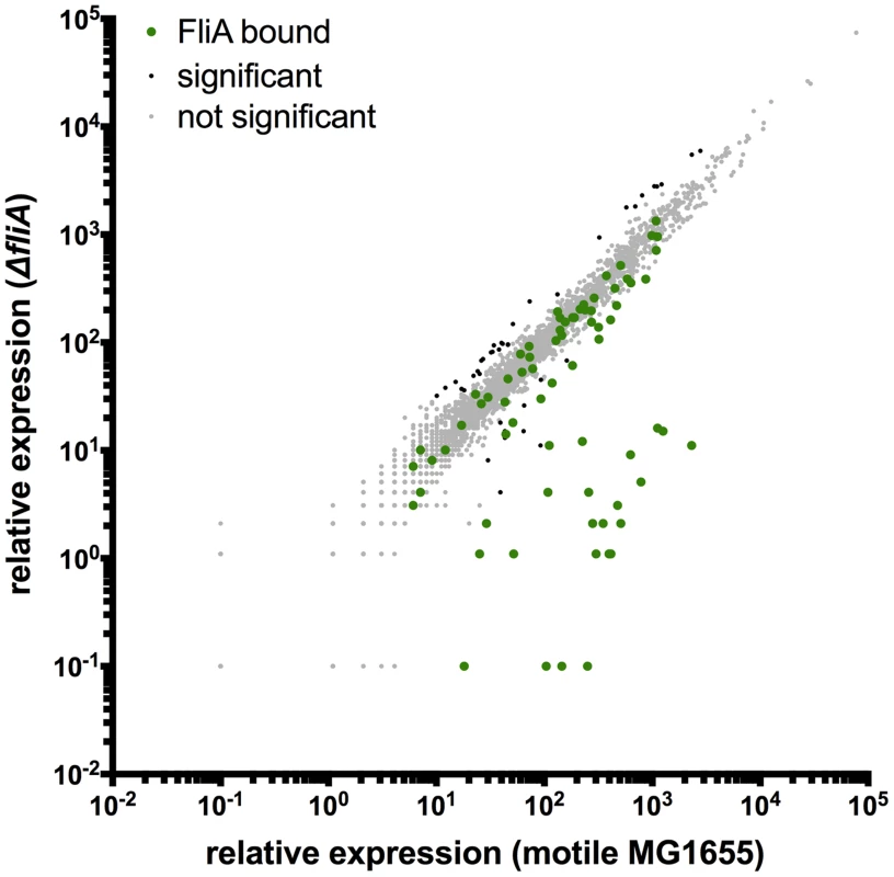
Relative expression of all genes in motile MG1655 versus relative expression in ΔfliA. Gene expression values represent normalized expression values calculated by Rockhopper. Green dots represent genes associated with FliA binding. Genes not associated with binding are color-coded according to whether they are significantly regulated (Rockhopper, q-value≤0.01) and changed at least 2-fold (black) or not (grey). Of the 52 FliA binding sites identified by ChIP-seq, 14 were associated with significant gene expression changes under these conditions (Table 2, Figure 7). These included some, but not all, Class 3 promoters, and 8 other previously reported FliA promoters. Interestingly, three of the intragenic FliA binding sites were also associated with significant regulation of surrounding genes. The promoters inside flhC, yjdA, and yafY drive transcription of the downstream genes (motA, yjcZ, and ykfB, respectively; Figure 7C, Table 2).
Fig. 7. FliA binding and regulation at known and novel targets. 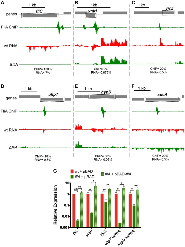
(A–F) Mapped reads from ChIP-seq and RNA-seq experiments. Genes and operons of interest are boxed (dotted black line) and labelled. Within each panel, both lanes of ChIP-seq data are scaled equivalently, and all three lanes of RNA-seq are scaled equivalently. Relative scales are indicated below each panel. Dotted gray lines indicate that mapped reads exceed the scale shown. (G) Gene expression, relative to mreB, measured by RT-PCR. Gene names are indicated on the x-axis and strain are indicated in the legend (n = 4–7). Statistical comparisons of were performed using two-sample T tests between the indicated groups: * p<0.05, ** p<0.01 (two-tailed). In addition to using Rockhopper to analyze differential gene expression, we visually inspected mapped RNA-seq data to find FliA-dependent transcripts that might otherwise have been overlooked. We identified an unusual transcript associated with the novel FliA binding site inside uhpT. Unlike the previously discussed examples, this promoter drives transcription of a purely intragenic RNA. RNA-seq detects a small (∼50 nt) RNA encoded in the same orientation as the gene (Figure 7D). Visual examination of the RNA-seq data also led to the discovery of an antisense orientation noncoding RNA (asRNA) associated with the novel intragenic FliA-promoter inside hypD (Figure 7E). This asRNA is contained entirely within the hypD gene and overlaps ∼500 nt of the 5′ end of the hypD open reading frame (ORF). The hypD antisense RNA is too weakly expressed to be detected by Rockhopper, despite the program's ability to identify novel antisense RNAs. The remaining novel FliA binding sites were not associated with detectable FliA-dependent changes in gene expression under these conditions (eg. speA, Figure 7F).
A variety of canonical and novel examples of FliA-dependent regulation were further validated using qRT-PCR (Figure 7G, Table S2). In all cases, including the two novel non-coding RNAs, the regulation identified in the RNA-seq experiment was confirmed in these targeted experiments. Additionally, overexpression of fliA in the ΔfliA strain resulted in higher than wild-type levels of expression of all FliA-dependent transcripts tested.
Dual regulation of complex promoters and overlapping operons
Complex promoters and overlapping operons were first evaluated using our ChIP-seq and RNA-seq data (Figure 8). The previously described (fliAZY [22] and fliLMNOPQR [22]) or predicted (fliDST [24]) complex promoters are supported by our findings (Figure 8A,B,D). The previously described overlapping flgAMN/flgMN operons are also supported by our data (Figure 8C). Finally, our data confirm that flgKL can be transcribed as part of the upstream FlhDC-dependent flgBCDEFGHIJ operon, in addition to being transcribed from its own Class 3 promoter (Figure 8C, Table 2). While these operons have been previously described in Salmonella [56], [57], this is the first experimental demonstration of the overlapping operons in E. coli. The novel flgBCDEFGHIJKL operon was further confirmed by using RT-PCR to amplify across the flgJ-flgK boundary (Figure S5A).
Fig. 8. FlhDC and FliA binding and regulation at dual regulated targets. 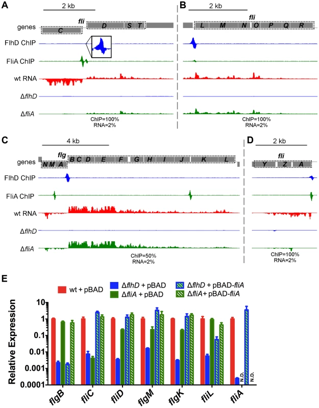
(A–D) Mapped reads from ChIP-seq and RNA-seq experiments. Genes and operons of interest are boxed (dotted black line) and labelled. Within each panel, both lanes of ChIP-seq data are scaled equivalently, and all three lanes of RNA-seq are scaled equivalently. Relative scales are indicated below each panel. Dotted gray lines indicate that mapped reads exceed the scale shown. (E) Gene expression, relative to mreB, measured by RT-PCR. Gene names are indicated on the x-axis and strain are indicated in the legend (n = 4–7). Due to the high number of potential comparisons, statistical analysis is presented in Table S2. In addition to confirming dual regulation of fliDST and discovering that flgKL can be transcribed as part of the upstream operon, the RNA-seq data from this study reveal a surprising pattern for all dual regulated flagellar genes. In the investigated conditions and growth phase, deletion of FliA has little or no effect on the expression levels of dual regulated genes (Figure 8A–D). This suggests that, while FliA occupancy is detected at all dual promoters by ChIP-seq, most transcription is coming from the FlhDC-dependent σ70 promoters under our conditions.
We used qRT-PCR to further investigate the regulatory input of FlhDC and FliA for dual regulated genes (Figure 8E). As seen in the RNA-seq experiments, deletion of fliA had a much less dramatic effect on gene expression compared to deletion of flhD. Overexpression of fliA in the ΔflhD strain significantly increased RNA levels of all dual targets (compared to ΔflhD) showing that the FliA promoters are capable of transcribing the target genes. However, this effect was smaller for fliL than for any other dual regulated gene. Furthermore, overexpression of fliA in the ΔfliA strain (hashed green bar) reduced fliL expression by 2.10-fold compared to the empty vector control (solid green bar; t-test, p-value 0.02), suggesting a possible repressive interaction (Figure 8E).
As dual regulation has been suggested for all Class 2 targets [23], we performed similar qRT-PCR experiments for these genes. Overexpression of fliA in the ΔflhD strain was not able to significantly increase the expression of any other Class 2 target tested (Figure S6). While not statistically significant, flhB expression does increase with fliA overexpression (Figure S6). Since flhB is downstream of the FliA-dependent meche operon, it is possible that the flhBAE operon is dual regulated and can be transcribed as part of the upstream operon. However, there is no evidence of co-transcription from the RNA-seq or RT-PCR targeting the cheZ-flhB intergenic region (Figure S5B) so we conclude that this read-through is likely an artifact of extreme FliA overexpression. It should also be noted that no FliA ChIP-seq signal was detected in the vicinity of the flhBAE, fliE, or fliFGHIJK operons, nor were any of these Class 2 genes differentially expressed in RNA-seq experiments (motile wild-type and ΔfliA strains). Therefore, with the exception of the dual targets already discussed, our data do not support widespread dual regulation of Class 2 genes.
Motility assessment of novel FlhDC and FliA targets
To determine if novel FlhDC and/or FliA targets were required for motility, knockouts were generated for ampH, ymdA, sbmA, gntR, ykfB, yjcZ, ynjH, hypD, and uhpT. Motility on semi-solid agar was determined for each deletion strain. None of the deletions resulted in a substantial change in motility (Figure S7), suggesting that these genes do not directly contribute to motility under the conditions tested.
Discussion
FlhDC regulon
To our knowledge, this study provides the first look at genome-wide in vivo binding of FlhD and FlhC. We have redefined the direct FlhDC regulon by identifying new targets and showing that many previously reported non-flagellar targets are incorrect or not bound under these conditions (Table 1, Figure S2). Our data support a surprisingly limited direct FlhDC regulon and show no evidence of either protein binding DNA independently. Under the conditions tested, FlhDC binds 11 loci and directly regulates 11 transcriptional units. However, it is important to note that some binding sites are associated with regulation of two divergent operons while others are not associated with any detectable regulation. In addition to the classical flagellar Class 2 binding sites, our study detects previously reported binding sites upstream of yecR and fliDST, and 4 novel FlhDC binding sites. Two of these novel binding sites were associated with detectable FlhDC-dependent regulation of adjacent genes (ymdA and yciK), while the remaining two were not (gntR and ampH/sbmA). It is likely that the two latter sites are functional, perhaps under different conditions, because the probability of two spurious sites occurring in intergenic regions is very small (only ∼11% of the genome is intergenic sequence). None of the genes surrounding the 4 novel binding sites have an obvious functional connection to flagellar synthesis, and deletion of these genes had no detectable effect on motility (Figure S7). The RNA-seq data provided little evidence of an extensive indirect FlhDC regulon. While 183 genes were differentially regulated but not associated with FlhDC or FliA binding, the magnitude of the indirect regulation is significantly smaller than for direct targets (Figure 2; Krustal-Wallis, p<0.0001). FliZ is the only transcription factor regulated directly by either FlhDC or FliA, and we do not see regulation of described FliZ targets [58]. Therefore, we doubt that FliZ-dependent regulation substantially contributes to the indirect FlhDC-dependent regulation detected in this study. Instead, it is likely that the indirectly regulated genes do not represent an additional level of the FlhDC transcriptional network but result from physiological and metabolic changes associated with flagellar motility.
Despite having a degenerate consensus motif, FlhDC appears to bind DNA highly specifically - at only 11 sites throughout the genome. The presence of a motif alone is insufficient for binding in vivo, even at sites that are bound in vitro (Figure S2). We propose that as-yet unidentified factors such as DNA conformation or competition with nucleoid-associated proteins play a role in the surprising specificity of FlhDC.
Although the presence of a motif and in vitro binding does not always predict in vivo FlhDC behavior, a striking pattern emerges for flagellar Class 2 sites. There is a perfect correlation between reported in vitro FlhDC affinities [54], expression kinetics [59], and in vivo FlhDC occupancy (this study). Coupled with the σ7°ChIP data presented here (Figure 4), this suggests a simple mechanism of transcriptional activation: σ70-RNAP is unable to bind these promoters in the absence of FlhDC, but as its concentration increases, FlhDC binds DNA and recruits σ70-RNAP to promoters in the order of relative FlhDC binding affinity. This model of affinity-based temporal ordering has been commonly predicted in transcriptional networks [60]. Furthermore, this suggests that for transcription factors with a similar recruitment-based mechanism of action, relative in vivo occupancy derived from ChIP-seq can be used to predict temporal expression patterns.
FliA regulon
By coupling ChIP-seq and RNA-seq, we have comprehensively identified FliA binding sites and matched some of these with FliA-dependent transcripts. We identified 52 FliA binding sites, 35 of which are novel. Furthermore, we have demonstrated that existing computational approaches [30], [32], [33], [51] are poor predictors of in vivo FliA binding. Our data confirm all the better-characterized members of the FliA regulon while discounting most promoters for which only bioinformatic predictions exist (Table S3). It should be noted that our findings largely agree with a previous microarray expression study of the FliA regulon [20]. However, we have greatly expanded the regulon and identified precise promoter positions, many of which are different from those predicted by Zhao et al. based on their ORF-based expression data [20].
Under the conditions used in our study, 14 out of 52 FliA binding sites were associated with significant changes in the RNA levels of one or more surrounding genes (wild-type vs. ΔfliA). This suggests, as will be discussed below, that a large number of promoter sequences bind FliA but are relatively inactive. Similar to FlhDC, the RNA-seq data provide little evidence for an extensive indirect FliA regulon. Only 40 genes were differentially regulated but not associated with FliA binding. The magnitude of indirect regulation was dramatically smaller than direct regulation (Figure 6; Mann-Whitney, p<0.0001). As with FlhDC, it seems likely that indirect regulation is due to secondary physiological and metabolic changes associated with flagellar motility. Consistent with this idea, 29 of the 40 indirectly regulated genes are also indirectly regulated by FlhDC.
Non-canonical FliA binding sites
The most striking finding from our FliA ChIP-seq experiments is that more than half of FliA binding sites are inside genes. While intragenic binding has been reported for other σ-factors and for many transcription factors, the proportion of intragenic FliA sites is remarkable. Similar to our findings, 40% of the sites bound by the Mycobacterium tuberculosis σ-factor, SigF, are intragenic. Some of these sites are associated with SigF-dependent transcription in the antisense orientation relative to the overlapping gene [61]. RpoH (σ32) has been reported to bind many intragenic sites, but these sites only account for ∼25% of the total sites bound (22 out of 87, [62]). Lastly, a recent study reported that σ70 can bind and initiate transcription at a large number of intragenic sites, but that this phenomenon is repressed at many locations by the nucleoid-associated protein H-NS [63]. Combined with our FliA results, it is clearly important to take intragenic transcription initiation into account when analyzing genome-wide data.
Of the 30 intragenic FliA sites identified in our study, only 5 are associated with detectable changes in RNA levels. Three intragenic promoters appear to drive transcription of canonical mRNAs for the downstream gene(s). Two of these promoters have been accurately predicted before (inside flhC driving motAB-cheAW [32], [64], and inside yafY driving ykfB [20]), but one is novel (inside yjdA driving yjcZ). A FliA promoter upstream of yjdA has been incorrectly predicted, based on expression data [20]. These three sites function as canonical promoters and are likely inside genes simply due to the spatial constraints of a small genome. Additionally, two novel intragenic FliA binding sites are associated with detectable small, intragenic RNAs. The FliA promoter inside uhpT transcribes a small RNA overlapping the 3′ end of the ORF in the sense orientation (Figure 7D), while the FliA promoter inside hypD drives transcription of an antisense RNA (Figure 7E). A FliA promoter upstream of uhpT has been predicted based on expression data [20], but our data clearly show that the FliA-dependent transcript is not an mRNA. While the FliA promoters inside uhpT and hypD are associated with detectable transcription, the functions of the corresponding RNAs are not known. Neither intragenic RNA shows evidence of regulating the overlapping mRNA. It is possible that one or both of these RNAs regulate trans-encoded targets.
Most intragenic FliA binding sites are not associated with detectable transcripts. Furthermore, most of these putative promoters are greater than 300 nucleotides upstream of the start codon to an adjacent gene making it improbable that they drive transcription of canonical mRNAs. More likely, these promoters, if active, transcribe non-coding RNAs. However, since no changes in local RNA levels were detected flanking these binding sites, it remains unclear whether these FliA binding sites represent functional FliA-dependent promoters. Many studies have reported widespread spurious transcription, most of which is rapidly terminated and produces highly unstable transcripts [65]. Therefore, these promoters may be active but the resulting RNAs are too unstable to detect by standard RNA-seq methods. Three of these intragenic promoters were previously predicted in E. coli and two of the three, those inside galK and speA, showed FliA-dependent activity when cloned upstream of the chloramphenicol acetyltransferase reporter gene [64]. This supports the hypothesis that at least some of the intragenic FliA binding sites are functional promoters, even if no transcripts were detectable in our study. Alternatively, these binding sites could represent promoter-like sequences where FliA∶RNAP can bind but cannot initiate transcription under the conditions tested, or potentially, ever. Regardless of their transcriptional activity, intragenic FliA sites could serve to alter the available pool of FliA, indirectly affecting transcription from canonical promoters, as has been proposed for some transcription factors [66], [67].
Network topology and the consequences of dual regulation
Over the last 15 years there has been a growing interest in modeling cellular processes as networks [60]. Several frequently occurring network motifs have been identified, with feed-forward loops (FFLs) being one of the most common. In FFLs, one regulator regulates another regulator, and both regulate a common target gene (or genes) [68]. A quantitative computational model of the flagellar network has been constructed [23] and is considered a seminal work in the field of network modeling [69]. A key aspect of the existing flagellar network model is that all Class 2 genes are modeled as FFLs with dual FlhDC/FliA input. However, our data clearly indicate that three Class 2 operons (10 genes) are not dual regulated (Figure S6, Figure 9A). This implies that the true behavior of the transcriptional network cannot be accurately predicted by the existing model.
Fig. 9. Updated flagellar transciption network and localization of dual-regulated targets. 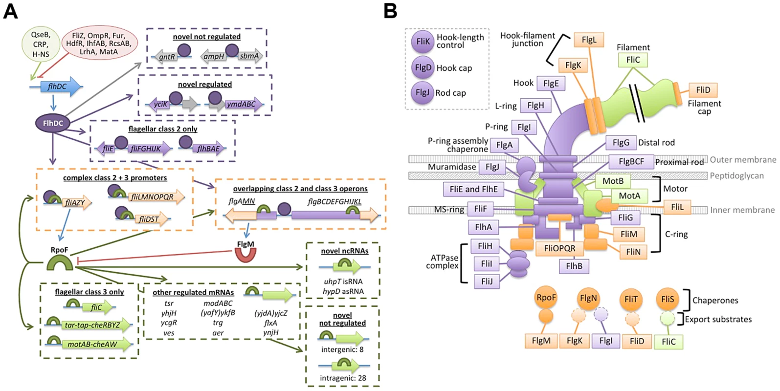
(A) The transcription network map has been updated to accurately depict dual-regulated targets and to incorporated novel regulon members. Ovals at top left represent positive (green) and negative (red) regulation of flhDC transcription. Blue arrows indicate translation of gene products. Purple ovals represent FlhDC and purple arrows indicate FlhDC regulatory interactions. Green cresents represent FliA and green arrows represent FliA regulatory interactions. Dotted arrows indicate potential regulation of genes associated with binding sites but without detectable regulation in this study. Target genes/operons are boxed according to their regulatory input: FlhDC only (green), FliA only (green), and dual FlhDC and FliA (orange). (B) Localization and function of flagellar gene products, color-coded by regulatory input as described for panel B. Gene products not physically associated with the flagellum are omitted. In addition to clarifying the overall topology of the network, our genome-wide and targeted experiments have revealed interesting information about the regulatory inputs for specific dual targets. One striking pattern that emerges is that, while FliA binding is readily detected at all dual targets, RNA levels detected by RNA-seq and qRT-PCR only change moderately between wild-type and ΔfliA. In most cases, expression of dual regulated genes is affected less than 4-fold by fliA deletion, but more than 100-fold by flhDC deletion (Figure 8). This suggests FliA-dependent transcription is contributing very little to the overall abundance of dual regulated transcripts under these conditions, despite FliA being present at the associated promoters. Despite the seemingly low level of activity of these FliA promoters, when fliA is overexpressed in a ΔflhD strain, expression of fliD, flgM, and flgK returns to wild-type or higher levels. This demonstrates that these promoters do have the capacity to be very active, but it remains to be seen if FliA levels and activity are ever high enough to trigger high-level promoter activity in wild-type cells. Interestingly, fliA overexpression results in lower fliL expression in both ΔflhD and ΔfliA strains compared to wild-type (Figure 8E). Since the FliA promoter is downstream of the FlhDC-dependent σ70 promoter, we speculate that high FliA∶RNAP occupancy yields little FliA-dependent transcription and actually competes with σ70:RNAP at the level of DNA binding to repress transcription from the upstream promoter. Overall, our data suggest that flagellar genes likely have diverse temporal expression patterns due to the diversity of regulatory inputs at each promoter (Figure 9A).
Now that we have systematically identified dual-regulated genes, it is tempting to surmise why certain genes are under dual control, and others are not. The correlation between the gene expression class (Class 2 or 3) and the developmental stage at which the resulting proteins are utilized has been described (reviewed [3]). However, dual-regulated gene products show less of a pattern in localization or correlation with a specific phase of assembly (Figure 9B). In Salmonella, the Class 3 promoters of fliDST and flgKL have been shown to be required for swarming and for rapid repair of sheared flagella [57]. The authors hypothesized that these genes utilize their Class 2 promoters during de novo flagellar assembly but are transcribed from the Class 3 promoters, independent of FlhDC activity, to repair flagella broken during swarming. Wozniak et al. also suggest that flgMN is dual regulated so that FlgM can fine-tune FliA activity throughout flagellar assembly and so that FlgN can assist in FlgK folding and transport during flagellar repair [57]. The final dual-regulated operon identified in our study, fliLMNOPQR, encodes components of the C-ring and secretion apparatus that localize to the proximal portion of the flagella (Figure 9B). Our results suggest that high levels of FliA may repress this operon (Figure 8E) but it is unclear why these components would be specifically targeted by negative feedback. Alternatively, FliA may positively regulate the fliL operon at some stage of flagellar development but it is equally unclear why this would be required. The specific roles of the Class 2 and Class 3 promoters of dual-regulated targets should be explored further to determine the physiological importance of the complex temporal gene expression patterns resulting from dual regulation.
Conclusions
We have identified many new targets of FlhDC and FliA, including many non-canonical FliA binding sites. Additionally we have shed new light on the complex topology of the network by systematically defining Class 2, Class 3, and dual-regulated targets. Finally, we have demonstrated that the combined application of ChIP-seq and RNA-seq to related regulators provides sufficient data to build a transcriptional network model from scratch or redefine even the best-characterized networks, such as the flagellar transcription network.
Materials and Methods
Strains and plasmids
Bacterial strains used in this work are listed in Table S4. Cells were grown in Lysogeny Broth (LB: 1% NaCl, 1% tryptone, 0.5% yeast extract) or Tryptone broth (TB: 1% tryptone, 0.5% NaCl). Strains harboring plasmids were cultured with the appropriate antibiotic, as listed in Table S4.
Primers used in strain construction are described in Table S5. Epitope-tagged strains (DMF11, DMF14, RPB081) were generated using the FRUIT method of recombineering [70]. All epitope-tagged strains originate from AMD052 (MG1655 ΔthyA), which has a poorly motile phenotype. FliA was N-terminally tagged by inserting a 3×FLAG tag after the third amino acid. FlhD and FlhC were tagged by inserting 3×FLAG tags into internal loop regions (Figure S1A). Internal sites were chosen since the termini of both proteins are known or predicted to participate in protein-protein and/or protein-DNA contacts [42], [43]. Following motility selection (see below), the region upstream of flhDC and the flhDC-coding region were sequenced in each strain (Wadsworth Center Applied Genomic Technologies Core; File S1). Strains DMF62–65 were also generated using FRUIT. In each strain, the thyA cassette was inserted (facing away from flhDC) at a precise location upstream of flhDC. thyA insertions were confirmed by PCR and sequencing (Wadsworth Center Applied Genomic Technologies Core).
Deletion strains (ΔflhD, ΔflhC, and ΔfliA) were also generated using recombineering. First, DMF35 was generated from AMD052 by motility selection, as described below. With DMF35 as the common parent strain, FRUIT was used to generate scarless deletions of flhD (DMF38) and fliA (DMF40). The flhC deletion strain (DMF58) was generated by amplifying the flhC::kan allele from the Keio collection [71], electroporating it into DMF35, and then removing the kan cassette by expressing FLP recombinase from pCP20 [72]. Following tag insertion or gene deletion, thyA was reintroduced at its native locus and strains were cured of all plasmids. Strains DMF50–57 were also generated from AMD052, using FRUIT to replace the genes of interest with the thyA gene. Note that these strains lack thyA at its native locus, but have a thyA+ phenotype. Overexpression plasmids were generated by cloning the ORF of interest into pBAD24 [73] cut with NheI and SphI (NEB) using the In-Fusion method (Clontech). All deletion strains and plasmids were verified by PCR and sequencing (Wadsworth Center Applied Genomic Technologies Core).
Motility selection to generate motile isolates
Our lab isolate of MG1655, and the related strain AMD052, displayed a poorly motile phenotype. PCR using JW3100 and JW5358 demonstrated that these strains lacked an IS element upstream of the flhDC, which is required for high-level motility. Epitope tagged strains were generated from AMD052, and were thus initially poorly motile as well. To isolate highly motile derivatives, saturated overnight cultures of strains AMD052 and preliminary epitope-tagged strains were spotted (5 µL) onto soft TB agar (0.3%) and incubated at 30°C. Motile subpopulations began emerging between 20 and 24 hours. We typically observed 1–5 motile subpopulations on each plate. Motile cells were collected from stabs, yielding motile strains DMF35, DMF11, DMF14, and RPB081. IS element insertion was verified by PCR and sequencing with oligonucleotides JW3100 and JW5358 (Wadsworth Center Applied Genomic Technologies Core; File S1).
Motility assays
Overnight cultures were grown in TB at 30°C. Saturated cultures (5 µL) were spotted onto 155 mm TB soft agar (0.2%) plates and incubated at 30°C. Each plate included DMF36 for reference and 1–4 other strains of interest. Each assay was performed 5 times from independent overnight cultures. Images were taken at hourly intervals from 4–6 hours post-inoculation. Representative images are shown in Figure S1 and S7.
ChIP-qPCR
Strains DMF11, DMF14, RPB081, and DMF36 were used for all ChIP experiments. Subcultures were grown in LB at 37°C with aeration to an OD600 of 0.5–0.7. Cultures were harvested, crosslinked, sonicated, and immunoprecipitated as previously described [74], with minor modifications. Anti-FLAG (2 µL per IP, M2 monoclonal; Sigma) or anti-σ70 (1 µL per IP; Neoclone) antibodies were used for all immunoprecipitations, both for ChIP-qPCR and ChIP-seq. ChIP and input DNA was purified using Zymo PCR Clean and Concentrate kit. Samples were analyzed using qPCR as previously described [74]. Oligonucleotides used to amplify the bglB control region and regions of interest are described in Table S6. ChIP-qPCR experiments were utilized 3–7 biological replicates per strain. Complete ChIP-qPCR data is presented in Table S1.
ChIP-seq library preparation and sequencing
Cultures for ChIP-seq experiments were grown as described for ChIP-qPCR. ChIP-seq libraries were constructed and sequenced as previously described [63] with the exception of one replicate of FlhC-FLAG, which was prepared as described [37]. Antibodies used were the same as those used for ChIP-qPCR. ChIP-seq libraries were constructed and sequenced for 2 biological replicates per strain. Sequencing was performed on an Illumina Hi-Seq instrument (University at Buffalo, SUNY).
ChIP-seq data analysis
Sequences were aligned to the MG1655 genome (NC_000913.2) using the CLC Genomics Workbench. Mapped reads were piled up and written to a .gff file using a custom Python script and viewed in SignalMap (Nimblegen). All ChIP-seq images presented in this study are captured from SignalMap and manipulated in the image editing software GIMP to highlight baselines (zero reads) and fill gaps in the data resulting from image artifacts.
Almost all ChIP-seq analysis programs have been designed and optimized for eukaryotic ChIP-seq data and, in our experience, do not perform well with bacterial ChIP-seq data. We have generated custom Python scripts to identify peaks in bacterial ChIP-seq data. First, all datasets were normalized to 100 million reads. Pairs of replicate datasets were considered together. For each replicate dataset in the pair, an appropriate threshold was determined. The plus and minus strands were considered separately. For the first replicate, for a given strand, a value T1 was selected as the threshold. For the second replicate, a value T2 was selected as the threshold. Values for T1 and T2 were considered between 1 and 1000. For each combination of values for T1 and T2, the number of genome positions with values ≥T1 in the first replicate and with values ≥T2 in the second replicate was determined. The false discovery rate was estimated using the null hypothesis that no regions are enriched. The combination of thresholds yielding the highest number of true positive positions, with an estimated false discovery rate of less than 0.01, was selected. Once T1 and T2 were chosen, peak calling was performed as previously described (Supplementary Material of [54]). Briefly, a region was identified as a peak if both replicates showed enrichment above the corresponding thresholds for each strand. For a peak to be called there must be a peak on the plus strand within a threshold distance of a peak on the minus strand, as previously described (Supplementary Material of [54]). To identify regions of artifactual enrichment, peaks identified in tagged strains were compared to those called in a control ChIP-seq experiment using an untagged strain (DMF35). For each factor, the calculated T values were adjusted to reflect the total number of reads in control experiment replicates and then applied for peak calling in the controls. Any regions for which a peak was called in the true ChIP-seq experiment and in the untagged control experiment within 50 bp of each other were considered potential artifacts and excluded from further analysis.
RNA isolation and RNA-seq library preparation
Strains DMF36, DMF38, DMF58, and DMF40 were used for RNA-seq experiments. Subcultures were grown in LB at 37°C with aeration to an OD600 of 0.5–0.7. RNA was purified using a modified hot phenol method, as previously described [37]. Following isolation, RNA was treated with DNase (TURBO DNA-free kit; Life Technologies) for 45 minutes at 37°C, followed by phenol extraction and ethanol precipitation. rRNA was removed using the RiboZero kit (Epicentre) and strand-specific DNA libraries were constructed using the ScriptSeq 2.0 kit (Epicentre). Sequencing was performed using an Illumina Hi-Seq instrument (University at Buffalo, SUNY).
RNA-seq data visualization and differential expression analysis
Sequence reads were mapped and visualized as described above. Differential expression analysis was performed using Rockhopper [36] with default parameters. As suggested by the developers, changes in gene expression were considered statistically significant if q-value≤0.01. Genes were required to be regulated at least 2-fold to be considered significantly differentially regulated. Ribosomal RNAs (rRNAs) were excluded from RNA-seq analysis since rRNA was removed during library preparation.
RNA isolation and qRT-PCR
Strains pDMF9–18 were used for qRT-PCR experiments. Subcultures were grown in LB+100 µg/ml ampicillin at 37°C with aeration to an OD600 of 0.4–0.6. Arabinose was added to a final concentration of 0.2% and cultures were incubated at 37°C for an additional 10 minutes. RNA was isolated as follows. 10 ml of culture were harvested by centrifugation, resuspended in 1 ml Trizol reagent (Life Technologies) and incubated at room temperature for 5 min. Samples were centrifuged, supernatants were transferred to new tubes, and 200 µL of chloroform was added. Samples were mixed well, incubated at room temperature for 3 minutes, and centrifuged. The aqueous layers were transferred to new tubes and mixed with 500 µL isopropanol. Samples were mixed well, incubated at room temperature for 10 minutes, and centrifuged to collect precipitated RNA. Pellets were washed once with 75% ethanol and then dried. RNA was resuspended in H2O and treated with TURBO DNase (Life Technologies) as described above. RNA was reverse transcribed using SuperScript III reverse transcriptase (Invitrogen) with 100 ng of random hexamer, according to the manufacturer's instructions. A control reaction lacking reverse transcriptase (no RT) was performed for each sample. cDNA and no RT samples were diluted and used as templates for qPCR. qPCR was performed with an ABI 7500 Fast real time PCR machine. Oligonucleotides used to amplify target genes, and the control (mreB) are listed in Table S7. Relative expression values were calculated using a modified 2−ΔΔCt method [75]. First target Ct values were normalized to the control (mreB) yielding ΔCt values, then 1.9−ΔCt (assuming imperfect PCR efficiency) was calculated for each target in each strain. Finally, expression values in all strains were normalized to the average 1.9−ΔCt value in motile MG1655+pBAD. qRT-PCR experiments utilized 3–4 biological replicates per strain and all qPCR reactions were performed in triplicate.
Supporting Information
Zdroje
1. MacnabRM (1992) Genetics and biogenesis of bacterial flagella. Annu Rev Genet 26 : 131–158 doi:10.1146/annurev.ge.26.120192.001023
2. ChilcottGS, HughesKT (2000) Coupling of flagellar gene expression to flagellar assembly in Salmonella enterica serovar typhimurium and Escherichia coli. Microbiol Mol Biol Rev 64 : 694–708 doi:10.1128/MMBR.64.4.694-708.2000.Updated
3. Chevance FFV, HughesKT (2008) Coordinating assembly of a bacterial macromolecular machine. Nat Rev Microbiol 6 : 455–465 doi:10.1038/nrmicro1887
4. SoutourinaOA, BertinPN (2003) Regulation cascade of flagellar expression in Gram-negative bacteria. FEMS Microbiol Rev 27 : 505–523 doi:10.1016/S0168-6445(03)00064-0
5. ShinS, ParkC (1995) Modulation of flagellar expression in Escherichia coli by acetyl phosphate and the osmoregulator OmpR. J Bacteriol 177 : 4696–4702.
6. LehnenD, BlumerC, PolenT (2002) LrhA as a new transcriptional key regulator of flagella, motility and chemotaxis genes in Escherichia coli. Mol Microbiol 45 : 521–532.
7. SperandioV, TorresA, KaperJ (2002) Quorum sensing Escherichia coli regulators B and C (QseBC): a novel two-component regulatory system involved in the regulation of flagella and motility by. Mol Microbiol 43 : 809–821.
8. SoutourinaO, KolbA (1999) Multiple control of flagellum biosynthesis in Escherichia coli: role of H-NS protein and the cyclic AMP-catabolite activator protein complex in transcription of the flhDC. J Bacteriol 181 : 7500–7508.
9. YakhninA, BakerC (2013) CsrA activates flhDC expression by protecting flhDC mRNA from RNase E-mediated cleavage. Mol Microbiol 87 : 851–866 doi:10.1111/mmi.12136
10. ThomasonM, FontaineF (2012) A small RNA that regulates motility and biofilm formation in response to changes in nutrient availability in Escherichia coli. Mol Microbiol 84 : 17–35 doi:10.1111/j.1365-2958.2012.07965.x
11. Lay NDe, GottesmanS (2012) A complex network of small non-coding RNAs regulate motility in Escherichia coli. Mol Microbiol 86 : 524–538 doi:10.1111/j.1365-2958.2012.08209.x
12. LiuX, FujitaN, IshihamaA, MatsumuraP (1995) The C-terminal region of the alpha subunit of Escherichia coli RNA polymerase is required for transcriptional activation of the flagellar level II operons by the FlhD/FlhC complex. J Bacteriol 177 : 5186–5188.
13. HelmannJD, ChamberlinMJ (1987) DNA sequence analysis suggests that expression of flagellar and chemotaxis genes in Escherichia coli and Salmonella typhimurium is controlled by an alternative sigma factor. Proc Natl Acad Sci U S A 84 : 6422–6424.
14. ArnostiDN, ChamberlinMJ (1989) Secondary sigma factor controls transcription of flagellar and chemotaxis genes in Escherichia coli. Proc Natl Acad Sci U S A 86 : 830–834.
15. ArnostiD (1990) Regulation of Escherichia coli sigma F RNA Polymerase by flhD and flhC Flagellar Regulatory Genes. J Bacteriol 172 : 4106–4108.
16. OhnishiK, KutsukakeK, SuzukiH, LinoT (1992) A novel transcriptional regulation mechanism in the flagellar regulon of Salmonella typhimurium: an anti-sigma factor inhibits the activity of the flagellum-specific Sigma factor, σF. Mol Microbiol 6 : 3149–3157 doi:10.1111/j.1365-2958.1992.tb01771.x
17. ZaslaverA, MayoAE, RosenbergR, BashkinP, SberroH, et al. (2004) Just-in-time transcription program in metabolic pathways. Nat Genet 36 : 486–491 doi:10.1038/ng1348
18. HollandsK, LeeDJ, LloydGS, BusbySJW (2010) Activation of sigma 28-dependent transcription in Escherichia coli by the cyclic AMP receptor protein requires an unusual promoter organization. Mol Microbiol 75 : 1098–1111 doi:10.1111/j.1365-2958.2009.06913.x
19. KunduTK, KusanoS, IshihamaA (1997) Promoter selectivity of Escherichia coli RNA polymerase sigmaF holoenzyme involved in transcription of flagellar and chemotaxis genes. J Bacteriol 179 : 4264–9.
20. ZhaoK, LiuM, BurgessRR (2007) Adaptation in bacterial flagellar and motility systems: from regulon members to “foraging”-like behavior in E. coli. Nucleic Acids Res 35 : 4441–4452 doi:10.1093/nar/gkm456
21. KoM, ParkC (2000) Two novel flagellar components and H-NS are involved in the motor function of Escherichia coli. J Mol Biol 303 : 371–382.
22. LiuX, MatsumuraP (1996) Differential regulation of multiple overlapping promoters in flagellar class II operons in Escherichia coii. Mol Microbiol 21 : 613–620.
23. KalirS, AlonU (2004) Using a quantitative blueprint to reprogram the dynamics of the flagella gene network. Cell 117 : 713–720 doi:10.1016/j.cell.2004.05.010
24. StaffordGP, OgiT, HughesC (2005) Binding and transcriptional activation of non-flagellar genes by the Escherichia coli flagellar master regulator FlhD2C2. Microbiology 151 : 1779–1788 doi:10.1099/mic.0.27879-0
25. PrüßB, CampbellJW, Van DykTK, ZhuC, KoganY, et al. (2003) FlhD/FlhC is a regulator of anaerobic respiration and the Entner-Doudoroff pathway through induction of the methyl-accepting chemotaxis protein Aer. J Bacteriol 185 : 534–543.
26. PrüßB, MatsumuraP (1996) A regulator of the flagellar regulon of Escherichia coli, flhD, also affects cell division. J Bacteriol 178 : 668–674.
27. PrüßB, MarkovicD, MatsumuraP (1997) The Escherichia coli flagellar transcriptional activator flhD regulates cell division through induction of the acid response gene cadA. J Bacteriol 179 : 3818–3821.
28. PrüßB, LiuX, HendricksonW, MatsumuraP, PruBM (2001) FlhD/FlhC-regulated promoters analyzed by gene array and lacZ gene fusions. FEMS Microbiol Lett 197 : 91–97.
29. SiegeleDA, BainS, MaoW (2010) Mutations in the flhD gene of Escherichia coli K-12 do not cause the reported effect on cell division. FEMS Microbiol Lett 309 : 94–99 doi:10.1111/j.1574-6968.2010.02021.x
30. YuHHY, KiblerD, TanM (2006) In Silico Prediction and Functional Validation of 28-Regulated Genes in Chlamydia and Escherichia coli. J Bacteriol 188 : 8206–8212 doi:10.1128/JB.01082-06
31. IdeN, KutsukakeK (1997) Identification of a novel Escherichia coli gene whose expression is dependent on the flagellum-specific sigma factor, FliA, but dispensable for motility development. Gene 199 : 19–23.
32. ParkK, ChoiS, KoM, ParkC (2001) Novel sigmaF-dependent genes of Escherichia coli found using a specified promoter consensus. FEMS Microbiol Lett 202 : 243–250.
33. HuertaAM, Collado-VidesJ (2003) Sigma70 Promoters in Escherichia coli: Specific Transcription in Dense Regions of Overlapping Promoter-like Signals. J Mol Biol 333 : 261–278 doi:10.1016/j.jmb.2003.07.017
34. GalaganJ, LyubetskayaA, GomesA (2013) ChIP-Seq and the Complexity of Bacterial Transcriptional Regulation. Curr Top Microbiol Immunol 363 : 43–68 doi:10.1007/82
35. WadeJT, StruhlK, BusbySJW, GraingerDC (2007) Genomic analysis of protein-DNA interactions in bacteria: insights into transcription and chromosome organization. Mol Microbiol 65 : 21–26 doi:10.1111/j.1365-2958.2007.05781.x
36. McClureR, BalasubramanianD, SunY, BobrovskyyM, SumbyP, et al. (2013) Computational analysis of bacterial RNA-Seq data. Nucleic Acids Res 41: e140 doi:10.1093/nar/gkt444
37. StringerAM, CurrentiS, BonocoraRP, BaranowskiC, PetroneBL, et al. (2014) Genome-Scale Analyses of Escherichia coli and Salmonella enterica AraC Reveal Noncanonical Targets and an Expanded Core Regulon. J Bacteriol 196 : 660–671 doi:10.1128/JB.01007-13
38. GalaganJ, MinchK, PetersonM (2013) The Mycobacterium tuberculosis regulatory network and hypoxia. Nature 499 : 178–183 doi:10.1038/nature12337
39. MyersK, YanH, OngI, ChungD, LiangK (2013) Genome-scale analysis of Escherichia coli FNR reveals complex features of transcription factor binding. PLoS Genet 9: e1003565 doi:10.1371/journal.pgen.1003565
40. DongT, MekalanosJ (2012) Characterization of the RpoN regulon reveals differential regulation of T6SS and new flagellar operons in Vibrio cholerae O37 strain V52. Nucleic Acids Res 40 : 7766–7775 doi:10.1093/nar/gks567
41. BlankaA, SchulzS, EckweilerD, BieleckaA, NicolaiT, et al. (2014) Identification of the Alternative Sigma Factor SigX Regulon and Its Implications for Pseudomonas aeruginosa Pathogenicity. J Bacteriol 196 : 345–356 doi:10.1128/JB.01034-13
42. CamposA, MatsumuraP (2001) Extensive alanine scanning reveals protein-protein and protein-DNA interaction surfaces in the global regulator FlhD from Escherichia coli. Mol Microbiol 39 : 581–594.
43. WangS, FlemingRT, WestbrookEM, MatsumuraP, McKayDB (2006) Structure of the Escherichia coli FlhDC complex, a prokaryotic heteromeric regulator of transcription. J Mol Biol 355 : 798–808 doi:10.1016/j.jmb.2005.11.020
44. BarkerCS, PrüßBM, MatsumuraP (2004) Increased Motility of Escherichia coli by Insertion Sequence Element Integration into the Regulatory Region of the flhD Operon. J Bacteriol 186 : 7529–7537 doi:10.1128/JB.186.22.7529
45. WangX, WoodTK (2011) IS 5 inserts upstream of the master motility operon flhDC in a quasi-Lamarckian way. ISME J 5 : 1517–1525 doi:10.1038/ismej.2011.27
46. LeeC, ParkC (2013) Mutations Upregulating the flhDC Operon in Escherichia coli K-12. J Microbiol 51 : 140–144.
47. KimD, HongJS, QiuY, NagarajanH, SeoJ, et al. (2012) Comparative Analysis of Regulatory Elements between Escherichia coli and Klebsiella pneumoniae by Genome - Wide Transcription Start Site Profiling. PLoS Genet 8: e1002867 doi:10.1371/journal.pgen.1002867
48. TeytelmanL, ThurtleDM, RineJ, OudenaardenA Van (2013) Highly expressed loci are vulnerable to misleading ChIP localization of multiple unrelated proteins. Proc Natl Acad Sci U S A 110 : 18602–18607 doi:10.1073/pnas.1316064110/-/DCSupplemental.www.pnas.org/cgi/doi/10.1073/pnas.1316064110
49. LiuX, BrutlagD, LiuJ (2001) BioProspector: discovering conserved DNA motifs in upstream regulatory regions of co-expressed genes. Pac Symp Biocomput 127–138.
50. LeeY, BarkerCS, MatsumuraP, BelasR (2011) Refining the binding of the Escherichia coli flagellar master regulator, FlhD4C2, on a base-specific level. J Bacteriol 193 : 4057–4068 doi:10.1128/JB.00442-11
51. SalgadoH, Peralta-GilM, Gama-CastroS, Santos-ZavaletaA, Muñiz-RascadoL, et al. (2013) RegulonDB v8.0: omics data sets, evolutionary conservation, regulatory phrases, cross-validated gold standards and more. Nucleic Acids Res 41: D203–13 doi:10.1093/nar/gks1201
52. BaileyTL, BodenM, BuskeFA, FrithM, GrantCE, et al. (2009) MEME SUITE: tools for motif discovery and searching. Nucleic Acids Res 37: W202–8 doi:10.1093/nar/gkp335
53. BaileyTL, MachanickP (2012) Inferring direct DNA binding from ChIP-seq. Nucleic Acids Res 40 : 1–10 doi:10.1093/nar/gks433
54. LiuX, MatsumuraP (1994) The FlhD/FlhC complex, a transcriptional activator of the Escherichia coli flagellar class II operons. J Bacteriol 176 : 7345–7351.
55. ChoB-K, KimD, KnightEM, ZenglerK, PalssonBO (2014) Genome-scale reconstruction of the sigma factor network in Escherichia coli: topology and functional states. BMC Biol 12 : 4 doi:10.1186/1741-7007-12-4
56. KutsukakeK, IdeN (1995) Transcriptional analysis of the flgK and fliD operons of Salmonella typhimurium which encode flagellar hook-associated proteins. Mol Gen Genet 247 : 275–281.
57. WozniakCE, Chevance FFV, HughesKT (2010) Multiple promoters contribute to swarming and the coordination of transcription with flagellar assembly in Salmonella. J Bacteriol 192 : 4752–4762 doi:10.1128/JB.00093-10
58. PesaventoC, HenggeR (2012) The global repressor FliZ antagonizes gene expression by σS-containing RNA polymerase due to overlapping DNA binding specificity. Nucleic Acids Res 40 : 4783–4793 doi:10.1093/nar/gks055
59. KalirS, McClureJ, PabbarajuK, SouthwardC, RonenM, et al. (2001) Ordering genes in a flagella pathway by analysis of expression kinetics from living bacteria. Science 292 : 2080–2083 doi:10.1126/science.1058758
60. AlonU (2007) Network motifs: theory and experimental approaches. Nat Rev Genet 8 : 450–461 doi:10.1038/nrg2102
61. HartkoornRC, SalaC, UplekarS, BussoP, RougemontJ, et al. (2012) Genome-wide definition of the SigF regulon in Mycobacterium tuberculosis. J Bacteriol 194 : 2001–2009 doi:10.1128/JB.06692-11
62. WadeJT, Castro RoaD, GraingerDC, HurdD, BusbySJW, et al. (2006) Extensive functional overlap between sigma factors in Escherichia coli. Nat Struct Mol Biol 13 : 806–814 doi:10.1038/nsmb1130
63. SinghSS, SinghN, BonocoraRP, FitzgeraldDM, WadeJT, et al. (2014) Widespread suppression of intragenic transcription initiation by H-NS. Genes Dev 28 : 214–219 doi:10.1101/gad.234336.113
64. IdeN, IkebeT, KutsukakeK (1999) Reevaluation of the promoter structure of the class 3 flagellar operons of Escherichia coli and Salmonella. Genes Genet Syst 74 : 113–116.
65. PetersJM, MooneyRA, GrassJA, JessenED, TranF, et al. (2012) Rho and NusG suppress pervasive antisense transcription in Escherichia coli. Genes Dev 26 : 2621–2633 doi:10.1101/gad.196741.112
66. BrewsterRC, WeinertFM, GarciaHG, SongD, RydenfeltM, et al. (2014) The Transcription Factor Titration Effect Dictates Level of Gene Expression. Cell 156 : 1312–1323 doi:10.1016/j.cell.2014.02.022
67. GöpelY, GörkeB (2014) Lies and deception in bacterial gene regulation: the roles of nucleic acid decoys. Mol Microbiol 92 : 641–647 doi:10.1111/mmi.12604
68. ManganS, AlonU (2003) Structure and function of the feed-forward loop network motif. Proc Natl Acad Sci U S A 100 : 11980–11985 doi:10.1073/pnas.2133841100
69. SeshasayeeASN, BertoneP, FraserGM, LuscombeNM (2006) Transcriptional regulatory networks in bacteria: from input signals to output responses. Curr Opin Microbiol 9 : 511–519 doi:10.1016/j.mib.2006.08.007
70. StringerAM, SinghN, YermakovaA, PetroneBL, AmarasingheJJ, et al. (2012) FRUIT, a scar-free system for targeted chromosomal mutagenesis, epitope tagging, and promoter replacement in Escherichia coli and Salmonella enterica. PLoS One 7: e44841 doi:10.1371/journal.pone.0044841
71. BabaT, AraT, HasegawaM, TakaiY, OkumuraY, et al. (2006) Construction of Escherichia coli K-12 in-frame, single-gene knockout mutants: the Keio collection. Mol Syst Biol 2 : 2006.0008 doi:10.1038/msb4100050
72. DatsenkoKA, WannerBL (2000) One-step inactivation of chromosomal genes in Escherichia coli K-12 using PCR products. Proc Natl Acad Sci U S A 97 : 6640–6645 doi:10.1073/pnas.120163297
73. GuzmanL, BelinD, CarsonMJ, BeckwithJ (1995) Tight regulation, modulation, and high-level expression by vectors containing the arabinose PBAD promoter. J Bacteriol 177 : 4121–4130.
74. BonocoraRP, FitzgeraldDM, StringerAM, WadeJT (2013) Non-canonical protein-DNA interactions identified by ChIP are not artifacts. BMC Genomics 14 : 254 doi:10.1186/1471-2164-14-254
75. SchmittgenT, LivakK (2008) Analyzing real-time PCR data by the comparative Ct method. Nat Protoc 3 : 1101–1108.
76. CrooksG, HonG (2004) WebLogo: a sequence logo generator. Genome Res 14 : 1188–1190 doi:10.1101/gr.849004.1
Štítky
Genetika Reprodukční medicína
Článek Oligoasthenoteratozoospermia and Infertility in Mice Deficient for miR-34b/c and miR-449 LociČlánek The Kinesin AtPSS1 Promotes Synapsis and is Required for Proper Crossover Distribution in MeiosisČlánek Payoffs, Not Tradeoffs, in the Adaptation of a Virus to Ostensibly Conflicting Selective PressuresČlánek Examination of Prokaryotic Multipartite Genome Evolution through Experimental Genome ReductionČlánek BMP-FGF Signaling Axis Mediates Wnt-Induced Epidermal Stratification in Developing Mammalian SkinČlánek Role of STN1 and DNA Polymerase α in Telomere Stability and Genome-Wide Replication in ArabidopsisČlánek RNA-Processing Protein TDP-43 Regulates FOXO-Dependent Protein Quality Control in Stress ResponseČlánek Integrating Functional Data to Prioritize Causal Variants in Statistical Fine-Mapping StudiesČlánek Salt-Induced Stabilization of EIN3/EIL1 Confers Salinity Tolerance by Deterring ROS Accumulation inČlánek Ethylene-Induced Inhibition of Root Growth Requires Abscisic Acid Function in Rice ( L.) SeedlingsČlánek Metabolic Respiration Induces AMPK- and Ire1p-Dependent Activation of the p38-Type HOG MAPK PathwayČlánek Signature Gene Expression Reveals Novel Clues to the Molecular Mechanisms of Dimorphic Transition inČlánek A Mouse Model Uncovers LKB1 as an UVB-Induced DNA Damage Sensor Mediating CDKN1A (p21) DegradationČlánek Dominant Sequences of Human Major Histocompatibility Complex Conserved Extended Haplotypes from to
Článek vyšel v časopisePLOS Genetics
Nejčtenější tento týden
2014 Číslo 10
-
Všechny články tohoto čísla
- An Deletion Is Highly Associated with a Juvenile-Onset Inherited Polyneuropathy in Leonberger and Saint Bernard Dogs
- Licensing of Yeast Centrosome Duplication Requires Phosphoregulation of Sfi1
- Oligoasthenoteratozoospermia and Infertility in Mice Deficient for miR-34b/c and miR-449 Loci
- Basement Membrane and Cell Integrity of Self-Tissues in Maintaining Immunological Tolerance
- The Kinesin AtPSS1 Promotes Synapsis and is Required for Proper Crossover Distribution in Meiosis
- Germline Mutations in Are Associated with Familial Gastric Cancer
- POT1a and Components of CST Engage Telomerase and Regulate Its Activity in
- Controlling Meiotic Recombinational Repair – Specifying the Roles of ZMMs, Sgs1 and Mus81/Mms4 in Crossover Formation
- Payoffs, Not Tradeoffs, in the Adaptation of a Virus to Ostensibly Conflicting Selective Pressures
- FHIT Suppresses Epithelial-Mesenchymal Transition (EMT) and Metastasis in Lung Cancer through Modulation of MicroRNAs
- Genome-Wide Mapping of Yeast RNA Polymerase II Termination
- Examination of Prokaryotic Multipartite Genome Evolution through Experimental Genome Reduction
- White Cells Facilitate Opposite- and Same-Sex Mating of Opaque Cells in
- BMP-FGF Signaling Axis Mediates Wnt-Induced Epidermal Stratification in Developing Mammalian Skin
- Genome-Wide Association Study of CSF Levels of 59 Alzheimer's Disease Candidate Proteins: Significant Associations with Proteins Involved in Amyloid Processing and Inflammation
- COE Loss-of-Function Analysis Reveals a Genetic Program Underlying Maintenance and Regeneration of the Nervous System in Planarians
- Fat-Dachsous Signaling Coordinates Cartilage Differentiation and Polarity during Craniofacial Development
- Identification of Genes Important for Cutaneous Function Revealed by a Large Scale Reverse Genetic Screen in the Mouse
- Sensors at Centrosomes Reveal Determinants of Local Separase Activity
- Genes Integrate and Hedgehog Pathways in the Second Heart Field for Cardiac Septation
- Systematic Dissection of Coding Exons at Single Nucleotide Resolution Supports an Additional Role in Cell-Specific Transcriptional Regulation
- Recovery from an Acute Infection in Requires the GATA Transcription Factor ELT-2
- HIPPO Pathway Members Restrict SOX2 to the Inner Cell Mass Where It Promotes ICM Fates in the Mouse Blastocyst
- Role of and in Development of Abdominal Epithelia Breaks Posterior Prevalence Rule
- The Formation of Endoderm-Derived Taste Sensory Organs Requires a -Dependent Expansion of Embryonic Taste Bud Progenitor Cells
- Role of STN1 and DNA Polymerase α in Telomere Stability and Genome-Wide Replication in Arabidopsis
- Keratin 76 Is Required for Tight Junction Function and Maintenance of the Skin Barrier
- Encodes the Catalytic Subunit of N Alpha-Acetyltransferase that Regulates Development, Metabolism and Adult Lifespan
- Disruption of SUMO-Specific Protease 2 Induces Mitochondria Mediated Neurodegeneration
- Caudal Regulates the Spatiotemporal Dynamics of Pair-Rule Waves in
- It's All in Your Mind: Determining Germ Cell Fate by Neuronal IRE-1 in
- A Conserved Role for Homologs in Protecting Dopaminergic Neurons from Oxidative Stress
- The Master Activator of IncA/C Conjugative Plasmids Stimulates Genomic Islands and Multidrug Resistance Dissemination
- An AGEF-1/Arf GTPase/AP-1 Ensemble Antagonizes LET-23 EGFR Basolateral Localization and Signaling during Vulva Induction
- The Proteomic Landscape of the Suprachiasmatic Nucleus Clock Reveals Large-Scale Coordination of Key Biological Processes
- RNA-Processing Protein TDP-43 Regulates FOXO-Dependent Protein Quality Control in Stress Response
- A Complex Genetic Switch Involving Overlapping Divergent Promoters and DNA Looping Regulates Expression of Conjugation Genes of a Gram-positive Plasmid
- ZTF-8 Interacts with the 9-1-1 Complex and Is Required for DNA Damage Response and Double-Strand Break Repair in the Germline
- Integrating Functional Data to Prioritize Causal Variants in Statistical Fine-Mapping Studies
- Tpz1-Ccq1 and Tpz1-Poz1 Interactions within Fission Yeast Shelterin Modulate Ccq1 Thr93 Phosphorylation and Telomerase Recruitment
- Salt-Induced Stabilization of EIN3/EIL1 Confers Salinity Tolerance by Deterring ROS Accumulation in
- Telomeric (s) in spp. Encode Mediator Subunits That Regulate Distinct Virulence Traits
- Ethylene-Induced Inhibition of Root Growth Requires Abscisic Acid Function in Rice ( L.) Seedlings
- Ancient Expansion of the Hox Cluster in Lepidoptera Generated Four Homeobox Genes Implicated in Extra-Embryonic Tissue Formation
- Mechanism of Suppression of Chromosomal Instability by DNA Polymerase POLQ
- A Mutation in the Mouse Gene Leads to Impaired Hedgehog Signaling
- Keeping mtDNA in Shape between Generations
- Targeted Exon Capture and Sequencing in Sporadic Amyotrophic Lateral Sclerosis
- TIF-IA-Dependent Regulation of Ribosome Synthesis in Muscle Is Required to Maintain Systemic Insulin Signaling and Larval Growth
- At Short Telomeres Tel1 Directs Early Replication and Phosphorylates Rif1
- Evidence of a Bacterial Receptor for Lysozyme: Binding of Lysozyme to the Anti-σ Factor RsiV Controls Activation of the ECF σ Factor σ
- Hsp40s Specify Functions of Hsp104 and Hsp90 Protein Chaperone Machines
- Feeding State, Insulin and NPR-1 Modulate Chemoreceptor Gene Expression via Integration of Sensory and Circuit Inputs
- Functional Interaction between Ribosomal Protein L6 and RbgA during Ribosome Assembly
- Multiple Regulatory Systems Coordinate DNA Replication with Cell Growth in
- Fast Evolution from Precast Bricks: Genomics of Young Freshwater Populations of Threespine Stickleback
- Mmp1 Processing of the PDF Neuropeptide Regulates Circadian Structural Plasticity of Pacemaker Neurons
- The Nuclear Immune Receptor Is Required for -Dependent Constitutive Defense Activation in
- Genetic Modifiers of Neurofibromatosis Type 1-Associated Café-au-Lait Macule Count Identified Using Multi-platform Analysis
- Juvenile Hormone-Receptor Complex Acts on and to Promote Polyploidy and Vitellogenesis in the Migratory Locust
- Uncovering Enhancer Functions Using the α-Globin Locus
- The Analysis of Mutant Alleles of Different Strength Reveals Multiple Functions of Topoisomerase 2 in Regulation of Chromosome Structure
- Metabolic Respiration Induces AMPK- and Ire1p-Dependent Activation of the p38-Type HOG MAPK Pathway
- The Specification and Global Reprogramming of Histone Epigenetic Marks during Gamete Formation and Early Embryo Development in
- The DAF-16 FOXO Transcription Factor Regulates to Modulate Stress Resistance in , Linking Insulin/IGF-1 Signaling to Protein N-Terminal Acetylation
- Genetic Influences on Translation in Yeast
- Analysis of Mutants Defective in the Cdk8 Module of Mediator Reveal Links between Metabolism and Biofilm Formation
- Ribosomal Readthrough at a Short UGA Stop Codon Context Triggers Dual Localization of Metabolic Enzymes in Fungi and Animals
- Gene Duplication Restores the Viability of Δ and Δ Mutants
- Selection on a Variant Associated with Improved Viral Clearance Drives Local, Adaptive Pseudogenization of Interferon Lambda 4 ()
- Break-Induced Replication Requires DNA Damage-Induced Phosphorylation of Pif1 and Leads to Telomere Lengthening
- Dynamic Partnership between TFIIH, PGC-1α and SIRT1 Is Impaired in Trichothiodystrophy
- Signature Gene Expression Reveals Novel Clues to the Molecular Mechanisms of Dimorphic Transition in
- Mutations in Moderate or Severe Intellectual Disability
- Multifaceted Genome Control by Set1 Dependent and Independent of H3K4 Methylation and the Set1C/COMPASS Complex
- A Role for Taiman in Insect Metamorphosis
- The Small RNA Rli27 Regulates a Cell Wall Protein inside Eukaryotic Cells by Targeting a Long 5′-UTR Variant
- MMS Exposure Promotes Increased MtDNA Mutagenesis in the Presence of Replication-Defective Disease-Associated DNA Polymerase γ Variants
- Coexistence and Within-Host Evolution of Diversified Lineages of Hypermutable in Long-term Cystic Fibrosis Infections
- Comprehensive Mapping of the Flagellar Regulatory Network
- Topoisomerase II Is Required for the Proper Separation of Heterochromatic Regions during Female Meiosis
- A Splice Mutation in the Gene Causes High Glycogen Content and Low Meat Quality in Pig Skeletal Muscle
- KDM5 Interacts with Foxo to Modulate Cellular Levels of Oxidative Stress
- H2B Mono-ubiquitylation Facilitates Fork Stalling and Recovery during Replication Stress by Coordinating Rad53 Activation and Chromatin Assembly
- Copy Number Variation in the Horse Genome
- Unifying Genetic Canalization, Genetic Constraint, and Genotype-by-Environment Interaction: QTL by Genomic Background by Environment Interaction of Flowering Time in
- Spinster Homolog 2 () Deficiency Causes Early Onset Progressive Hearing Loss
- Genome-Wide Discovery of Drug-Dependent Human Liver Regulatory Elements
- Developmentally-Regulated Excision of the SPβ Prophage Reconstitutes a Gene Required for Spore Envelope Maturation in
- Protein Phosphatase 4 Promotes Chromosome Pairing and Synapsis, and Contributes to Maintaining Crossover Competence with Increasing Age
- The bHLH-PAS Transcription Factor Dysfusion Regulates Tarsal Joint Formation in Response to Notch Activity during Leg Development
- A Mouse Model Uncovers LKB1 as an UVB-Induced DNA Damage Sensor Mediating CDKN1A (p21) Degradation
- Notch3 Interactome Analysis Identified WWP2 as a Negative Regulator of Notch3 Signaling in Ovarian Cancer
- An Integrated Cell Purification and Genomics Strategy Reveals Multiple Regulators of Pancreas Development
- Dominant Sequences of Human Major Histocompatibility Complex Conserved Extended Haplotypes from to
- The Vesicle Protein SAM-4 Regulates the Processivity of Synaptic Vesicle Transport
- A Gain-of-Function Mutation in Impeded Bone Development through Increasing Expression in DA2B Mice
- Nephronophthisis-Associated Regulates Cell Cycle Progression, Apoptosis and Epithelial-to-Mesenchymal Transition
- Beclin 1 Is Required for Neuron Viability and Regulates Endosome Pathways via the UVRAG-VPS34 Complex
- The Not5 Subunit of the Ccr4-Not Complex Connects Transcription and Translation
- Abnormal Dosage of Ultraconserved Elements Is Highly Disfavored in Healthy Cells but Not Cancer Cells
- Genome-Wide Distribution of RNA-DNA Hybrids Identifies RNase H Targets in tRNA Genes, Retrotransposons and Mitochondria
- The Chromosomal Association of the Smc5/6 Complex Depends on Cohesion and Predicts the Level of Sister Chromatid Entanglement
- Cell-Autonomous Progeroid Changes in Conditional Mouse Models for Repair Endonuclease XPG Deficiency
- PLOS Genetics
- Archiv čísel
- Aktuální číslo
- Informace o časopisu
Nejčtenější v tomto čísle- The Master Activator of IncA/C Conjugative Plasmids Stimulates Genomic Islands and Multidrug Resistance Dissemination
- A Splice Mutation in the Gene Causes High Glycogen Content and Low Meat Quality in Pig Skeletal Muscle
- Keratin 76 Is Required for Tight Junction Function and Maintenance of the Skin Barrier
- A Role for Taiman in Insect Metamorphosis
Kurzy
Zvyšte si kvalifikaci online z pohodlí domova
Současné možnosti léčby obezity
nový kurzAutoři: MUDr. Martin Hrubý
Všechny kurzyPřihlášení#ADS_BOTTOM_SCRIPTS#Zapomenuté hesloZadejte e-mailovou adresu, se kterou jste vytvářel(a) účet, budou Vám na ni zaslány informace k nastavení nového hesla.
- Vzdělávání


