-
Články
Top novinky
Reklama- Vzdělávání
- Časopisy
Top články
Nové číslo
- Témata
Top novinky
Reklama- Videa
- Podcasty
Nové podcasty
Reklama- Kariéra
Doporučené pozice
Reklama- Praxe
Top novinky
ReklamaDNA Ligase III Promotes Alternative Nonhomologous End-Joining during Chromosomal Translocation Formation
Nonhomologous end-joining (NHEJ) is the primary DNA repair pathway thought to underlie chromosomal translocations and other genomic rearrangements in somatic cells. The canonical NHEJ pathway, including DNA ligase IV (Lig4), suppresses genomic instability and chromosomal translocations, leading to the notion that a poorly defined, alternative NHEJ (alt-NHEJ) pathway generates these rearrangements. Here, we investigate the DNA ligase requirement of chromosomal translocation formation in mouse cells. Mammals have two other DNA ligases, Lig1 and Lig3, in addition to Lig4. As deletion of Lig3 results in cellular lethality due to its requirement in mitochondria, we used recently developed cell lines deficient in nuclear Lig3 but rescued for mitochondrial DNA ligase activity. Further, zinc finger endonucleases were used to generate DNA breaks at endogenous loci to induce translocations. Unlike with Lig4 deficiency, which causes an increase in translocation frequency, translocations are reduced in frequency in the absence of Lig3. Residual translocations in Lig3-deficient cells do not show a bias toward use of pre-existing microhomology at the breakpoint junctions, unlike either wild-type or Lig4-deficient cells, consistent with the notion that alt-NHEJ is impaired with Lig3 loss. By contrast, Lig1 depletion in otherwise wild-type cells does not reduce translocations or affect microhomology use. However, translocations are further reduced in Lig3-deficient cells upon Lig1 knockdown, suggesting the existence of two alt-NHEJ pathways, one that is biased toward microhomology use and requires Lig3 and a back-up pathway which does not depend on microhomology and utilizes Lig1.
Published in the journal: . PLoS Genet 7(6): e32767. doi:10.1371/journal.pgen.1002080
Category: Research Article
doi: https://doi.org/10.1371/journal.pgen.1002080Summary
Nonhomologous end-joining (NHEJ) is the primary DNA repair pathway thought to underlie chromosomal translocations and other genomic rearrangements in somatic cells. The canonical NHEJ pathway, including DNA ligase IV (Lig4), suppresses genomic instability and chromosomal translocations, leading to the notion that a poorly defined, alternative NHEJ (alt-NHEJ) pathway generates these rearrangements. Here, we investigate the DNA ligase requirement of chromosomal translocation formation in mouse cells. Mammals have two other DNA ligases, Lig1 and Lig3, in addition to Lig4. As deletion of Lig3 results in cellular lethality due to its requirement in mitochondria, we used recently developed cell lines deficient in nuclear Lig3 but rescued for mitochondrial DNA ligase activity. Further, zinc finger endonucleases were used to generate DNA breaks at endogenous loci to induce translocations. Unlike with Lig4 deficiency, which causes an increase in translocation frequency, translocations are reduced in frequency in the absence of Lig3. Residual translocations in Lig3-deficient cells do not show a bias toward use of pre-existing microhomology at the breakpoint junctions, unlike either wild-type or Lig4-deficient cells, consistent with the notion that alt-NHEJ is impaired with Lig3 loss. By contrast, Lig1 depletion in otherwise wild-type cells does not reduce translocations or affect microhomology use. However, translocations are further reduced in Lig3-deficient cells upon Lig1 knockdown, suggesting the existence of two alt-NHEJ pathways, one that is biased toward microhomology use and requires Lig3 and a back-up pathway which does not depend on microhomology and utilizes Lig1.
Introduction
Recurrent reciprocal chromosomal translocations are hallmarks of several tumor types [1]. Breakpoint junction analysis indicates that translocations arise primarily through a nonhomologous end-joining (NHEJ) mechanism of double-strand break (DSB) repair in a process that results in a variety of DNA end modifications, including deletions and insertions. Notably, DNA ends frequently join at short sequence homologies of one or a few bases (microhomology) which may promote the joining reaction [2], [3].
A set of NHEJ factors has been defined based on their requirement both for cellular resistance to ionizing radiation and during V(D)J recombination for antigen receptor formation and diversity [4], [5]. These canonical NHEJ factors include the end protection protein Ku, DNA end processing enzymes, and the DNA ligase complex Lig4-XRCC4. Des pite the observation that translocation breakpoint junctions exhibit characteristics of NHEJ, the canonical pathway is not required for translocation formation; rather, this pathway is known to suppress translocations, as evidenced by the increased number of translocations arising in mouse cells deficient in components of this pathway. For example, canonical NHEJ deficiency in the context of p53 loss leads to pro-B cell lymphomas with Igh-Myc amplification and chromosomal translocation [6], [7]. Further, translocations involving induced DSBs on two different chromosomes are increased in frequency in either Ku or Lig4-XRCC4-deficient mouse embryonic stem (ES) cells, with breakpoint junctions showing similar end modifications and microhomology as in wild-type cells [8], [9], suggesting that canonical NHEJ does not play an important role in the joining events.
Although studies of NHEJ have focused on canonical NHEJ in the context of V(D)J recombination, the existence of an alternative pathway(s) of NHEJ has been evident from the earliest analyses of canonical NHEJ-deficient cells, using either plasmid or chromosomal substrates for DSB repair in rodent and human cells [10]–[16]. This alternative pathway, termed alt-NHEJ, is poorly defined, although recently several candidate components of this pathway have been proposed, including the Mre11 complex [17]–[19], the end resection protein CtIP [20]–[22], and poly (ADP-ribose) polymerases (PARPs) [23], [24]. The increase in translocations in canonical NHEJ-deficient mouse cells implies that alt-NHEJ is primarily responsible for their formation and, moreover, that alt-NHEJ leading to translocations is suppressed by the canonical pathway. Given that translocations appear to arise by alt-NHEJ even in the presence of the canonical pathway [9], they provide a good model with which to characterize components of the alt-NHEJ pathway.
As NHEJ ultimately involves DNA ligation and Lig4 is not required for translocation formation, one (or both) of the other two known DNA ligases present in mammalian cells, Lig1 and Lig3 [25], must be required for this process. Both of these ligases have essential cellular functions – the primary cellular role of Lig1 is for Okazaki fragment ligation during DNA replication [25], while Lig3 is essential for mitochondrial DNA metabolism [26], [27]. Lig3 interacts with the single-strand break repair protein XRCC1 via its C-terminal BRCT domain [28], [29]. Regarding DSB repair, Lig3 has been shown to have an end-joining activity in cell extracts [23] and in Lig4-deficient cells depleted for Lig3 using plasmid substrates, implicating Lig3 in a backup pathway of NHEJ [30].
Given that chromosomal translocations have been shown to arise by alt-NHEJ in mouse cells, we investigated the role of the three DNA ligases in this process. We demonstrate that in the absence of nuclear Lig3, translocations are reduced in frequency and that the residual translocation breakpoint junctions show less microhomology, demonstrating that Lig3 has a preference for joining ends at pre-existing microhomology. Lig3-dependent events do not require the C-terminal BRCT domain, indicating that interaction with XRCC1 is dispensable for these alt-NHEJ events. Knockdown of Lig1, but not Lig4, in the nuclear Lig3-deficient cells further reduces translocation formation, while having no effect in wild-type cells, indicating that it acts as a backup to Lig3 for these events. These experiments define Lig3 as having a primary role in this alt-NHEJ process even in the presence of canonical NHEJ and suggest the existence of multiple alt-NHEJ pathways.
Results
Chromosomal translocations are reduced in Lig3-deficient mouse cells
Lig3 is essential to cells [31] due to its ligase activity in mitochondria [26], [27]. We were able to rescue Lig3KO/KO mouse embryonic stem (ES) cells through pre-emptive complementation by expressing DNA ligases targeted to mitochondria [26]. In this approach, a Lig3KO/cKOneo+ cell line, which contains one Lig3 null allele and a second conditional allele with an intronic neomycin selection marker, was constructed (Figure S1). Transgenes expressing various DNA ligase forms fused to GFP were stably integrated into the Lig3KO/cKOneo+ cells, which were then treated with Cre recombinase to transform the conditional Lig3 allele to a second null allele. Cells specifically deficient for nuclear Lig3 or altogether deleted for Lig3 were constructed by this approach (Figure 1A) [26]. Viable Lig3 null cells were generated through expression of Lig1 fused to a mitochondrial leader sequence (MtLig1). MtLig1 was expressed at a fraction of the level of endogenous Lig1 in these cells [26], but to diminish the possibility that it contributes to nuclear ligation activity, we also deleted the Lig1 nuclear localization signal, generating MtLig1-ΔNLS. Additionally, nuclear Lig3-deficient cells were created by expressing a highly modified form of Lig3 (MtLig3-ΔBRCT-NES), where the nuclear translation initiation site was mutated, the BRCT domain implicated in nuclear transport [32] was deleted, and a potent nuclear export signal (NES) [33] was fused to the C-terminus. Lig3 null and nuclear Lig3-deficient cells are not sensitive to ionizing radiation [26], [27], suggesting that Lig3 is not required for global DSB repair, in contrast to the canonical NHEJ ligase, Lig4 [3].
Fig. 1. Lig3-deficient mouse cells have fewer chromosomal translocations and reduced microhomology at junctions. 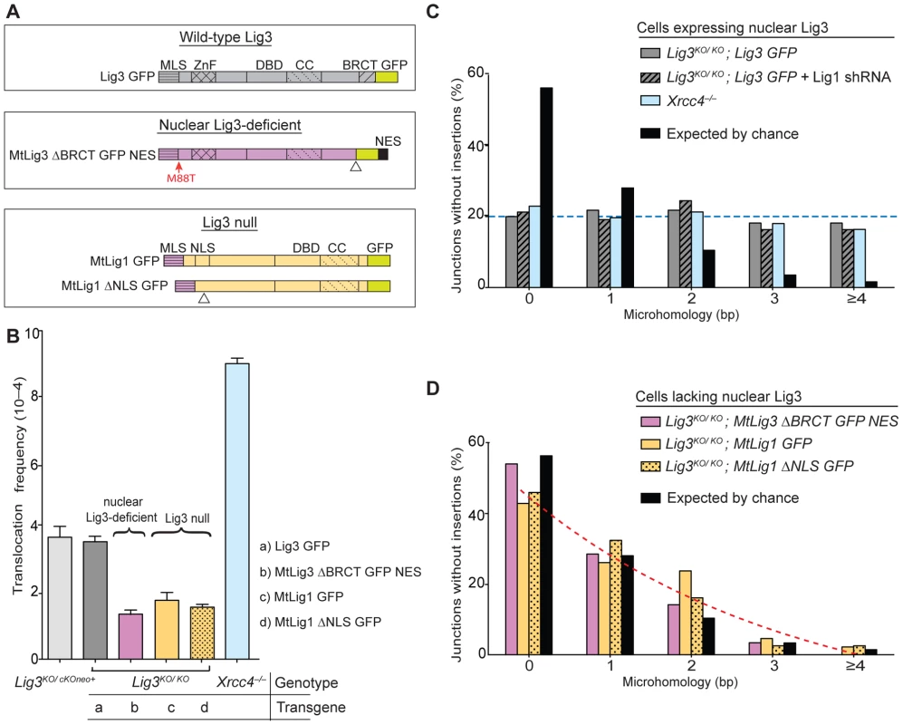
(A) DNA ligases expressed from transgenes in Lig3KO/KO cells. Wild-type Lig3 is expressed in control cells. Nuclear Lig3-deficient cells express mitochondria-restricted Lig3 (MtLig3-ΔBRCT-GFP-NES), while Lig3 null cells express mitochondria-targeted Lig1 (MtLig1 or MtLig1-ΔNLS). All transgenes are GFP tagged. MLS, mitochondrial leader sequence from Lig3 for mitochondrial localization; ZnF, zinc finger domain; BRCT, BRCA1 C-terminal related domain; DBD, DNA-binding domain; CC, catalytic core; *M88T, mutation of the Lig3 nuclear translation initiation site which disrupts translation of a nuclear-specific form of Lig3; NES, nuclear export signal derived from MAPKK to exclude Lig3 from the nucleus; ΔNLS, deletion of Lig1 nuclear localization signal (amino acids 135 to 147) to decrease nuclear localization of Lig1; ΔBRCT, deletion of theLig3 BRCT domain (amino acids 934 to 1009); GFP, green fluorescent protein. Triangles denote position of deletions of the indicated domains. A color scheme is used throughout the figures to assist in tracking various ligase forms (gray, wild-type Lig3; purple, mitochondria-only Lig3; orange, Lig1 forms). (B) Translocations are reduced in cell lines without nuclear Lig3. By contrast, translocations increase in cells deficient for Lig4–XRCC4 complex. (C) Microhomology length distributions for der(6) breakpoint junctions are similar for control cells, cells depleted for Lig1, and cells deficient for Lig4–XRCC4, and differ from that expected by the chance presence of microhomology. Only junctions with simple deletions (i.e., without an insertion) are included. The probability that a junction will have X nucleotides of microhomology by chance assumes an unbiased base composition and is calculated as previously described [9]. The transgenes present in the Lig3KO/KO cells are indicated in italics. (D) Microhomology length distributions in cells without nuclear Lig3 are shifted to a distribution similar to that expected by chance. To address whether Lig3 plays a role in alt-NHEJ during translocation formation, DSBs were introduced at two loci in Lig3KO/KO rescued and parental cells at the Rosa26 and H3f3b loci on chromosomes (chr) 6 and 11, respectively, by expressing zinc finger nucleases (ZFNs) [34] (Figure 2A and Figure S2). After allowing for translocation formation for 60 hours, translocation breakpoint junctions were amplified for both derivative chromosomes, der(6) and der(11), by nested PCR (Figure 2B), similar to an approach recently developed in human cells [35]. Using this approach, we quantified der(6) junctions in rescued and control cells and, for comparison, cells defective in the canonical NHEJ component XRCC4, the required Lig4 cofactor [25].
Fig. 2. Induction of chromosomal translocations at endogenous mouse loci. 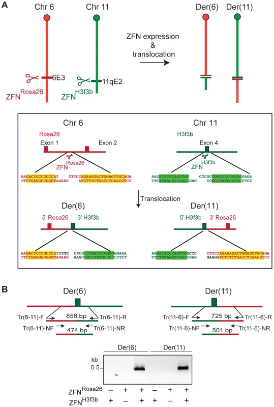
(A) DSBs are induced at cleavage sites for the zinc finger nucleases (ZFNs) ZFNRosa26 and ZFNH3f3b on chromosomes 6 and 11, respectively. Joining of DNA ends from 2 different chromosomes can lead to the translocations generating derivative chromosomes der(6) and der(11). Sequences surrounding the DSBs generated by the ZFNs, together with sequences of the resultant translocation chromosomes, are shown. The latter assumes fill-in of the 5′ overhangs and no loss of sequence information from the DNA ends. (B) Nested PCR is used to identify translocation breakpoint junctions for der(6) and der(11), as shown, which is dependent on expression of both ZFNRosa26 and ZFNH3f3b. Translocations were readily detected in both parental cells and increased by a factor of 2.4 in Xrcc4−/− cells (3.7 vs 9×10⁻⁴; Figure 1B, Table 1), confirming previous results obtained with a different translocation system [9]. Lig3KO/KO cells rescued by wild-type Lig3 expression had a similar frequency of translocations as the parental cells expressing Lig3 from the endogenous locus. By contrast, translocations were substantially reduced in frequency in the absence of nuclear Lig3: translocations in both Lig3 null cells (Lig3KO/KO complemented with mitochondrial Lig1 transgenes) and nuclear Lig3-deficient cells (Lig3KO/KO complemented with the modified mitochondrial Lig3 transgene) were reduced by a factor of ∼2.3, ranging from 1.4 to 1.8×10⁻⁴ (Figure 1B, Table 1). Similar levels of intrachromosomal repair were observed in all cell lines, indicating that the reduced frequency of translocations was not due to reduced cleavage of the chromosomal loci (Figure S3A and S3B).
Tab. 1. Translocation frequencies for various cell lines tested. 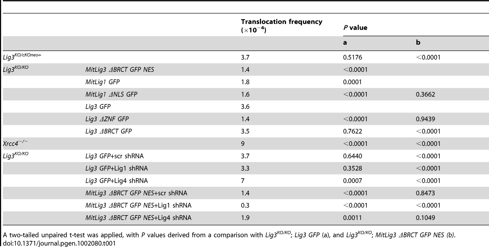
A two-tailed unpaired t-test was applied, with P values derived from a comparison with Lig3KO/KO; Lig3 GFP (a), and Lig3KO/KO; MitLig3 ΔBRCT GFP NES (b). Translocation junctions demonstrate less microhomology in the absence of Lig3
We next examined translocation breakpoint junctions. Similar microhomology, deletion, and insertion distributions were observed for cells expressing wild-type Lig3 and Xrcc4−/− cells (Figure 1C and Figure 3, Figures S4, S5, S6, Table 2), recapitulating what has been observed in another translocation system [9]. Notably, in both cell lines the microhomology distribution was different from that expected by chance (p<0.0001, two-tailed Mann-Whitney test; Figure S4), suggesting that microhomology drives many of these alt-NHEJ events between the two chromosomes. This contrasts with intrachromosomal joining which does not show a clear microhomology bias, except in the absence of the canonical NHEJ components [9],[14].
Fig. 3. Deletion lengths for der(6) breakpoint junctions. 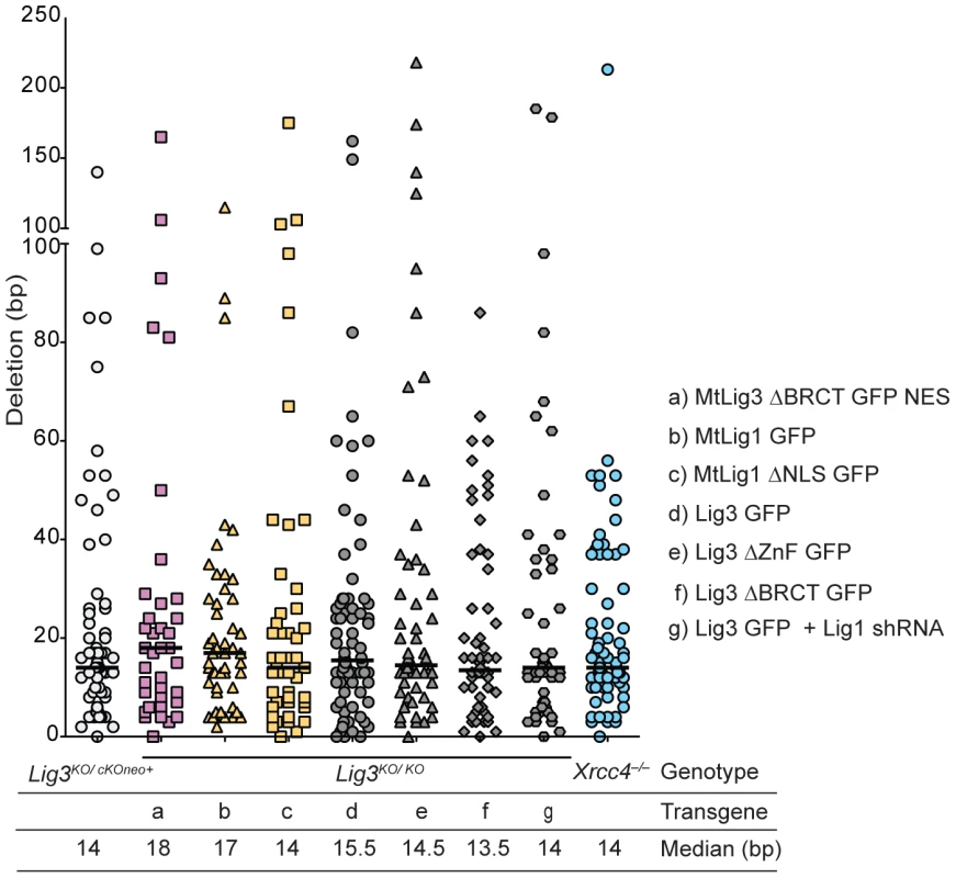
Deletion lengths for the indicated genotypes do not differ significantly from each other. Each value represents the combined deletion from both ends of an individual junction. The median deletion length for each genotype is indicated by a bar on the graph and the value is given below the graph. Tab. 2. Percent of translocation junctions without microhomology and with insertions. 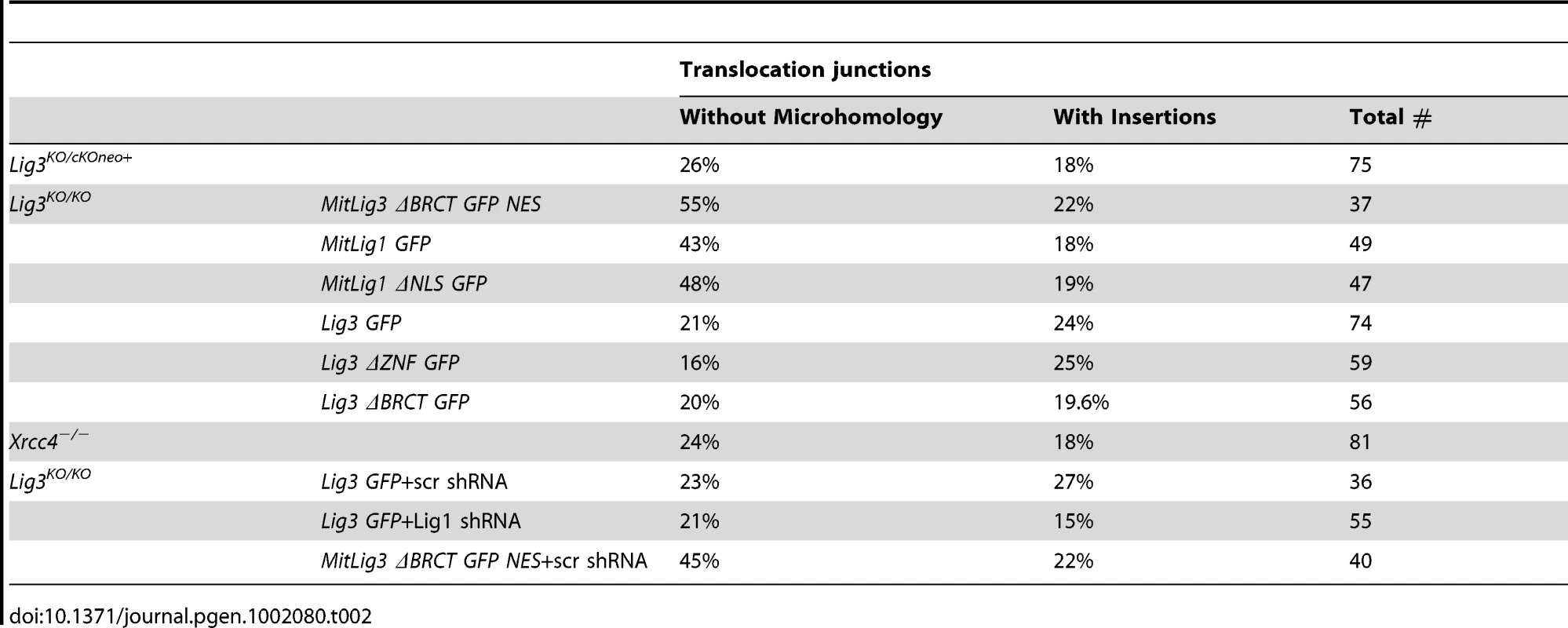
By contrast, translocation junctions from Lig3 null and nuclear-deficient cell lines showed significantly reduced microhomology (compare red and blue dashed lines in Figure 1C and 1D). Notably, the microhomology distribution for junctions from each of the Lig3 null and nuclear Lig3-deficient cells was not significantly different from that expected by chance (Figure S4), indicating that pre-existing microhomology does not drive NHEJ events in the absence of Lig3. Thus, both the reduced frequency and the reduced microhomology at junctions demonstrate a role for Lig3 in alt-NHEJ leading to translocation formation. Unlike microhomology, no significant difference in the deletion and insertion distributions were observed (Figure 3, Figures S5 and S6, Table 2), suggesting that Lig3 does not affect the processing of the ends prior to ligation.
Lig1, but not Lig4, is a backup DNA ligase for translocation formation
As translocations were not completely abolished in the absence of Lig3, we next addressed which of the other two DNA ligases was responsible for the remaining translocations. For this, we performed shRNA knockdowns for Lig1 or Lig4 (Figure 4). As expected, Lig4 depletion significantly increased translocations in cells expressing wild-type Lig3 (7×10⁻⁴; Figure 4, Table 1), similar to Xrcc4−/− cells (9×10⁻⁴; Figure 1B). By contrast, Lig4 depletion had little effect in nuclear Lig3-deficient cells (1.9 vs 1.4×10⁻⁴; Figure 4, Table 1), indicating that in the absence of Lig3 translocations did not occur by canonical NHEJ. While loss of Lig3 reduces translocations in otherwise wild-type cells (2.3-fold, ∼1.6 vs 3.7×10⁻⁴; Figure 4, Table 1), loss of Lig3 in Lig4-depleted cells leads to an even more severe decrease in translocations (3.7-fold, 1.9 vs 7×10⁻⁴, Table 1). These results reinforce the conclusion that Lig3 acts in the alt-NHEJ pathway to promote translocations whether or not Lig4 is present, whereas Lig4 acts in the canonical pathway to suppress translocations.
Fig. 4. Lig1, but not Lig4, is a backup DNA ligase for translocation formation. 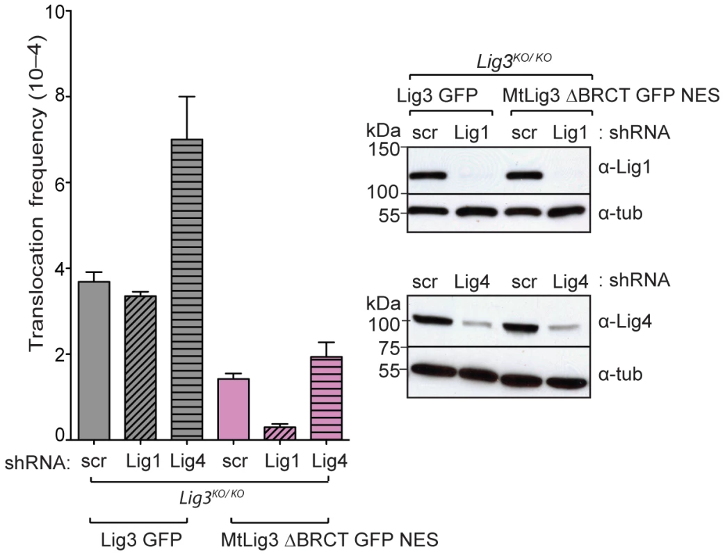
Lig1 depletion results in reduced translocation frequency in nuclear Lig3-deficient cells (Lig3KO/KO; MtLig3 ΔBRCT GFP NES), but not in control cells expressing wild-type Lig3 (Lig3KO/KO; Lig3 GFP). By contrast, Lig4 depletion increases translocations significantly in wild-type cells but not nuclear Lig3-deficient cells. Lig1 and Lig4 lentiviral shRNA knockdown are similar in both nuclear Lig3-deficient cells and cells expressing wild-type Lig3. scr, shRNA with a scrambled sequence. Next we examined the role of Lig1. Short-term depletion of Lig1 had no effect on survival of cells expressing wild-type Lig3, although survival was reduced about 20% in nuclear Lig3-deficient cells. Lig1 depletion in cells expressing wild-type Lig3 did not alter translocation frequency (3.3 vs 3.7×10⁻⁴; Figure 4, Table 1), indicating that Lig1 does not normally play a role in this alt-NHEJ pathway. Notably in nuclear Lig3-deficient cells, very few translocations were recovered upon Lig1 depletion. Thus, translocations were reduced by a factor of ∼12 in nuclear Lig3-deficient cells upon Lig1 depletion compared with cells expressing wild-type Lig3 (0.3 vs 3.7×10⁻⁴; Figure 4, Table 1). These results indicate that Lig1 can back-up Lig3 in alt-NHEJ for translocation formation, but that the participation of Lig1 is prominent only in the absence of Lig3.
Consistent with Lig1 having no effect on translocation frequency, we also observed that microhomology was not affected by Lig1 depletion in cells expressing wild-type Lig3 (p = 0.7859; Figure 1C, Figures S4 and S5). Given the low level of translocations, sufficient numbers of junctions could not be obtained for analysis from nuclear Lig3-deficient cells depleted for Lig1. Upon Lig1 depletion, similar levels of intrachromosomal repair were observed as in wild-type cells, indicating that the reduced frequency of translocations in nuclear Lig3-deficient cells was not due to reduced cleavage of the chromosomal loci (Figure S3C).
Deletion of the ZnF domain of Lig3, but not the XRCC1-interacting BRCT domain, affects translocation frequency and outcome
Having established a role for Lig3 in alt-NHEJ, we next sought to determine which domains of Lig3 are important in this process. The BRCT domain of Lig3 interacts with the scaffold protein XRCC1 [28], which has been implicated in the recruitment of Lig3 to DNA damage foci [32]. When we delete this domain, the protein is still present in the nucleus, although at reduced levels (Figure S7) [32]. Lig3-ΔBRCT Lig3KO/KO cells were found to have indistinguishable translocation frequencies from cells expressing wild-type Lig3 (3.5 vs 3.6×10⁻⁴; Figure 5A, Table 1). Furthermore, translocation junctions showed very similar characteristics (Figure 3, Figures S5 and S6, Table 2), including microhomology (p = 0.9112; Figure 5B, Figure S4). Lig3 interaction with XRCC1, therefore, appears to be dispensable for alt-NHEJ leading to translocations.
Fig. 5. Analysis of Lig3 domain requirements in translocation formation. 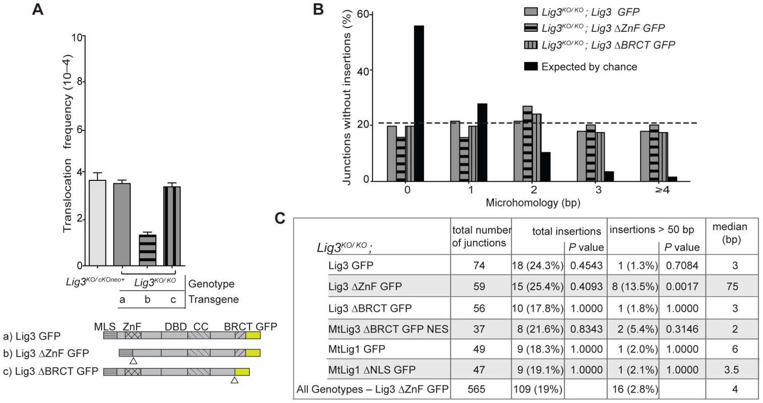
(A) Deletion of the Lig3 BRCT domain has no effect on translocation frequency, while deletion of the Lig3 ZnF domain reduces translocations. Triangles denote the deletions of the indicated domains. ΔZnF, deletion of Lig3 amino acids 89 to 258 including the ZnF itself and additional sequences which corresponds to a previously described Lig3 ΔZnF mutant [37], [38]; ΔBRCT, deletion of Lig3 amino acids 934 to 1009. (B) Microhomology length distributions in translocation junctions are similar in cells expressing wild-type Lig3 and those expressing either ZnF or BRCT deletions of Lig3. (C) Large, complex insertions are more frequent in cells expressing Lig3 deleted for the ZnF domain. The summary “All Genotypes – Lig3 ΔZnF” denotes all junctions analyzed except those from Lig3KO/KO; Lig3 ΔZnF cells. P values are calculated relative to this group with a Fisher's exact test. Lig3 has an N-terminal zinc finger (ZnF) domain which interacts with PARP1 [36] and which has been reported to be critical for its intermolecular ligation activity in vitro [37], [38]. Lig3-ΔZnF Lig3KO/KO cells had a reduced translocation frequency compared with wild-type cells (1.4 vs 3.6×10⁻⁴; Figure 5A, Table 1). Microhomology in Lig3-ΔZnF Lig3KO/KO cells, as well as deletions, were not obviously different from wild-type cells (p = 0.3426; Figure 3 and Figure 5B, Figures S4 and S5, Table 2), in contrast to what was observed with nuclear Lig3-deficient cells. However, a significant fraction of the junctions were unique to the Lig3-ΔZnF Lig3KO/KO cells in that they had long insertions of >50 bp (13.5% vs 2.8% for all other genotypes) (Figure S6), such that the median insertion length was 75 bp compared with 4 bp for all other genotypes (Figure 5C). Taken together, these results suggest that the ZnF domain promotes efficient joining but is not required for microhomology use.
Discussion
The lack of Lig3 in model organisms like yeast and the lack of Lig3 mutant mammalian cells had limited functional studies of Lig3 in vivo. The recent discovery that the cellular viability requirement for Lig3 depends on its role in mitochondria [26], [27] led to the development of cell lines that are deficient for Lig3 in the nucleus [26], allowing us to address the role of Lig3 in chromosomal translocation formation and alt-NHEJ. Using ZFNs, DSBs were introduced into endogenous loci in these cells without prior integration of reporter substrates; nested PCR allowed the recovery of chromosomal translocation junctions within 60 hours. This system works as efficiently in mouse ES cells as in human ES cells [35]. With this approach, we were able to systematically induce and analyze translocations in a variety of ligase deficient backgrounds in mouse cells. Given that they arise by alt-NHEJ even in the presence of the canonical NHEJ [9], translocations provide a good model with which to characterize components of the alt-NHEJ pathway.
Here, we establish that Lig3 is a component of the alt-NHEJ pathway leading to translocations in mouse cells, as Lig3 deficiency leads to a >2-fold decrease in translocation frequency. By examining an extensive number of translocation breakpoint junctions, we observed a redistribution of microhomology with Lig3 loss to that expected by chance, implying that Lig3 favors the use of microhomology during joining. Microhomologies are short, as would be expected from limited end resection which exposes single-strands for annealing [21] and from analysis of translocation junctions found in patients [2]. The use of short microhomologies in chromosomal rearrangements is further underscored by recent genome-wide analysis of breast cancers [39].
We also demonstrate that the role of Lig3 in translocation formation is independent of XRCC1, as deletion of the XRCC1-interacting BRCT domain does not affect either translocation frequency or breakpoint junction characteristics. Although Lig3 and XRCC1 have been suggested to work in a complex [25], our results are consistent with recent studies that have differentiated the roles of Lig3 and XRCC1 in DNA damage repair [26], [27]. In contrast, deletion of the ZnF domain results in a decrease in translocation frequency. The ZnF domain may promote intermolecular ligation in this context, joining two translocation partners, in agreement with a role for this domain in the ligation of oligomers in vitro [38]. The lack of a shift in microhomology use as seen in the absence of Lig3 argues that the ZnF domain is not required for microhomology use in translocations. However, an unusual class of junctions – those with long insertions (>50 bp) – was more prominent in cells expressing this protein. It is conceivable that the deletion of the ZnF domain results in slower kinetics of joining in vivo, which could allow for longer polymerization giving rise to insertions in a subset of junctions.
Consistent with previous results obtained in mouse cells [9], [14], [40], the loss of XRCC4 or Lig4 increases translocation formation, highlighting once again that the canonical NHEJ ligase suppresses translocations. Lig4–XRCC4 could act as a physical barrier together with Ku to prevent access of Lig3 to DNA ends for translocation formation; alternatively, this complex, together with other canonical NHEJ components, could promote the efficient joining of ends to narrow the kinetic window for translocation formation. Importantly, depletion of Lig4 in nuclear Lig3-deficient cells did not increase the translocation frequency as it did in wild-type cells, such that the relief of the translocation suppression by Lig4–XRCC4 is specifically related to Lig3 access to ends. Thus, Lig3 loss leads to an even greater fold decrease in translocation frequency in Lig4-depleted cells (3.7-fold) than it does in otherwise wild-type cells (2.3-fold). This suggests a more dominant role for Lig3 for translocation formation in the absence of the canonical NHEJ ligase.
We further found that Lig1 depletion in wild-type mouse cells does not have any effect on translocation frequency, whereas Lig1 depletion in nuclear Lig3-deficient cells nearly abolishes translocations. This implies that Lig3 has a primary role in alt-NHEJ resulting in translocations, but that Lig1 can function in the absence of Lig3 as a back-up ligase to provide limited activity, suggesting the existence of at least two alt-NHEJ pathways with these ligases operating in a hierarchy. Recent results examining base excision repair have also indicated that Lig1 and Lig3 can act in a hierarchy, although in this case Lig1 appears to be the primary ligase whereas Lig3 acts as the back-up ligase [27]. That these two ligases function in distinct pathways in translocations, as opposed to substituting for each other within one pathway, is supported by the different microhomology distributions: breakpoint junctions formed by Lig3 show a preference for pre-existing microhomology, whereas those formed by Lig1 do not (Figure 6). We cannot exclude that microhomology is generated by short polymerization (polymerase-generated microhomology) [3] that would promote Lig1-dependent joining. Polymerase-generated microhomology could also account for the 0 bp microhomology class of joining events that occur in the presence on Lig3; alternatively, there may not be a strict dependence on microhomology for joining by Lig3. The short polymerization may be template-dependent (Figure 6) or arise by chance in a template-independent manner (not shown). Although polymerase-generated microhomology cannot be scored, the existence of short insertions at translocation breakpoint junctions (either template dependent or independent) provides evidence for polymerization at DNA ends [9].
Fig. 6. Model for the role of the three mammalian DNA ligases in chromosomal translocation formation in mouse cells. 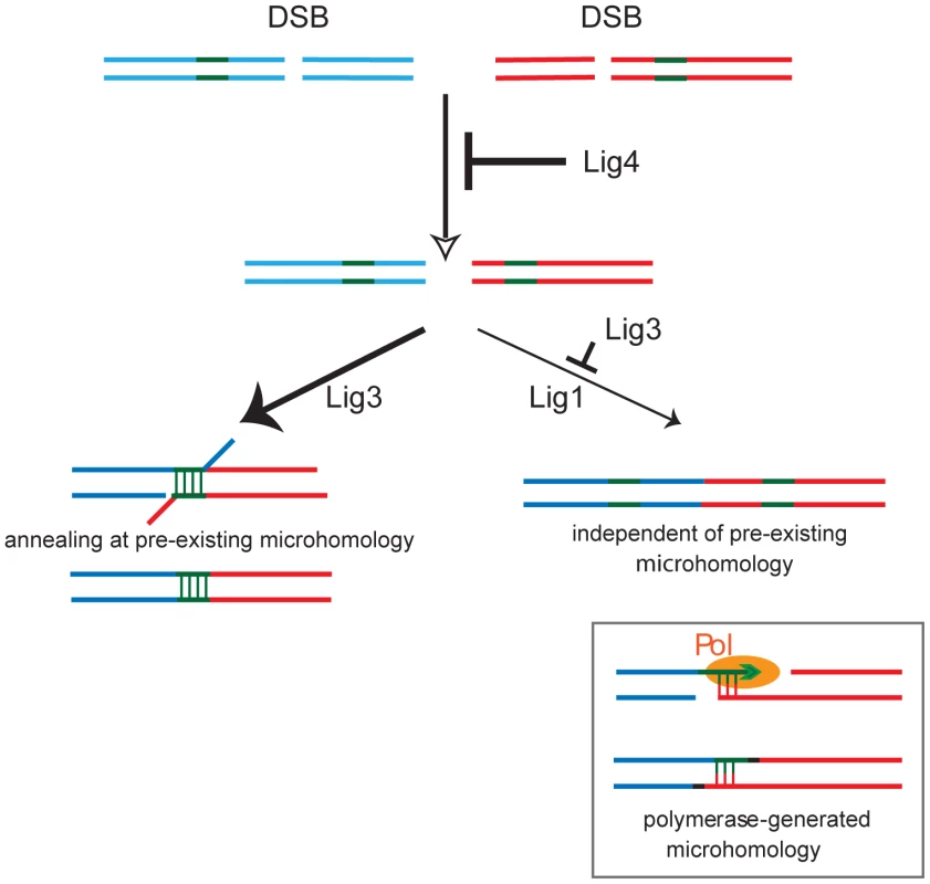
Upon DSB formation on two chromosomes (blue and red lines), Lig4 promotes intrachromosomal joining to maintain genomic integrity and suppresses translocations. Translocations are formed by Lig3, preferentially at short microhomologies (green lines). DNA end resection, which is likely suppressed by Lig4, forms single-stranded DNA to expose microhomologies which can anneal. In the absence of Lig3, Lig1 can form translocations independent of pre-existing microhomology. However, microhomology can also be generated by polymerase (Pol) activity at a DNA end. In the end, NHEJ is a DNA ligation process [41], and here we establish the intricate interplay of the three DNA ligases in alt-NHEJ leading to translocations in mouse cells. Lig3 promotes alt-NHEJ, but in its absence Lig1 can also function in this process. The roles for Lig3 and Lig1 contrast with that of Lig4, which suppresses chromosomal translocations.
Materials and Methods
Western blotting
Whole cell extracts were prepared with Nonidet-P40 buffer and were run on a 7.5% (w/v) Tris-HCl SDS page gel, blotted, and then probed with Lig3 antibody clone 7 (BD Transduction Labs), which recognizes both the human and mouse Lig3 proteins, or Lig1 antibody N-13 (Santa Cruz). α-tubulin (Sigma) was used as a loading control.
Translocation analysis
The ZFNRosa26 pair (Sigma-Aldrich) was designed and tested as described using an archive of validated 2-finger modules [42], [43]. ZFNRosa26 was obtained from Sigma-Aldrich; ZFNH3f3b was previously described [44]. For translocation frequency analysis, a similar approach to the method recently described in human cells was used [35]. Basically, for transfection with ZFN plasmids, 1×10⁶ ES cells were plated in each well of a 6-well plate and 4 hours later, the ZFNRosa26 pair (0.5 µg 18473 and 2.5 µg 18477 plasmids) and ZFNH3f3b pair (0.5 µg 25000 and 0.5 µg 25001) were transfected with Lipofectamine 2000 (Invitrogen), according to manufacturer's instructions. After 6 hours, cells were plated in a 96-well plate at a density of 2×10⁴ cells per plate. For translocation analysis, cells were lysed 60 hours after transfection directly in the 96-well plate in 40 µl lysis buffer (10 mM Tris pH 8.0, 0.45% (v/v) Nonidet P-40, 0.45% (v/v) Tween20) per well. The lysate was incubated with 100 µg/ml Proteinase K at 55°C for 2 hours and then incubated at 95°C for 10 min before PCR. The first round of PCR primers used for der(11) are: Tr(11-6) - F 5′-TTGACGCCTTCCTTCTTCTG-3′ and Tr(11-6) - R 5′ - GCACGTTTCCGACTTGAGTT -3′, used at an annealing temperature of 62°C. The second round, nested PCR primers are: Tr(11-6) - NF 5′ - CTGCCATTCCAGAGATTGGT -3′ and Tr(11-6) - NF 5′ - TCCCAAAGTCGCTCTGAGTT -3′, used at an annealing temperature of 62°C. For PCR quantification of der(6) translocation frequency, the primers indicated in Figure 2 were: Tr(6-11) - F 5′-GCGGGAGAAATGGATATGAA-3′, Tr(6-11) - R 5′-AACCTTTGAAAAAGCCCACA-3′, Tr(6-11) - NF 5′-GGCGGATCACAAGCAATAAT-3′, and Tr(6-11) - NR 5′-AGCCACAGTGCTCACATCAC-3′. The first round of PCR was performed with 7 µl cell lysate from each well in a total of 50 µl per well (24 cycles, annealing temperature of 60°C). Then, 0.5 µl of the first PCR was used in a second, nested PCR (40 cycles, annealing temperature of 60°C) with SYBR Green for qPCR (Stratagene MX3005). The PCR cycle contains a denaturation curve cycle. Nested PCR fragments corresponding to translocation junctions are ∼503 bp, and have melting temperature between 87–90°C. For each experiment, cells were counted at the time of lysis to ensure that there was no growth perturbation in the experiment. For breakpoint sequence analysis, PCR reactions positive for translocation formation were purified using a PCR purification kit (Invitrogen) and sent for sequencing with Tr(6-11)-1NF and Tr(6-11)-1NR primers.
Lentiviral Lig1 and Lig4 knock down
Purified pLKO.1-puro plasmids containing shRNA (Sigma) were transfected into 293T cells, using Mission Lentiviral packaging mix (Sigma, SHP001) and Fugene 6 (Roche). Infectious lentiviruses were harvested at 48 hours posttransfection and filtered through a 0.45 µm filter. 4×10⁴ ES-cells per well were seeded in 12-well plates with medium containing 4 µg/ml polybrene (Sigma). After 16 hours, cells were incubated with 750 µl of lentiviral particles with 4 µg/ml polybrene. After 4 hours, 750 µl of ES cell medium was added. After 24 hours, medium was changed with 1.6 µg/ml puromycin (Sigma) and after 3.5 days in puromycin selection cells are plated for translocation analysis.
Surveyor nuclease assay
Surveyor nuclease assay was performed as previously described [42]. Basically, 1×10⁶ ES-cells transfected with none or both of the ZFNRosa26 and ZFNH3f3b were used as templates to amplify either Rosa26 or H3f3b regions. These samples were further used to quantify translocations. The Rosa26 or H3f3b region was amplified in the presence of 32P labelled dNTPs using AccuPrime taq DNA polymerase (Invitrogen) with the following primers, respectively: Rosa26FW1 5′ - TAAAACTCGGGTGAGCATGT -3′ and RosaRv1 5′ - GGAGTTCTCTGCTGCCTCCTG -3′ with an annealing temperature of 61°C; H3f3bFw 5′ - GCGGCGGCTTGATTGCTCCAG -3′ and H3Rv1 5′ - AGCAACTTGTCACTCCTGAGCCAC -3′ with an annealing temperature of 61°C. 2 µL of each PCR was mixed with 1× Accuprime buffer II and incubated as follows: 95°C for 5 min, 95–85°C at −2°C/s, 85–25°C at −0.1°C/s; hold at 4°C. This step melts and randomly reanneals the amplicons, which converts any mutations into mismatched duplex DNA. 1 µL Surveyor nuclease (Transgenomic) was incubated for 20 min at 42°C and the sample was run in a 10% acrylamide (BioRad). The cleaved bands were quantified by ImageQuant 5.1. Imprecise NHEJ percent is calculated using the formula: % indel = 100×(1−(1−fraction cleaved)1/2).
Single-break NHEJ by bacterial colony hybridization
Mouse ES cells transfected with none or both of the ZFNRosa26 and ZFNH3f3b were used as templates to amplify either the Rosa26 or H3f3b region. The same primers used in the Surveyor nuclease assay were used. Amplicons were cloned by TOPO-TA (Invitrogen) and transformed into bacteria, similar to an approach recently described [35]. The probes used to determine precise NHEJ for Rosa26 or H3f3b loci are as follows, respectively: ProbeRosa26Nor - 5′ - CGCCCATCTTCTAGAAAG-3′; ProbeH3Nor - 5′-CCAGTTGGCTCGCCGGAT-3′.
Supporting Information
Zdroje
1. ManiRSChinnaiyanAM 2010 Triggers for genomic rearrangements: insights into genomic, cellular and environmental influences. Nat Rev Genet 11 819 829
2. WeinstockDMElliottBJasinM 2006 A model of oncogenic rearrangements: differences between chromosomal translocation mechanisms and simple double-strand break repair. Blood 107 777 780
3. LieberMR 2010 The Mechanism of Double-Strand DNA Break Repair by the Nonhomologous DNA End-Joining Pathway. Annu Rev Biochem
4. JeggoPA 1998 Identification of genes involved in repair of DNA double-strand breaks in mammalian cells. Radiat Res 150 S80 91
5. LieberMRMaYPannickeUSchwarzK 2003 Mechanism and regulation of human non-homologous DNA end-joining. Nat Rev Mol Cell Biol 4 712 720
6. DifilippantonioMJPetersenSChenHTJohnsonRJasinM 2002 Evidence for replicative repair of DNA double-strand breaks leading to oncogenic translocation and gene amplification. J Exp Med 196 469 480
7. ZhuCMillsKDFergusonDOLeeCManisJ 2002 Unrepaired DNA breaks in p53-deficient cells lead to oncogenic gene amplification subsequent to translocations. Cell 109 811 821
8. WeinstockDMBrunetEJasinM 2007 Formation of NHEJ-derived reciprocal chromosomal translocations does not require Ku70. Nat Cell Biol 9 978 981
9. SimsekDJasinM 2010 Alternative end-joining is suppressed by the canonical NHEJ component Xrcc4-ligase IV during chromosomal translocation formation. Nat Struct Mol Biol 17 410 416
10. LiangFJasinM 1996 Ku80-deficient cells exhibit excess degradation of extrachromosomal DNA. J Biol Chem 271 14405 14411
11. DelacoteFHanMStamatoTDJasinMLopezBS 2002 An xrcc4 defect or Wortmannin stimulates homologous recombination specifically induced by double-strand breaks in mammalian cells. Nucleic Acids Res 30 3454 3463
12. Guirouilh-BarbatJHuckSBertrandPPirzioLDesmazeC 2004 Impact of the KU80 pathway on NHEJ-induced genome rearrangements in mammalian cells. Mol Cell 14 611 623
13. Guirouilh-BarbatJRassEPloIBertrandPLopezBS 2007 Defects in XRCC4 and KU80 differentially affect the joining of distal nonhomologous ends. Proc Natl Acad Sci U S A 104 20902 20907
14. YanCTBoboilaCSouzaEKFrancoSHickernellTR 2007 IgH class switching and translocations use a robust non-classical end-joining pathway. Nature 449 478 482
15. CorneoBWendlandRLDerianoLCuiXKleinIA 2007 Rag mutations reveal robust alternative end joining. Nature 449 483 486
16. FattahFLeeEHWeisenselNWangYLichterN 2010 Ku regulates the non-homologous end joining pathway choice of DNA double-strand break repair in human somatic cells. PLoS Genet 6 e1000855
17. RassEGrabarzAPloIGautierJBertrandP 2009 Role of Mre11 in chromosomal nonhomologous end joining in mammalian cells. Nat Struct Mol Biol 16 819 824
18. DinkelmannMSpehalskiEStonehamTBuisJWuY 2009 Multiple functions of MRN in end-joining pathways during isotype class switching. Nat Struct Mol Biol 16 808 813
19. XieAKwokAScullyR 2009 Role of mammalian Mre11 in classical and alternative nonhomologous end joining. Nat Struct Mol Biol 16 814 818
20. BennardoNChengAHuangNStarkJM 2008 Alternative-NHEJ is a mechanistically distinct pathway of mammalian chromosome break repair. PLoS Genet 4 e1000110
21. ZhangYJasinM 2011 An essential role for CtIP in chromosomal translocation formation through an alternative end-joining pathway. Nat Struct Mol Biol 18 80 84
22. Lee-TheilenMMatthewsAJKellyDZhengSChaudhuriJ 2011 CtIP promotes microhomology-mediated alternative end joining during class-switch recombination. Nat Struct Mol Biol 18 75 79
23. AudebertMSallesBCalsouP 2004 Involvement of poly(ADP-ribose) polymerase-1 and XRCC1/DNA ligase III in an alternative route for DNA double-strand breaks rejoining. J Biol Chem 279 55117 55126
24. WangMWuWRosidiBZhangLWangH 2006 PARP-1 and Ku compete for repair of DNA double strand breaks by distinct NHEJ pathways. Nucleic Acids Res 34 6170 6182
25. EllenbergerTTomkinsonAE 2008 Eukaryotic DNA ligases: structural and functional insights. Annu Rev Biochem 77 313 338
26. SimsekDFurdaAGaoYArtusJBrunetE 2011 Crucial role for DNA ligase III in mitochondria but not in Xrcc1-dependent repair. Nature 471 245 248
27. GaoYKatyalSLeeYZhaoJRehgJE 2011 DNA ligase III is critical for mtDNA integrity but not Xrcc1-mediated nuclear DNA repair. Nature 471 240 244
28. CaldecottKWMcKeownCKTuckerJDLjungquistSThompsonLH 1994 An interaction between the mammalian DNA repair protein XRCC1 and DNA ligase III. Mol Cell Biol 14 68 76
29. NashRACaldecottKWBarnesDELindahlT 1997 XRCC1 protein interacts with one of two distinct forms of DNA ligase III. Biochemistry 36 5207 5211
30. WangHRosidiBPerraultRWangMZhangL 2005 DNA ligase III as a candidate component of backup pathways of nonhomologous end joining. Cancer Res 65 4020 4030
31. Puebla-OsorioNLaceyDBAltFWZhuC 2006 Early embryonic lethality due to targeted inactivation of DNA ligase III. Mol Cell Biol 26 3935 3941
32. MortusewiczORothbauerUCardosoMCLeonhardtH 2006 Differential recruitment of DNA Ligase I and III to DNA repair sites. Nucleic Acids Res 34 3523 3532
33. HendersonBREleftheriouA 2000 A comparison of the activity, sequence specificity, and CRM1-dependence of different nuclear export signals. Exp Cell Res 256 213 224
34. UrnovFDRebarEJHolmesMCZhangHSGregoryPD 2010 Genome editing with engineered zinc finger nucleases. Nat Rev Genet 11 636 646
35. BrunetESimsekDTomishimaMDeKelverRChoiVM 2009 Chromosomal translocations induced at specified loci in human stem cells. Proc Natl Acad Sci U S A 106 10620 10625
36. LeppardJBDongZMackeyZBTomkinsonAE 2003 Physical and functional interaction between DNA ligase IIIalpha and poly(ADP-Ribose) polymerase 1 in DNA single-strand break repair. Mol Cell Biol 23 5919 5927
37. TaylorRMWhitehouseCJCaldecottKW 2000 The DNA ligase III zinc finger stimulates binding to DNA secondary structure and promotes end joining. Nucleic Acids Res 28 3558 3563
38. Cotner-GoharaEKimIKTomkinsonAEEllenbergerT 2008 Two DNA-binding and nick recognition modules in human DNA ligase III. J Biol Chem 283 10764 10772
39. StephensPJMcBrideDJLinMLVarelaIPleasanceED 2009 Complex landscapes of somatic rearrangement in human breast cancer genomes. Nature 462 1005 1010
40. BoboilaCJankovicMYanCTWangJHWesemannDR 2010 Alternative end-joining catalyzes robust IgH locus deletions and translocations in the combined absence of ligase 4 and Ku70. Proc Natl Acad Sci U S A 107 3034 3039
41. LieberMRWilsonTE SnapShot: Nonhomologous DNA end joining (NHEJ). Cell 142 496 496 e491
42. HockemeyerDSoldnerFBeardCGaoQMitalipovaM 2009 Efficient targeting of expressed and silent genes in human ESCs and iPSCs using zinc-finger nucleases. Nat Biotechnol 27 851 857
43. UrnovFDMillerJCLeeYLBeausejourCMRockJM 2005 Highly efficient endogenous human gene correction using designed zinc-finger nucleases. Nature 435 646 651
44. GoldbergADBanaszynskiLANohKMLewisPWElsaesserSJ 2010 Distinct factors control histone variant H3.3 localization at specific genomic regions. Cell 140 678 691
Štítky
Genetika Reprodukční medicína
Článek Local Absence of Secondary Structure Permits Translation of mRNAs that Lack Ribosome-Binding SitesČlánek Independent Chromatin Binding of ARGONAUTE4 and SPT5L/KTF1 Mediates Transcriptional Gene SilencingČlánek Trade-Off between Bile Resistance and Nutritional Competence Drives Diversification in the Mouse GutČlánek FGF Signaling Regulates the Number of Posterior Taste Papillae by Controlling Progenitor Field SizeČlánek Mammalian BTBD12 (SLX4) Protects against Genomic Instability during Mammalian SpermatogenesisČlánek Identification of Nine Novel Loci Associated with White Blood Cell Subtypes in a Japanese PopulationČlánek Differential Effects of and Risk Variants on Association with Diabetic ESRD in African AmericansČlánek Dynamic Chromatin Localization of Sirt6 Shapes Stress- and Aging-Related Transcriptional Networks
Článek vyšel v časopisePLOS Genetics
Nejčtenější tento týden
2011 Číslo 6
-
Všechny články tohoto čísla
- Local Absence of Secondary Structure Permits Translation of mRNAs that Lack Ribosome-Binding Sites
- Statistical Inference on the Mechanisms of Genome Evolution
- Revisiting Heterochromatin in Embryonic Stem Cells
- A Two-Stage Meta-Analysis Identifies Several New Loci for Parkinson's Disease
- Identification of a Sudden Cardiac Death Susceptibility Locus at 2q24.2 through Genome-Wide Association in European Ancestry Individuals
- Genomic Prevalence of Heterochromatic H3K9me2 and Transcription Do Not Discriminate Pluripotent from Terminally Differentiated Cells
- Epistasis between Beneficial Mutations and the Phenotype-to-Fitness Map for a ssDNA Virus
- Recurrent Chromosome 16p13.1 Duplications Are a Risk Factor for Aortic Dissections
- Telomere DNA Deficiency Is Associated with Development of Human Embryonic Aneuploidy
- Genome-Wide Association Study of White Blood Cell Count in 16,388 African Americans: the Continental Origins and Genetic Epidemiology Network (COGENT)
- Unexpected Role for DNA Polymerase I As a Source of Genetic Variability
- Transportin-SR Is Required for Proper Splicing of Genes and Plant Immunity
- How Chromatin Is Remodelled during DNA Repair of UV-Induced DNA Damage in
- Independent Chromatin Binding of ARGONAUTE4 and SPT5L/KTF1 Mediates Transcriptional Gene Silencing
- Two Evolutionary Histories in the Genome of Rice: the Roles of Domestication Genes
- Natural Allelic Variation Defines a Role for : Trichome Cell Fate Determination
- Multiple Common Susceptibility Variants near BMP Pathway Loci , , and Explain Part of the Missing Heritability of Colorectal Cancer
- Pathogenic Mechanism of the FIG4 Mutation Responsible for Charcot-Marie-Tooth Disease CMT4J
- A Functional Variant in Promoter Modulates Its Expression and Confers Disease Risk for Systemic Lupus Erythematosus
- Drift and Genome Complexity Revisited
- Chromosomal Macrodomains and Associated Proteins: Implications for DNA Organization and Replication in Gram Negative Bacteria
- Trade-Off between Bile Resistance and Nutritional Competence Drives Diversification in the Mouse Gut
- Pathways of Distinction Analysis: A New Technique for Multi–SNP Analysis of GWAS Data
- Web-Based Genome-Wide Association Study Identifies Two Novel Loci and a Substantial Genetic Component for Parkinson's Disease
- Chk2 and p53 Are Haploinsufficient with Dependent and Independent Functions to Eliminate Cells after Telomere Loss
- Exome Sequencing Identifies Mutations in High Myopia
- Distinct Functional Constraints Partition Sequence Conservation in a -Regulatory Element
- CorE from Is a Copper-Dependent RNA Polymerase Sigma Factor
- A Single Sex Pheromone Receptor Determines Chemical Response Specificity of Sexual Behavior in the Silkmoth
- FGF Signaling Regulates the Number of Posterior Taste Papillae by Controlling Progenitor Field Size
- Maps of Open Chromatin Guide the Functional Follow-Up of Genome-Wide Association Signals: Application to Hematological Traits
- Increased Susceptibility to Cortical Spreading Depression in the Mouse Model of Familial Hemiplegic Migraine Type 2
- Differential Gene Expression and Epiregulation of Alpha Zein Gene Copies in Maize Haplotypes
- Parallel Adaptive Divergence among Geographically Diverse Human Populations
- Genetic Analysis of Genome-Scale Recombination Rate Evolution in House Mice
- Mechanisms for the Evolution of a Derived Function in the Ancestral Glucocorticoid Receptor
- Mammalian BTBD12 (SLX4) Protects against Genomic Instability during Mammalian Spermatogenesis
- Interferon Regulatory Factor 8 Regulates Pathways for Antigen Presentation in Myeloid Cells and during Tuberculosis
- High-Resolution Analysis of Parent-of-Origin Allelic Expression in the Arabidopsis Endosperm
- Specific SKN-1/Nrf Stress Responses to Perturbations in Translation Elongation and Proteasome Activity
- Graded Nodal/Activin Signaling Titrates Conversion of Quantitative Phospho-Smad2 Levels into Qualitative Embryonic Stem Cell Fate Decisions
- Genome-Wide Analysis Reveals PADI4 Cooperates with Elk-1 to Activate Expression in Breast Cancer Cells
- Trait Variation in Yeast Is Defined by Population History
- Meiosis-Specific Loading of the Centromere-Specific Histone CENH3 in
- A Genome-Wide Survey of Imprinted Genes in Rice Seeds Reveals Imprinting Primarily Occurs in the Endosperm
- Multiple Regulatory Mechanisms to Inhibit Untimely Initiation of DNA Replication Are Important for Stable Genome Maintenance
- SIRT1 Promotes N-Myc Oncogenesis through a Positive Feedback Loop Involving the Effects of MKP3 and ERK on N-Myc Protein Stability
- Bacteriophage Crosstalk: Coordination of Prophage Induction by Trans-Acting Antirepressors
- Role of the Single-Stranded DNA–Binding Protein SsbB in Pneumococcal Transformation: Maintenance of a Reservoir for Genetic Plasticity
- Genomic Convergence among ERRα, PROX1, and BMAL1 in the Control of Metabolic Clock Outputs
- Genome-Wide Association of Bipolar Disorder Suggests an Enrichment of Replicable Associations in Regions near Genes
- Identification of Nine Novel Loci Associated with White Blood Cell Subtypes in a Japanese Population
- DNA Ligase III Promotes Alternative Nonhomologous End-Joining during Chromosomal Translocation Formation
- Differential Effects of and Risk Variants on Association with Diabetic ESRD in African Americans
- Finished Genome of the Fungal Wheat Pathogen Reveals Dispensome Structure, Chromosome Plasticity, and Stealth Pathogenesis
- Dynamic Chromatin Localization of Sirt6 Shapes Stress- and Aging-Related Transcriptional Networks
- Extracellular Matrix Dynamics in Hepatocarcinogenesis: a Comparative Proteomics Study of Transgenic and Null Mouse Models
- Integrating 5-Hydroxymethylcytosine into the Epigenomic Landscape of Human Embryonic Stem Cells
- Vive La Différence: An Interview with Catherine Dulac
- Multiple Loci Are Associated with White Blood Cell Phenotypes
- Nuclear Accumulation of Stress Response mRNAs Contributes to the Neurodegeneration Caused by Fragile X Premutation rCGG Repeats
- A New Mutation Affecting FRQ-Less Rhythms in the Circadian System of
- Cryptic Transcription Mediates Repression of Subtelomeric Metal Homeostasis Genes
- A New Isoform of the Histone Demethylase JMJD2A/KDM4A Is Required for Skeletal Muscle Differentiation
- Genetic Determinants of Lipid Traits in Diverse Populations from the Population Architecture using Genomics and Epidemiology (PAGE) Study
- A Genome-Wide RNAi Screen for Factors Involved in Neuronal Specification in
- PLOS Genetics
- Archiv čísel
- Aktuální číslo
- Informace o časopisu
Nejčtenější v tomto čísle- Recurrent Chromosome 16p13.1 Duplications Are a Risk Factor for Aortic Dissections
- Statistical Inference on the Mechanisms of Genome Evolution
- Genome-Wide Association Study of White Blood Cell Count in 16,388 African Americans: the Continental Origins and Genetic Epidemiology Network (COGENT)
- Chromosomal Macrodomains and Associated Proteins: Implications for DNA Organization and Replication in Gram Negative Bacteria
Kurzy
Zvyšte si kvalifikaci online z pohodlí domova
Současné možnosti léčby obezity
nový kurzAutoři: MUDr. Martin Hrubý
Všechny kurzyPřihlášení#ADS_BOTTOM_SCRIPTS#Zapomenuté hesloZadejte e-mailovou adresu, se kterou jste vytvářel(a) účet, budou Vám na ni zaslány informace k nastavení nového hesla.
- Vzdělávání


