-
Články
Top novinky
Reklama- Vzdělávání
- Časopisy
Top články
Nové číslo
- Témata
Top novinky
Reklama- Videa
- Podcasty
Nové podcasty
Reklama- Kariéra
Doporučené pozice
Reklama- Praxe
Top novinky
ReklamaPreventing Sepsis through the Inhibition of Its Agglutination in Blood
Staphylococcus aureus infection is a frequent cause of sepsis in humans, a disease associated with high mortality and without specific intervention. When suspended in human or animal plasma, staphylococci are known to agglutinate, however the bacterial factors responsible for agglutination and their possible contribution to disease pathogenesis have not yet been revealed. Using a mouse model for S. aureus sepsis, we report here that staphylococcal agglutination in blood was associated with a lethal outcome of this disease. Three secreted products of staphylococci - coagulase (Coa), von Willebrand factor binding protein (vWbp) and clumping factor (ClfA) – were required for agglutination. Coa and vWbp activate prothrombin to cleave fibrinogen, whereas ClfA allowed staphylococci to associate with the resulting fibrin cables. All three virulence genes promoted the formation of thromboembolic lesions in heart tissues. S. aureus agglutination could be disrupted and the lethal outcome of sepsis could be prevented by combining dabigatran-etexilate treatment, which blocked Coa and vWbp activity, with antibodies specific for ClfA. Together these results suggest that the combined administration of direct thrombin inhibitors and ClfA-antibodies that block S. aureus agglutination with fibrin may be useful for the prevention of staphylococcal sepsis in humans.
Published in the journal: . PLoS Pathog 7(10): e32767. doi:10.1371/journal.ppat.1002307
Category: Research Article
doi: https://doi.org/10.1371/journal.ppat.1002307Summary
Staphylococcus aureus infection is a frequent cause of sepsis in humans, a disease associated with high mortality and without specific intervention. When suspended in human or animal plasma, staphylococci are known to agglutinate, however the bacterial factors responsible for agglutination and their possible contribution to disease pathogenesis have not yet been revealed. Using a mouse model for S. aureus sepsis, we report here that staphylococcal agglutination in blood was associated with a lethal outcome of this disease. Three secreted products of staphylococci - coagulase (Coa), von Willebrand factor binding protein (vWbp) and clumping factor (ClfA) – were required for agglutination. Coa and vWbp activate prothrombin to cleave fibrinogen, whereas ClfA allowed staphylococci to associate with the resulting fibrin cables. All three virulence genes promoted the formation of thromboembolic lesions in heart tissues. S. aureus agglutination could be disrupted and the lethal outcome of sepsis could be prevented by combining dabigatran-etexilate treatment, which blocked Coa and vWbp activity, with antibodies specific for ClfA. Together these results suggest that the combined administration of direct thrombin inhibitors and ClfA-antibodies that block S. aureus agglutination with fibrin may be useful for the prevention of staphylococcal sepsis in humans.
Introduction
The Gram-positive bacterium Staphylococcus aureus is the causative agent of human skin and soft tissue infections, invasive disease and bacteremia [1]. Staphylococcal bacteremia leads to endocarditis and sepsis, diseases that, even under antibiotic therapy, are associated with high mortality [2]. Community - and hospital-acquired infections are frequently caused by antibiotic (methicillin)-resistant S. aureus (MRSA) [3], resulting in poor disease outcomes following the failure of antibiotic therapy [4]. A preventive strategy that can reduce the burden and improve the outcomes of S. aureus sepsis is therefore urgently needed [5].
S. aureus is a unique disease pathogen owing to its multiple interactions with fibrinogen [6], [7], [8], a highly abundant host protein responsible for the formation of fibrin clots following cleavage by thrombin [9]. Fibrinogen is a glycoprotein with Mr ∼340,000, formed by three pairs of Aα-, Bβ-, and γ-chains covalently linked to form a “dimer of trimers,” where A and B designate the fibrinopeptides released by thrombin cleavage [10]. The elongated molecule folds into three separate domains, a central domain E that contains the N-termini of all six chains and two flanking domains D formed mainly by the C-termini of the Bβ - and γ-chains [9]. These globular domains are connected by long triple-helical structures [9].
S. aureus secretes two coagulases, Coa and von-Willebrand factor binding protein (vWbp), polypeptides that also promote cleavage of the Aα and Bß chains of fibrinogen to generate fibrin clots [11]. Coagulases conformationally activate the central coagulation zymogen prothrombin [10]. The crystal structure of the active complex revealed binding of the D1 and D2 domains of coagulases to prothrombin and insertion of their Ile1-Val2 N-terminus into the Ile16 pocket of the zymogen, inducing a functional active site through conformational change [11]. Exosite I of α-thrombin, the fibrinogen recognition site, and proexosite I on prothrombin are blocked by the D2 of Coa [11]. Nevertheless, association of the tetrameric (Coa·prothrombin)2 complex enables fibrinogen binding at a new site with high affinity [10]. This model explains the coagulant properties and efficient fibrinogen conversion by coagulases [10]. S. aureus mutants lacking both coagulases, coa and vwb, are unable to form abscesses in a mouse model of staphylococcal diseases [12]. When used as a purified antigen, Coa and vWbp elicit protective immune responses that prevent the formation of abscesses in the same model [12].
The coagulation of calcium-chelated plasma following incubation with bacteria [12] is still used in clinical laboratories to distinguish S. aureus isolates from non-pathogenic staphylococci (coagulase test) [13]. Another diagnostic tool, the slide agglutination test, monitors the agglutination of S. aureus immersed in calcium-chelated plasma [14]. The biochemical attributes and physiological relevance of staphylococcal agglutination are not yet known. S. aureus strains express clumping factor A (ClfA) [15], a surface protein that promotes precipitation of staphylococci through association with soluble fibrinogen (clumping reaction) [16], [17], [18]. The N2 and N3 domains of ClfA (residues 229–545) bind to the C-terminal end of the fibrinogen γ-chains (residues 395–411) [19], [20]. S. aureus mutants lacking functional clfA display virulence defects in mouse models for septic arthritis or endocarditis, phenotypes that have been attributed to the loss of staphylococcal binding to fibrinogen deposited on inflamed joint tissues or on mechanically damaged heart valves [21], [22]. ClfA also contributes to staphylococcal escape from phagocytic killing, which involves its binding to complement regulatory factor I [23]. A ClfA-specific monoclonal antibody has been isolated that blocks staphylococcal association with the fibrinogen γ-chain [24]. A phase II clinical trial with bacteremic patients compared the efficacy of monoclonal antibody (Tefibazumab) and antibiotic treatment with placebo and antibiotic. However, composite clinical end point analysis did not detect differences between placebo and antibody [25].
Birch-Hirschfeld employed a biochemical approach to elucidate S. aureus agglutination in citrate-plasma and proposed a reaction pathway involving both fibrinogen and prothrombin [26]. This work suggests a considerably more complex mechanism for S. aureus agglutination rather than the direct association of bacteria with fibrinogen (clumping). To explore this possibility, we have searched for staphylococcal mutants that are defective for agglutination and/or sepsis with the purpose of identifying new preventive strategies for this disease.
Results
Surface proteins contribute to staphylococcal sepsis
We previously developed an animal model to examine the genetic requirements for staphylococcal sepsis [27]. Briefly, S. aureus Newman, 1×10⁸ CFU, is injected into the retro-orbital plexus of BALB/c mice, resulting in 100% lethality over a ten day observation period [27]. This model was used to examine the contribution of secreted coagulases to staphylococcal sepsis [12]. S. aureus Newman mutants lacking the coa and vwb genes displayed increased time-to-death and increased survival phenotypes [12](Table 1). Earlier work identified sortase A (SrtA), an enzyme that links surface proteins to the staphylococcal cell wall envelope [28], as an essential virulence factor for sepsis [27]. Nevertheless, these studies left unresolved which surface protein(s) play a key role in this disease process. S. aureus mutants with insertional lesions in any one of eighteen surface protein genes [29] were tested for their role in sepsis (Table 1). These experiments identified clumping factor A (ClfA) as the single most important contributor (Table 1). Although mutations in clfA diminished the severity of clinical disease and improved the outcome of sepsis, clfA mutants retained significant virulence and were still capable of killing infected animals, unlike srtA variants (Table 1).
Tab. 1. Surface protein genes and their contribution to S. aureus sepsis. 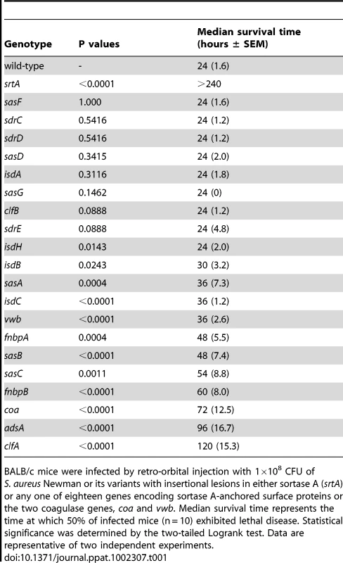
BALB/c mice were infected by retro-orbital injection with 1×10⁸ CFU of S. aureus Newman or its variants with insertional lesions in either sortase A (srtA) or any one of eighteen genes encoding sortase A-anchored surface proteins or the two coagulase genes, coa and vwb. Median survival time represents the time at which 50% of infected mice (n = 10) exhibited lethal disease. Statistical significance was determined by the two-tailed Logrank test. Data are representative of two independent experiments. Genetic requirements for staphylococcal agglutination
S. aureus Newman mutants with defined genetic lesions [29] were screened for defects in agglutination (Fig. 1A). Mutations that abrogated the secretion of only one of the two coagulases, Coa [30] or vWbp [31], had little or no effect on agglutination (Fig. 1AB). In contrast, a mutant lacking both genes (coa/vwb) was severely impaired for agglutination, similar to a clfA variant (Fig. 1AB). A mutant lacking all three genes - coa, vwb, and clfA - was unable to agglutinate in plasma (Fig. 1AB). Mutants with insertional lesions in other known fibrinogen binding proteins, efb [32], [33] and clfB [34], did not cause large defects in agglutination (Fig. 1AB). The phenotypic agglutination defects of coa/vwb as well as clfA mutants could be restored by transformation of staphylococci with pcoa-vwb and pclfA, respectively, plasmids encoding wild-type alleles to the corresponding mutational lesions (Fig. 1C). Thus, unlike ClfA-mediated clumping of staphylococci via binding to fibrinogen [15], S. aureus agglutination appears to be a multi-factorial process involving coagulases, ClfA, as well as fibrinogen and prothrombin [26].
Fig. 1. Staphylococcus aureus agglutination in citrate-plasma is a multi-factorial process and essential for the pathogenesis of sepsis in mice. 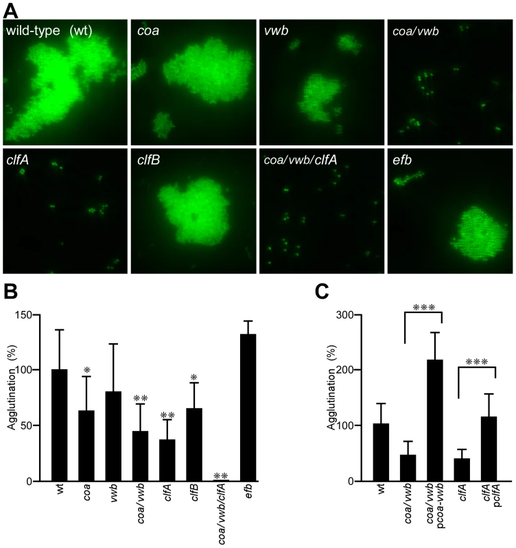
(A) Agglutination in EDTA-plasma of Syto-9 stained S. aureus Newman wild-type (wt) or its isogenic mutants with insertional lesions in single or multiple genes: coa (coagulase), vwb (von Willebrand factor binding protein), clfA (clumping factor A), clfB and efb (extracellular fibrinogen binding protein). (B) Quantification of agglutination for staphylococcal mutants (A) expressed as the percent relative to wt (100%). Average and standard error of the means were calculated from sixteen fields of microscopic view and statistical significance was assessed in pairwise comparison between wt and mutant with the two-tailed Student's t-test: *P<0.01, **P<0.0001. (C) Complementation studies of staphylococcal agglutination using the slide agglutination test. S. aureus Newman variants coa/vwb and clfA were transformed with plasmids pcoa-vwb and pclfA, respectively. Statistical significance was analyzed by two-tailed Student's t-test; ***P<0.0001. Staphylococcal agglutination in septic mice
To test whether staphylococcal agglutination occurred in mice with sepsis, the hearts of animals that had succumbed to S. aureus Newman challenge were examined for histopathology (Fig. 2). Deposits of large numbers of staphylococci, mostly without immune cell infiltrates, were identified in hematoxylin-eosin stained heart tissue twelve hours after infection (Fig. 2A–D). The appearance of these staphylococcal agglutinations is consistent with the general concept of thromboembolic deposition of S. aureus during sepsis [35] (Fig. 2A). Immuno-histochemical staining was used to detect specific agglutination factors (Fig. 2E). These experiments identified prothrombin and fibrinogen (fibrin) in the immediate vicinity of staphylococcal agglutinations (Fig. 2E). In agreement with the hypothesis that agglutination contributes to the pathogenesis of sepsis, fewer heart lesions were observed when mice were challenged with either clfA or coa/vwb variants (Fig. 3). Of note, heart tissues of animals necropsied twelve hours after intravenous challenge harbored considerable loads of staphylococci, irrespective of the challenge strain. Nevertheless, histopathology features of heart lesions associated with clfA or coa/vwb variants revealed immune cell infiltrates in the absence of staphylococcal agglutinations (Fig. 3B). A mutant lacking all three agglutination factors - clfA, coa and vwb - failed to generate either immune cell infiltrates or S. aureus agglutinations in heart tissues (Fig. 3B) and appeared avirulent in the mouse sepsis model (Fig. 3C).
Fig. 2. Staphylococcus aureus agglutination occurs during the pathogenesis of sepsis in mice. 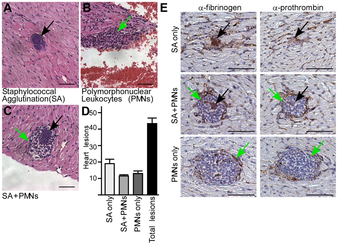
(A–D) Quantification of heart lesions in BALB/c mice (n = 10) 12 hours post-infection with S. aureus Newman. Three types of lesions were observed with either (A) staphylococcal agglutination (SA) without immune cell infiltrates (PMNs, polymorphonuclear leukocytes), (B) immune cell infiltrates without SAs (PMNs only) or (C) SA with surrounding granulocytes (SA+PMNs). Heart tissues were stained with hematoxylin-eosin and lesions enumerated (D). Error bars represent standard error of the mean of tissue samples. Data are representative of two independent experiments. (E) Immuno-histochemical analysis of heart tissues from BALB/c mice (n = 10) 12 hours following intravenous challenge with S. aureus Newman. Samples were stained with antibodies directed against mouse fibrinogen (α-fibrinogen) or mouse prothrombin (α-prothrombin). Arrows point to staphylococcal agglutinations (black) or immune cell infiltrates (green); scale bars represent 1 µm. Fig. 3. Staphylococcal agglutination in heart tissues is required for the pathogenesis of sepsis. 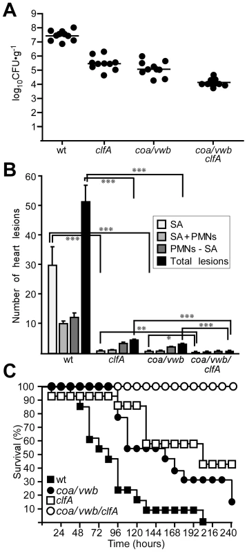
(A) Staphylococcal load, enumerated as colony forming units (CFU), in heart tissues of BALB/c mice (n = 10) 12 hours after retro-orbital inoculation with 108 CFU of S. aureus Newman (wt) or its variant strains (clfA, coa/vwb and coa/vwb/clfA). Horizontal lines represent mean CFU. Statistical analysis was performed with the Mann-Whitney test: wt vs. clfA, P = 0.0002; wt vs. coa/vwb, P = 0.0002; wt vs. coa/vwb/clfA, P = 0.0002; coa/vwb vs. coa/vwb/clfA, P = 0.0007; clfA vs. coa/vwb/clfA, P = 0.0002. Data are representative of two independent experiments. (B) Summary of histopathology findings in thin-sectioned and hematoxylin-eosin stained heart tissue from BALB/c mice (n = 10) 12 hours after retro-orbital injection of S. aureus Newman wild-type (wt) or its clfA, coa/vwb as well as clfA/coa/vwb variants. Representative lesions in heart tissues included staphylococcal agglutination without PMNs (SA), with PMNs (SA+PMNs), and PMN accumulation without staphylococcal agglutination (PMNs–SA). Error bars represent standard error of the mean from 10 hearts. Statistical significance of lesions for each mutant compared to wt infection was determined by Student's t test: *P<0.05, **P<0.01, ***P<0.001. Data are representative of two independent experiments. (C) Survival of cohorts of BALB/c mice (n = 20) following intravenous injection with S. aureus Newman (wt) or variants lacking coa, vwb or clfA. Data are representative of three independent experiments. Statistical significance was assessed with the logrank test: wt vs. coa/vwb (P<0.01), wt vs. clfA (P<0.001), and wt vs. coa/vwb/clfA (P<0.0001). Clumping factor A tethers staphylococci to fibrin cables
Staphylococcal agglutination requires coagulase catalyzed conversion of fibrinogen to fibrin as well as ClfA-mediated attachments. If so, ClfA may bind not only fibrinogen but also fibrin. This prediction was tested by measuring the binding of purified recombinant ClfA to either fibrinogen or fibrin immobilized in wells of polystyrene plates (Fig. 4A). Using non-linear regression analyses, we calculated a dissociation constant (Kd) of 395.2 nM (±51.82) for ClfA binding to fibrinogen, comparable to earlier affinity measurements [18]. The Kd of ClfA binding to fibrin was calculated as 661.9 nM (±80.32), which is not significantly different from the affinity of ClfA for fibrinogen (Fig. 4A). To further investigate S. aureus Newman interactions with fibrin, staphylococci were examined by scanning electron microscopy (SEM), which revealed agglutinated wild-type bacteria enmeshed in fibrin cables (Fig. 4B). SEM analysis of the staphylococcal variants coa/vwb and coa/vwb/clfA identified bacteria without fibrin cables (Fig. 4B). The clfA mutant continued to convert fibrinogen to fibrin, however clfA variant staphylococci did not agglutinate with fibrin cables (Fig. 4B). Plasmids pcoa-vwb and pclfA complemented the phenotypes caused by mutations in the corresponding genes and restored staphylococcal agglutination to wild-type levels (Fig. 4B). These data are in agreement with our general hypothesis that Coa/vWbp-derived fibrin cables provide a tether for ClfA-mediated staphylococcal agglutination (Fig. 4B).
Fig. 4. ClfA enables staphylococcal agglutination with fibrin cables in vitro and in vivo. 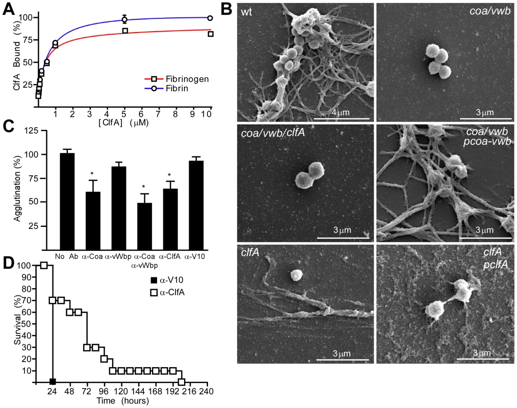
(A) The association of purified recombinant ClfA with immobilized fibrinogen or fibrin was assessed by ELISA and analyzed as the percentage of maximal binding. Average and standard error of the means were calculated from three independent experiments. Curves represent nonlinear regression for one-site binding saturation performed with GraphPad Prism, Fbgn R2 = 0.9876; Fibrin R2 = 0.9876. (B) Scanning electron micrographs of S. aureus Newman (wt) and its isogenic mutants immersed in plasma. (C) Affinity-purified rabbit IgG specific for Coa (α-Coa), vwb (α-vWb), ClfA (α-ClfA) or the plague protective antigen V10 (α-V10) was analyzed for its ability to prevent staphylococcal agglutination. Statistical significance of antibody effects compared to a mock treated control was assessed with the Student's t test: *P<0.05. (D) BALB/c mice (n = 10) were passively immunized by intraperitoneal injection with affinity-purified antibodies against V10 or ClfA and disease protection assessed by intravenous challenge with S. aureus Newman. Data represent one of three independent experiments. Statistical significance was assessed with the logrank test: P<0.01. Antibodies that prevent staphylococcal agglutination and sepsis
To further explore the contributions of Coa, vWbp and ClfA to staphylococcal agglutination, we raised rabbit antibodies against affinity purified recombinant proteins [12], [36]. Affinity purified rabbit antibodies specific for Coa, vWbp or ClfA inhibited S. aureus Newman agglutination in plasma (Fig. 4C). Passive transfer of ClfA-specific rabbit antibodies (85 µg purified antigen-specific IgG) into the peritoneal cavity of mice reduced the deposition of S. aureus Newman agglutinations in heart tissues of infected animals (Fig. 5A). Active immunization of mice with purified Coa and vWbp or ClfA raised specific IgG antibodies and reduced the frequency of heart lesions in animals challenged for twelve hours with wild-type S. aureus Newman (Fig. 5B). In particular, the abundance of staphylococcal agglutinations without immune cell infiltrates was reduced (Fig. 5B). Active immunization of mice with all three antigens – Coa, vWbp and ClfA – eliminated staphylococcal agglutination in heart tissues and caused the largest reduction of all types of pathological lesions (Fig. 5B). Similar to Coa - and vWbp-specific immunoglobulin [12], passive transfer of ClfA-specific rabbit antibodies into the peritoneal cavity of mice increased the survival time in the sepsis model of infection (Fig. 4D). These data corroborate the concept that ClfA-specific antibodies can improve the outcome of S. aureus Newman sepsis [24].
Fig. 5. Neutralization of coagulases and ClfA prevents staphylococcal agglutination in heart tissues of septic mice. 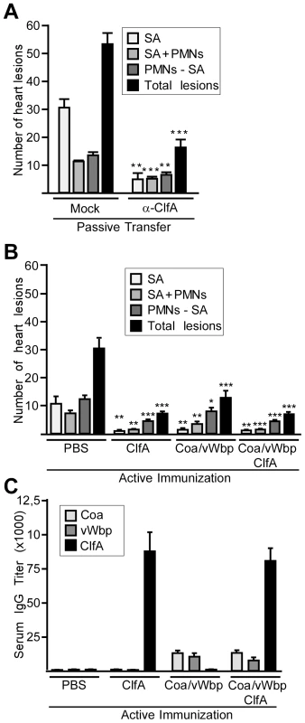
(A) Quantification of histopathology lesions in heart tissues of BALB/c mice (n = 10) passively immunized with affinity-purified V10 control antibodies (which neutralize the plague protective antigen LcrV) or ClfA antibodies prior to lethal infection. Hearts were removed during necropsy 12 hours after retro-orbital inoculation of staphylococci. Tissues were thin-sectioned, stained with hematoxylin-eosin and histopathology lesions enumerated. Error bars represent standard error of the mean from cohorts of ten mice. Statistical analysis was performed by two-tailed Student's t-test comparing same lesion types between mock-immunized and vaccinated animals: *P<0.05, **P<0.01, ***P<0.001. (B) Quantification of three types of histopathology lesions in heart tissues from mice actively immunized with recombinant Coa, vWbp, or ClfA. Hearts were removed during necropsy 12 hours after retro-orbital inoculation of staphylococci into BALB/c mice (n = 10). Tissues were thin-sectioned, stained with hematoxylin-eosin and histopathology lesions enumerated. Error bars represent standard error of the mean from cohorts of ten mice. Statistical analysis was performed by Student's two-tailed t-test comparing same lesion types between mock-immunized and vaccinated animals: *P<0.05, **P<0.01, ***P<0.001. Data are representative of two independent experiments. (C) Half maximal IgG antibody titer specific for Coa, vWb or ClfA antigens in serum following active vaccination of BALB/c mice (n = 5). Blood samples were drawn at the time of challenge. Error bars represent standard deviation of serum IgG titers. The limit of detection is 100. Direct thrombin inhibitors and staphylococcal sepsis
Univalent direct thrombin inhibitors, e.g. argatroban and dabigatran, inhibit the proteolytically active Coa·prothrombin complex [37], [38]. We examined whether these inhibitors also block the catalytic activity of vWbp·prothrombin. As a control, conversion of fibrinogen to fibrin by thrombin was monitored as an increase in sample absorbance at 450 nm. Compared to a mock control, this reaction was blocked with 200 ng argatroban (Fig. 6A). Treatment of fibrinogen with either Coa·prothrombin or vWbp·prothrombin led to fibrin conversion, whereas incubation with prothrombin alone did not (Fig. 6A). Incubation of both Coa·prothrombin or vWbp·prothrombin with 200 ng argatroban blocked the conversion of fibrinogen to fibrin (Fig. 6A). Argatroban treatment also interfered with the agglutination of S. aureus Newman in plasma (Fig. 6B).
Fig. 6. Direct thrombin inhibitors block a key step in staphylococcal pathogenesis. 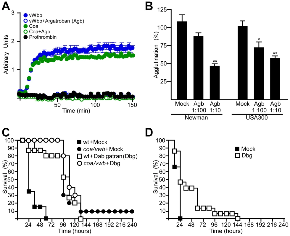
(A) Conversion of fibrinogen to fibrin by prothrombin, Coa·prothrombin or vwb·prothrombin was detected in the presence or absence of 200 ng argatroban (Agb). Arbitrary units are defined as A450*100. Average and standard error of the means were calculated from three independent measurements. (B) Agglutination of S. aureus Newman or S. aureus USA300 LAC in plasma in the presence of increasing concentrations of Agb. Average and standard error of the means were calculated from three independent measurements and statistical significance was assessed with the Student's two-tailed t-test: *P<0.05, **P<0.0001. (C) Survival of cohorts of BALB/c mice (n = 15) treated with saline (mock) or dabigatran-etexilate (Dbg) and infected with either S. aureus Newman or the coa/vwb mutant strain. Statistical significance was analyzed with the logrank test: mock vs. Dbg with wt challenge: P <0.0001; mock vs. Dbg with coa/vwb challenge: P = 0.43. Data are representative of three independent experiments. (D) Survival of cohorts of BALB/c mice (n = 15) treated with saline (mock) or dabigatran (Dbg) and challenged by intravenous inoculation with S. aureus USA300 LAC. Statistical significance was analyzed with the logrank test: mock vs. Dbg, P<0.01. Data are representative of three independent experiments. To evaluate the efficacy of direct thrombin inhibitors on the outcome of S. aureus Newman sepsis, mice received intraperitoneal injections with 10 mg/kg dabigatran-etexilate in 12 hour intervals. Dabigatran-etexilate is converted in mammalian tissues to its active form, dabigatran, which acts as a direct inhibitor of thrombin [39]. To assess dabigatran activity, mouse blood samples were drawn by cardiac puncture and the dilute thrombin time was determined (Fig. S1). Following challenge of mice via blood stream injection of 1×10⁸ CFU S. aureus Newman, mock treated animals died of sepsis within 60 hours post challenge (Fig. 6C). In contrast, dabigatran-etexilate treated animals survived up to 132 hours, albeit that all animals in this cohort eventually succumbed to the challenge (Fig. 6C). To determine whether direct thrombin inhibitors specifically block Coa and vWbp, mock or dabigatran-etexilate treated animals were challenged with the S. aureus coa/vwb mutant. In these experiments, dabigatran-etexilate treatment had no effect on survival or time-to-death (Fig. 6C). Mock or dabigatran-etexilate treated mice were also infected with lethal doses of S. aureus USA300 LAC, the current clone responsible for the epidemic of community-acquired MRSA infections in the United States [5]. Dabigatran-etexilate treatment prolonged the survival of septic mice (Fig. 6D).
Inhibiting multiple staphylococcal factors improves the outcome of sepsis
If clfA, coa and vwb act together to promote S. aureus Newman agglutination, dabigatran-etexilate treatment would be expected to improve the outcome of sepsis caused by clfA mutant staphylococci (Fig. 7A). Indeed, dabigatran-etexilate treatment increased the survival and time-to-death of mice with sepsis caused by clfA mutant S. aureus compared to a control strain harboring the complementing plasmid pclfA (Fig. 7A). Dabigatran-etexilate treatment further improved the disease outcome of animals challenged with clfA mutant staphylococci compared to a cohort of mock treated mice (Fig. 7A). Injection of clfA mutants carrying pclfA into the blood stream of mice resulted in reduced time-to-death compared to the wild-type parent, S. aureus Newman (Fig. 7A). Nevertheless, animals infected with the clfA (pclfA) variant also benefited from dabigatran-etexilate treatment (Fig. 7A).
Fig. 7. Additive protective effects of direct thrombin inhibitors and ClfA-specific antibodies against S. aureus sepsis. 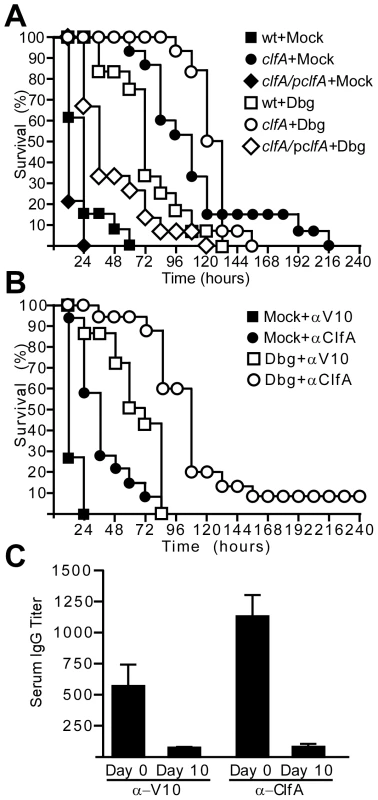
(A) Survival of cohorts of BALB/c mice (n = 15) treated with saline (mock) or dabigatran (Dbg) followed by intravenous inoculation with S. aureus Newman (wt), clfA or clfA (pClfA) variants. Data are representative of three independent experiments. (B) Survival of cohorts of BALB/c mice (n = 15) treated with saline (mock) or Dbg and passively immunized (5 mg·kg−1) with affinity-purified antibodies against V10 or ClfA. Animals were challenged by intravenous inoculation with S. aureus Newman. Statistical analysis was assessed with the logrank test: mock-V10 vs. mock-ClfA, P<0.001; Dbg-mock vs. Dbg-ClfA, P< 0.001. Data are representative of three independent experiments. (C) Half-maximal IgG titer of α-V10 or α-ClfA in serum of passively immunized mice (n = 5) was determined by ELISA. Blood was drawn on day 0, six hours post-immunization and at day 10, when the experiment was terminated. To test whether combining dabigatran-etexilate and ClfA-specific antibodies can improve the outcome of staphylococcal sepsis, animals received both treatments followed by challenge with a lethal dose of S. aureus (Fig. 7B). As compared to mock-treated animals or mice receiving either dabigatran or ClfA-specific antibodies, the combination of dabigatran and ClfA-specific antibodies led to increased time-to-death and survival of staphylococcal sepsis (Fig. 7BC).
We wondered whether the use of thrombin inhibitors and ClfA-specific antibodies could aid in the prevention of sepsis caused by clinical S. aureus isolates. To test this, we used the community-acquired MRSA isolate MW2, which was isolated from a fatal case of septicemia [40], as well as the hospital-acquired MRSA isolate N315 [41]. S. aureus strains N315 and MW2 both agglutinated when suspended in EDTA-plasma (Fig. 8A). These reactions were inhibited by treatment with argatroban (Fig. 8A) or with ClfA-specific antibodies (Fig. 8B). Treatment of mice with both dabigatran and ClfA-specific antibodies led to increased time-to-death during sepsis caused by either S. aureus N315 or S. aureus MW2 (Fig. 8CD). In contrast, the use of either dabigatran or ClfA-specific antibodies alone did not prolong the survival of mice receiving a lethal challenge of S. aureus N315 or S. aureus MW2 (Fig. 8CD).
Fig. 8. Direct thrombin inhibitors and ClfA-specific antibodies increase the time-to-death of MRSA sepsis in mice. 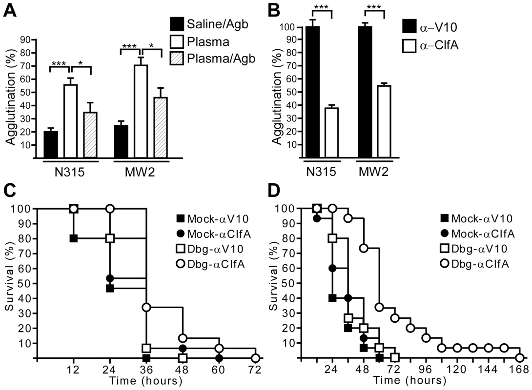
(A) Agglutination of methicillin-resistant S. aureus isolates N315 or MW2 in plasma in the presence or absence of argatroban (Agb). Average and standard error of the means were calculated from at least five independent measurements and statistical significance was assessed with the Student's two-tailed t-test: *P<0.05, ***P<0.0001. (B) Affinity-purified rabbit IgG specific for ClfA (α-ClfA) or the plague protective antigen V10 (α-V10) was analyzed for its ability to prevent agglutination of MRSA strains N315 and MW2 in plasma. Average and standard error of the mean were calculated from 16 fields of view from two independent experiments. Statistical significance of antibody effects compared to a mock treated control was assessed with the Student's two-tailed t test: ***P<0.001. (C) Survival of cohorts of BALB/c mice (n = 15) treated with saline (mock) or Dbg and passively immunized (5 mg·kg−1) with affinity-purified antibodies against V10 or ClfA. Animals were challenged by intravenous inoculation with MRSA strain N315. Statistical analysis was assessed with the logrank test: mock-V10 vs. mock-ClfA, not significant; mock-V10 vs. Dbg-ClfA, P<0.05; mock-ClfA vs. Dbg-ClfA, P<0.05; Dbg-mock vs. Dbg-ClfA, P<0.01. Data are representative of two independent experiments. (D) Survival of cohorts of BALB/c mice (n = 15) treated as described in panel (C) and challenged by intravenous inoculation with MRSA strain MW2. Statistical analysis was assessed with the logrank test: mock-V10 vs. mock-ClfA, not significant; mock-ClfA vs. Dbg-ClfA, P<0.001; Dbg-mock vs. Dbg-ClfA, P<0.001. Data are representative of two independent experiments. Discussion
Sepsis is a clinical condition in response to severe bacterial infection, which is associated with high mortality due to continued activation and apoptosis of immune cells and malperfusion of organ systems [42]. During sepsis, the physiological coordination between hemostasis and inflammation is dysregulated, triggering intravascular fibrin deposits [disseminated intravascular coagulation (DIC)] [43]. Dysregulated clotting places sepsis patients at high risk for systemic bleeding and loss of perfusion for vital organ systems [42]. Most anticoagulants, including the thrombin inhibitors heparin, hirudin and antithrombin, cannot alter the outcome of sepsis, likely because these compounds can halt an advancing coagulopathy but are unable to reverse the detrimental effects of established disease [43], [44], [45]. Activated protein C is a serine protease that inactivates the clotting cascade factors Va and VIIIa, without which thrombin-mediated coagulation, i.e. the conversion of fibrinogen to fibrin, is slowed by 2–3 orders of magnitude [46]. Recombinant human activated protein C (drotrecogin alfa) is currently the only FDA-licensed adjunctive therapy in patients with severe sepsis [47], [48]. Clinical trial data indicate reduced mortality in 6.1% of all cases but also considerable bleeding risks. Activated protein C may be particularly useful in preventing sepsis caused by Escherichia coli or other Gram-negative bacteria [49]. The ability of activated protein C treatment to affect the outcome of sepsis caused by different bacterial or fungal pathogens is not known [50].
Invasive S. aureus infections are frequently associated with bacteremia and may rapidly advance to sepsis [51]. The use of β-lactam antibiotics is obsolete for the treatment of sepsis with drug-resistant S. aureus strains (MRSA)[51]. Patients with MRSA infections typically receive vancomycin [51], a glycopeptide antibiotic that blocks bacterial cell wall synthesis [52]. Due to significant nephrotoxicity, vancomycin therapy must be carefully monitored to exceed the minimal inhibitory concentration for staphylococci in host tissues yet avoid the detrimental effects of this compound on kidney function [53]. Even with intensive clinical care, the annual survival of patients with MRSA sepsis is low (<50%) [54]. Thus, preventive measures or therapeutics that improve the outcome of MRSA sepsis represent a pressing public health issue in the United States [51].
S. aureus sepsis isolates coagulate blood and/or agglutinate in citrate-plasma [2]. Genome sequencing revealed that all clinical S. aureus isolates harbor functional coa, vwb and clfA genes [55]. In contrast, only some staphylococcal strains, those carrying hlb-converting phages, harbor the sak gene [56], whose secreted product staphylokinase associates with plasminogen to promote fibrinolysis [57], [58] in addition to cleaving human antimicrobial peptides (defensins)[59]. We wondered whether the unique attributes of S. aureus to generate fibrin deposits contribute also to the pathogenesis of sepsis. Using a mouse model for this disease, we observed that sortase A mutants, unable to anchor any one of nineteen different surface proteins in the staphylococcal envelope [60], were unable to cause sepsis [27]. ClfA, a surface protein associated with bacterial binding to fibrinogen [17], is the most important sortase A anchored virulence factor for sepsis (Table 1). Mutations in two coagulases, coa and vwb [12], further diminish the virulence of clfA mutants to a level that resembles that of sortase A variants (Fig. 1). To investigate the physiological role of coagulases and ClfA, we studied staphylococcal agglutination, a clinical microbiological assay requiring fibrinogen and prothrombin for bacterial association with fibrin fragments [26]. All three staphylococcal products - Coa, vWbp and ClfA – were required to agglutinate the pathogen in blood and cause lethal disease in mice. We presume that staphylococcal agglutination in vivo promotes the formation of thromboembolic lesions that contribute to the rapid lethality of S. aureus Newman [61] or USA300 LAC infections [62] into the bloodstream of mice.
Pretreatment of animals with dabigatran-etexilate, which blocks cleavage of fibrinogen by Coa·prothrombin and vWbp·prothrombin, as well as administration of ClfA-specific antibodies both interfere with S. aureus agglutination and reduce the mortality of sepsis. Nevertheless, combining direct thrombin inhibitors with ClfA-specific antibodies or combining anti-ClfA with anti-Coa/anti-vWbp can generate an even higher level of protection against sepsis. The recent licensure of direct thrombin inhibitors (e.g. dabigatran) and the availability of ClfA-specific monoclonal antibody provide an opportunity for the rapid testing of such regimen to reduce the incidence and/or the mortality of S. aureus sepsis.
ClfA binding to the C-terminal residues of the fibrinogen γ-chain may not be involved in S. aureus agglutination; this portion of the polypeptide is thought to be buried within polymerized fibrin cables [63], [64]. If so, staphylocoagulase mediated cleavage of fibrinogen may reveal another binding site for ClfA on the surface of fibrin cables, enabling staphylococcal agglutination in a manner that can be inhibited with ClfA-specific antibodies. Direct thrombin inhibitors block coagulase (Coa and vWbp) mediated cleavage of fibrinogen and thereby hinder the formation of ClfA binding sites on the surface of fibrin cables. These compounds also mimic the phenotype of S. aureus Newman coagulase mutants (Δcoa, vwb) in the murine sepsis model and presumably exert a similar effect in preventing the formation of staphylococcal abscess.
S. aureus is a frequent cause of human wound infections [1], however the contribution of coagulases towards the establishment of this disease is not known. Of note, physiological hemostasis during wound healing generates fibrin and platelet deposits within wounded tissues [65]. Thus, it would be interesting to explore whether preventive treatment with direct thrombin inhibitors as well as ClfA-specific antibodies can reduce the incidence of hospital-acquired sepsis and/or wound infections.
Materials and Methods
Ethics statement
Animal experiments involving S. aureus challenge followed protocols that were reviewed, approved and performed under the regulatory supervision of The University of Chicago's Institutional Biosafety Committee (IBC) and the Institutional Animal Care and Use Committee (IACUC). Animals were managed by the University of Chicago Animal Resource Center, which is accredited by the American Association for Accreditation of Laboratory Animal Care and the Department of Health and Human Services (DHHS number A3523-01). Animals were maintained in accordance with the applicable portions of the Animal Welfare Act and the DHHS “Guide for the Care and Use of Laboratory Animals”. Veterinary Care was under the direction of full-time resident veterinarians boarded by the American College of Laboratory Animal Medicine. BALB/c mice and New Zealand white rabbits were purchased from Charles River Laboratories and Harlan Sprague Dawley, respectively. After confirming that the data sets abide by a normal distribution, the statistical analysis of staphylococcal sepsis was analyzed using the two-tailed Logrank test. Quantification of mouse heart tissue histopathology was analyzed for statistical significance using the unpaired two-tailed Student's t-test. The bacterial load (CFU) in heart tissue from mice infected with staphylococcal variants was analyzed with the Mann Whitney test. The results of all animal experiments were examined for reproducibility.
Bacterial strains and growth of cultures
S. aureus strains Newman [61], USA300 LAC [62], MW2 [40] and N315 [41] were cultured on tryptic soy agar or broth at 37°C. E. coli strains DH5α and BL21 (DE3) were cultured on Luria Bertani agar or broth at 37°C. Ampicillin (100 µg/ml) and chloramphenicol (10 µg/ml) were used for pET15b and pOS1 selection [66], respectively.
Transposon mutants and plasmids
Insertional mutations carrying the bursa aurealis transposon with an erthyromycin resistance cassette from the Phoenix library [29] were transduced with bacteriophage into S. aureus Newman or the coa/vwb mutant [12]. Mutations were verified by PCR with specific primer pairs for coa (CGCGGATCCATAGTAACAAAGGATTATAGTGGGAAATCACAAG and TCCCCCGGGTTATTTTGTTACTCTAGGCCCATATGTCGC), vwb (CGCGGATCCGTGGTTTCTGGGGAGAAGAATCC and TCCCCCGGGTTTGCAGCCATGCATTAATTATTTGCC) and clfA (CGCGGATCC-AAGGTCAAATCGACCGTT and CGGGGTACC-TTATTTCTTATCTTTATTTTCTTTTTTTC) as well as by immunoblotting with specific rabbit antibodies [12], [36]. Complementing plasmids pcoa-vWbp and pclfA were described previously [12], [67]. For immunoblot analysis, 1 mL of staphylococcal overnight cultures grown in tryptic soy broth (Difco) were centrifuged at 8,000×g for 3 min in a table top centrifuge and the supernatant was recovered. Proteins in culture supernatants were precipitated with 10% trichloroacetic acid on ice for 20 minutes. Pellets were washed once in 1 mL TSM (100 mM Tris-HCl, pH 7.5, 0.5 M sucrose, 10 mM MgCl2), suspended in 500 µL TSM, incubated with 50 µg lysostaphin for 15 minutes at 37°C for 15 minutes. 10% TCA was added and samples were incubated on ice for 10 min. All samples were centrifuged and washed with 1 mL ice-cold 100% acetone. Samples were air dried and solubilized in 75 µL sample buffer (4% SDS, 50 mM Tris-HCl, pH 8.0, 10% glycerol, and bromophenol blue).
Scanning electron microscopy
Staphylococcal strains were grown to mid-log phase (OD600 0.5), washed twice and suspended in PBS to a final OD600 1. Bacteria were mixed with EDTA-chelated rabbit plasma (1 : 1) and incubated for 15 minutes. Samples were fixed for 60 minutes in 2% glutaraldehyde in phosphate buffered saline (PBS) at room temperature onto freshly prepared poly-L-lysine coated glass coverslips. Samples were washed twice with PBS and subsequently serially dehydrated by consecutive incubations in 25% and 50% ethanol/PBS, 75% and 90% ethanol/H2O, 2× 100% ethanol, followed by 50% ethanol/hexamethyldisilazane (HDMS) and finally with 100% HDMS. After overnight evaporation of HDMS at room temperature, samples were mounted onto specimen mounts (Ted Pella, Inc.) and coated with 80% Pt/20% Pd to 8 nm using a Cressington 208HR Sputter Coater at 20mA prior to examination with a Fei Nova NanoSEM 200 scanning electron microscope. The SEM was operated with an acceleration voltage of 5 kV and samples were viewed at a distance of 5 mm.
Protein purification
E. coli BL21(DE3) harboring expression vectors containing coa, vwb, or clfA were grown at 37°C and induced with 1 mM IPTG after two hours. Three hours following induction, cells were centrifuged at 7,000×g, suspended in column buffer (0.1 M Tris-HCl, pH 7.5, 0.5 M NaCl) and lysed in a French pressure cell at 14,000 lb/in2. Lysates were subjected to ultracentrifugation at 40,000 ×g for 30 min and the supernatant was subjected to Ni-NTA chromatography, washed with column buffer containing 10 mM imidazole, followed by elution with 500 mM imidazole. Eluates were dialyzed against PBS. To remove endotoxin, 1∶100 Triton-X114 was added and the solution was chilled for 10 min, incubated at 37°C for 10 min, and centrifuged at 13,000 ×g. This was repeated twice. Supernatant was loaded onto a HiTrap desalting column to remove remnants of Triton-X114. Purity was verified by SDS-PAGE analysis and Coomassie Brilliant Blue staining.
Rabbit antibodies
Protein concentration was determined using a BCA kit (Pierce). Purity was verified by SDS-PAGE analysis and Coomassie Brilliant Blue staining. Six month old New-Zealand white female rabbits were immunized with 500 µg protein emulsified in CFA (Difco) for initial immunization or IFA for booster immunizations on day 24 and 48. On day 60, rabbits were bled and serum recovered for immunoblotting or passive transfer experiments. For antibody purification, recombinant His6-Coa [12], His6-vWbp [12], or His6-ClfA (5 mg) [36] was covalently linked to HiTrap NHS-activated HP columns (GE Healthcare). This antigen-matrix was then used for affinity chromatography of 10–20 ml of rabbit serum raised against Coa [12], vWbp [12] or ClfA at 4°C. Charged matrix was washed with 50 column volumes of PBS, antibodies eluted with elution buffer (1 M glycine pH 2.5, 0.5 M NaCl) and immediately neutralized with 1 M Tris-HCl, pH 8.5. Purified antibodies were dialyzed overnight against PBS, 0.5 M NaCl at 4°C.
Agglutination assay
Overnight cultures of staphylococcal strains were washed in 1 mL 0.85% NaCl and suspended to a final concentration of OD600 4.0 in 1 mL. Bacteria were incubated with 1∶500 Syto9 (Invitrogen) for 15 minutes, washed with 1 mL 0.85% NaCl, and suspended in 1 mL saline. Bacteria were mixed 1∶1 with EDTA-chelated rabbit plasma (Becton, Dickinson) on a glass microscope slide and incubated for 15 minutes. Samples were viewed and images captured on an Olympus Provis microscope using a 40× objective. For quantification of agglutination, plasma and bacteria were inoculated onto polystyrene C-Chip disposable hemocytometer slides (IN-CYTO). Brightfield images from sixteen fields of view were taken of bacterial strains using a Nikon TE2000 U with a 20× objective. To determine the degree of agglutination, the Threshold Function in ImageJ software was used to convert the image into a binary image, in which staphylococci are black and the background is white. The mean intensity of the image was measured. The average mean intensity of S. aureus Newman in saline without plasma was subtracted from all values and percent agglutination was calculated by normalizing all mean intensity values to S. aureus Newman in plasma. To assess the inhibitory affect of antibodies on agglutination, affinity-purified antibodies were incubated with staphylococci to a final concentration of 3 µM for 10 minutes prior to mixture with plasma. To assess the inhibitory affect of argatroban on agglutination, argatroban was diluted 1∶10 and 1∶100 in plasma and incubated for 10 minutes prior to mixture with bacteria. Percent agglutination was measured compared to bacteria in plasma without argatroban. For experiments using S. aureus N315 and MW2, agglutination was measured as percent change in OD550 following two hours incubation of bacteria with saline containing argatroban (1 mg/mL), plasma, or plasma containing argatroban (1 mg/mL). Error bars represent standard error of the mean from at least three independent experiments to ensure reproducibility.
Sepsis
Overnight cultures of staphylococcal strains were diluted 1∶100 into fresh TSB and grown until they reached an OD600 of 0.4. Bacteria were centrifuged at 7,000 ×g, washed, and suspended in the one-tenth volume of PBS. Six week-old female BALB/c mice (n = 15) (Charles River) were injected retro-orbitally with 1×10⁸ CFU (S. aureus Newman, MW2, and N315) or 5×10⁷ CFU (S. aureus USA300) suspensions in 100 µl of PBS. Mice were monitored for survival over 10 days. To enumerate staphylococcal load in heart tissue twelve hours post-infection, mice were euthanized by CO2 asphyxyation and hearts were removed during necropsy. Heart tissue was homogenized in PBS, 0.1% Triton X-100. Serial dilutions of homogenate were spread on TSA and incubated for colony formation. The bacterial load in organ tissue was analyzed in pairwise comparisons between wild-type and mutant strains with the unpaired two-tailed Student's t-test. For histopathology, mice infected with S. aureus were euthanized 12 hours after infection. Hearts were removed during necropsy and fixed in 10% formalin for 24 hours at room temperature. Tissues were embedded in paraffin, thin-sectioned, stained with hematoxylin and eosin, and examined by light microscopy to enumerate pathological lesions per organ. Data were analyzed in pairwise comparisons between wild-type and mutant strains with the unpaired two-tailed Student's t-test. For immunohistochemical analysis, thin-sectioned heart tissues were stained with polyclonal antibodies against mouse prothrombin (Haematologic Technologies) or mouse fibrinogen (Haematologic Technologies).
ClfA binding to fibrinogen and fibrin
MaxSorb 96-well ELISA plates (Nunc) were coated with human fibrinogen (Sigma) overnight. Wells were washed and solutions of PBS or alpha-thrombin (Innovative Research), 100 nM in 1% sodium-citrate/PBS were added for one hour at room temperature to generate fibrinogen and fibrin wells respectively. As controls, the same conditions were generated in Eppendorf tubes. Following incubation with or without alpha-thrombin, samples were centrifuged at 13,000 ×g for 10 min and supernatants were recovered. The sediment was dissolved in 8 M urea. Running buffer (3 M urea, 4% SDS, 10% BME) was added 1 : 1. Proteins in supernatants and pellets were separated by SDS-PAGE (15%) and stained with Coomassie Brilliant Blue to analyze soluble fibrinogen in the supernatant fraction and fibrin in the sediment. Purified recombinant ClfA in 1% sodium-citrate was added at increasing concentrations to 96-well plates and incubated for one hour. Samples were incubated with polyclonal anti-ClfA (1 : 1,000) to detect bound-ClfA followed by goat anti-rabbit-HRP (1 : 10,000). The wells were developed using an OptEIA kit (BD Lifesciences) and absorbance at 450 nm was measured. Non-linear regression assuming one-site saturation kinetics was performed using GraphPad Prism.
Active immunization
Three week-old BALB/c mice (n = 10) were injected with 50 µg protein emulsified in 100 µl complete Freund's adjuvant. Eleven days post vaccination these mice were boosted with 50 µg protein each emulsified in 100 µl incomplete Freund's adjuvant. On day 21, mice were injected with 1×10⁸ CFU of S. aureus challenge strains.
Passive transfer of antibodies
Six hours prior to infection, six week old BALB/c mice (n = 15) were injected intraperitoneally with specific rabbit antibodies affinity-purified on ClfA - or V10-coupled resin (control IgG specific for the LcrV plague antigen) at a dose of 5 mg/kg body weight. Control mice (n = 5) that received the same antibody via passive transfer were anesthetized and bled retro-orbitally at the time of infection and again at the end of the experiment. Blood was collected using micro-hematocrit capillary tubes (Fisher) in Z-Gel microtubes (Sarstedt). Tubes were centrifuged at 8,000 ×g for three minutes, and serum was collected. Antibody titer was measured by ELISA as previously described [27].
Coagulase activity
Purified recombinant Coa or vWbp (100 nM) were mixed with human prothrombin (Innovative Research) in 1% sodium-citrate/PBS. After an initial reading, fibrinogen (3 µM) (Sigma) was added and conversion of fibrinogen to fibrin was measured as an increase in turbidity at 450 nm in a plate reader (BioTek) at 2.5 min intervals. As controls, the enzymatic activity of human alpha-thrombin (Innovative Research) or prothombin alone were measured. Argatroban (200 ng, Novaplus) was added to reactions prior to the addition of fibrinogen.
Dabigatran etexilate treatment
Dabigatran (Boehringer Ingelheim) tablets were dissolved in 0.9 N saline and doses of 10 mg/kg in 100 µL were administered. Mice (n = 15) were injected intraperitoneally starting 24 hours prior to infection and continuing every twelve hours during the course of the infection. Control mice received injections of 0.9 N saline. To measure dilute thrombin time, mice (n = 5) received saline or dabigatran treatment and were euthanized by CO2 asphyxiation at the time of infection. Blood was drawn by cardiac puncture, diluted in sodium-citrate (1%), centrifuged at 1,500 ×g for 5 minutes, and plasma diluted in pooled fresh human plasma 1 : 6. Thrombin time was measured on a STA-R analyzer (Diagnostica Stago).
Supporting Information
Zdroje
1. LowyFD 1998 Staphylococcus aureus infections. New Engl J Med 339 520 532
2. KlevensRMMorrisonMANadleJPetitSGershmanK 2007 Invasive methicillin-resistant Staphylococcus aureus infections in the United States. JAMA 298 1763 1771
3. DeLeoFRChambersHF 2009 Waves of resistance: Staphylococcus aureus in the antibiotic era. Nat Rev Microbiol 7 629 641
4. FowlerVGJMiroJMHoenBCabellCHAbrutynE 2005 Staphylococcus aureus endocarditis: a consequence of medical progress. JAMA 293 3012 3021
5. DeLeoFROttoMKreiswirthBNChambersHF 2010 Community-associated meticillin-resistant Staphylococcus aureus. Lancet 375 1557 1568
6. FosterTJ 2005 Immune evasion by staphylococci. Nat Rev Microbiol 3 948 958
7. WalshEJMiajlovicHGorkunOVFosterTJ 2008 Identification of the Staphylococcus aureus MSCRAMM clumping factor B (ClfB) binding site in the alphaC-domain of human fibrinogen. Microbiology 154 550 558
8. ChengAGDeDentACSchneewindOMissiakasDM 2011 A play in four acts: Staphylococcus aureus abscess formation. Trends Microbiol 19 225 232
9. DoolittleRF 2003 Structural basis of the fibrinogen-fibrin transformation: contributions from X-ray crystallography. Blood Rev 17 33 41
10. PanizziPFriedrichRFuentes-PriorPRichterKBockPE 2006 Fibrinogen substrate recognition by staphylocoagulase (pro)thrombin complexes. J Biol Chem 281 1179 1187
11. FriedrichRPanizziPFuentes-PriorPRichterKVerhammeI 2003 Staphylocoagulase is a prototype for the mechanism of cofactor-induced zymogen activation. Nature 425 535 539
12. ChengAGMcAdowMKimHKBaeTMissiakasDM 2010 Contribution of coagulases towards Staphylococcus aureus disease and protective immunity. PLoS Pathog 6 e1001036
13. LoebL 1903 The influence of certain bacteria on the coagulation of blood. J Med Res 10 407
14. KolleWOttoR 1902 Die Differenzierung der Staphylokokken mittelst der Agglutination. Z Hygiene 41 369 379
15. McDevittDFrancoisPVaudauxPFosterTJ 1994 Molecular characterization of the clumping factor (fibrinogen receptor) of Staphylococcus aureus. Mol Microbiol 11 237 248
16. HawigerJTimmonsSStrongDDCottrellBARileyM 1982 Identification of a region of human fibrinogen interacting with staphylococcal clumping factor. Biochemistry 21 1407 1413
17. McDevittDFrancoisPVaudauxPFosterTJ 1995 Identification of the ligand-binding domain of the surface-located fibrinogen receptor (clumping factor) of Staphylococcus aureus. Mol Microbiol 16 895 907
18. McDevittDNanavatyTHouse-PompeoKBellETurnerN 1997 Characterization of the interaction between the Staphylococcus aureus clumping factor (ClfA) and fibrinogen. Eur J Biochem 247 416 424
19. StrongDDLaudanoAPHawigerJDoolittleRF 1982 Isolation, characterization and synthesis of peptides from human fibrinogen that block the staphylococcal clumping reaction and construction of a synthetic clumping particle. Biochemistry 21 1414 1420
20. GaneshVKRiveraJJSmedsEKoY-PBowdenMG 2008 A structural model of the Staphylococcus aureus ClfA-fibrinogen interaction opens new avenues for the design of anti-staphylococcal therapeutics. PLoS Pathog 4 e1000226
21. JosefssonEHartfordOO'BrienLPattiJMFosterTJ 2001 Protection against experimental Staphylococcus aureus arthritis by vaccination with clumping factor A, a novel virulence determinant. J Infect Dis 184 1572 1580
22. MoreillonPEntenzaJMFrancioliPMcDevittDFosterTJ 1995 Role of Staphylococcus aureus coagulase and clumping factor in pathogenesis of experimental endocarditis. Infect Immun 63 4738 4743
23. HairPSEchagueCGShollAMWatkinsJAGeogheganJA 2010 Clumping factor A interaction with complement factor I increases C3b cleavage on the bacterial surface of Staphylococcus aureus and decreases complement-mediated phagocytosis. Infect Immun 78 1717 1727
24. HallAEDomanskiPJPatelPRVernachioJHSyribeysPJ 2003 Characterization of a protective monoclonal antibody recognizing Staphylococcus aureus MSCRAMM protein clumping factor A. Infect Immun 71 6864 6870
25. WeemsJJJrSteinbergJPFillerSBaddleyJWCoreyGR 2006 Phase II, randomized, double-blind, multicenter study comparing the safety and pharmacokinetics of Tefibazumab to placebo for treatment of Staphylococcus aureus bacteremia. Antimicrob Agents Chemother 50 2751 2755
26. Birch-HirschfeldL 1934 Über die Agglutination von Staphylokokken durch Bestandteile des Säugetierblutplasmas. Klinische Woschenschrift 13 331
27. KimHKDeDentAChengAGMcAdowMBagnoliF 2010 IsdA and IsdB antibodies protect mice against Staphylococcus aureus abscess formation and lethal challenge. Vaccine 28 6382 6392
28. MazmanianSKLiuGTon-ThatHSchneewindO 1999 Staphylococcus aureus sortase, an enzyme that anchors surface proteins to the cell wall. Science 285 760 763
29. BaeTBangerAKWallaceAGlassEMAslundF 2004 Staphylococcus aureus virulence genes identified by bursa aurealis mutagenesis and nematode killing. Proc Natl Acad Sci U S A 101 12312 12317
30. KaidaSMiyataTYoshizawaYKawabataSMoritaT 1987 Nucleotide sequence of the staphylocoagulase gene: its unique COOH-terminal 8 tandem repeats. J Biochem 102 1177 1186
31. BjerketorpJNilssonMLjunghAFlockJIJacobssonK 2002 A novel von Willebrand factor binding protein expressed by Staphylococcus aureus. Microbiology 148 2037 2044
32. PalmaMNozohoorSSchenningsTHeimdahlAFlockJI 1996 Lack of the extracellular 19-kilodalton fibrinogen-binding protein from Staphylococcus aureus decreases virulence in experimental wound infection. Infect Immun 64 5284 5289
33. PalmaMWadeDFlockMFlockJI 1998 Multiple binding sites in the interaction between an extracellular fibrinogen-binding protein from Staphylococcus aureus and fibrinogen. J Biol Chem 273 13177 13181
34. Ní EidhinDPerkinsSFrancoisPVaudauxPHöökM 1998 Clumping factor B (ClfB), a new surface-located fibrinogen-binding adhesin of Staphylococcus aureus. Mol Microbiol 30 245 257
35. HawigerJHammondDKTimmonsS 1975 Human fibrinogen possesses binding sites for staphylococci on Aalpha and Bbeta polypeptide chains. Nature 258 643 645
36. Stranger-JonesYKBaeTSchneewindO 2006 Vaccine assembly from surface proteins of Staphylococcus aureus. Proc Nat Acad Sci U S A 103 16942 16947
37. Hijikata-OkunomiyaAKataokaN 2003 Argatroban inhibits staphylothrombin. J Thromb Haemost 1 2060 2061
38. VanasscheTVerhaegenJPeetermansWEHoylaertsMFVerhammeP 2010 Dabigatran inhibits Staphylococcus aureus coagulase activity. J Clin Microbiol 48 4248 4250
39. HaulNHNarHPriepkeHRiesUStassenJM 2002 Structure-based design of novel potent nonpeptide thrombin inhibitors. J Med Chem 45 1757 1766
40. BabaTTakeuchiFKurodaMYuzawaHAokiK 2002 Genome and virulence determinants of high-virulence community acquired MRSA. Lancet 359 1819 1827
41. KurodaMOhtaTUchiyamaIBabaTYuzawaH 2001 Whole genome sequencing of meticillin-resistant Staphylococcus aureus. Lancet 357 1225 1240
42. Stearns-KurosawaDJOsuchowskiMFValentineCKurosawaSRemickDG 2010 The pathogenesis of sepsis. Annu Rev Pathol Mech Dis 6 19 48
43. WarrenHSSuffrediniAFEichackerPQMunfordRS 2002 Risks and benefits of activated protein C treatment for severe sepsis. N Engl J Med 347 1027 1030
44. JaimesFDe La RosaGMoralesCFortichFArangoC 2009 Unfractioned heparin for treatment of sepsis: A randomized clinical trial (The HETRASE Study). Crit Care Med 37 1185 1196
45. Di NisioMMiddeldorpSBullerHR 2005 Direct thrombin inhibitors. N Engl J Med 353 1028 1040
46. MarlarRAKleissAJGriffinJH 1981 Human protein C: inactivation of factors V and VIII in plasma by the activated molecule. Ann NY Acad Sci 370 303 310
47. BernardGRVincentJLLaterrePFLaRosaSPDhainautJF 2001 Efficacy and safety of recombinant human activated protein C for severe sepsis. N Engl J Med 344 699 709
48. LevyMMDellingerRPTownsendSRLinde-ZwirbleWTMarshallJC 2010 The Surviving Sepsis Campaign: results of an international guideline-based performance improvement program targeting severe sepsis. Intensive Care Med 36 222 231
49. TaylorFBJChangAEsmonCTD'AngeloAVigano-D'AngeloS 1987 Protein C prevents the coagulopathic and lethal effects of Escherichia coli infusion in the baboon. J Clin Invest 79 918 925
50. MartinGSManninoDMEatonSMossM 2003 The epidemiology of sepsis in the United States from 1979 through 2000. N Engl J Med 348 1546 1554
51. LiuCBayerASCosgroveSEDaumRSFridkinSK 2011 Clinical practice guidelines by the infectious diseases society of america for the treatment of methicillin-resistant Staphylococcus aureus infections in adults and children: executive summary. Clin Infect Dis 52 285 292
52. WalshCT 1993 Vancomycin resistance: decoding the molecular logic. Science 261 308 309
53. FowlerVGJrBoucherHWCoreyGRAbrutynEKarchmerAW 2006 Daptomycin versus standard therapy for bacteremia and edocarditis caused by Staphylococcus aureus. N Engl J Med 355 653 665
54. KlevensRMEdwardsJRGaynesRPSystemNNIS 2008 The impact of antimicrobial-resistant, health care-associated infections on mortality in the United States. Clin Infect Dis 47 927 930
55. McCarthyAJLindsayJA 2010 Genetic variation in Staphylococcus aureus surface and immune evasion genes is lineage associated: implications for vaccine design and host-pathogen interactions. BMC Microbiology 10 173
56. ColemanDCSullivanDJRussellRJArbuthnottJPCareyBF 1989 Staphylococcus aureus bacteriophages mediating the simultaneous lysogenic conversion of beta-lysin, staphylokinase and enterotoxin A: a molecular mechanism of triple conversion. J Gen Microbiol 135 1679 1697
57. LijnenHRVan HoefBDe CockFOkadaKUeshimaS 1991 On the mechanism of fibrin-specific plasminogen activation by staphylokinase. J Biol Chem 266 11826 11832
58. MölkänenTTyyneläJHelinJKalkkinenNKuuselaP 2002 Enhanced activation of bound plasminogen on Staphylococcus aureus by staphylokinase. FEBS Lett 517 72 78
59. JinTBokarewaMFosterTMitchellJHigginsJ 2004 Staphylococcus aureus resists human defensins by production of staphylokinase, a novel bacterial evasion mechanism. J Immunol 172 1169 1176
60. MazmanianSKLiuGJensenERLenoyESchneewindO 2000 Staphylococcus aureus mutants defective in the display of surface proteins and in the pathogenesis of animal infections. Proc Natl Acad Sci U S A 97 5510 5515
61. BabaTBaeTSchneewindOTakeuchiFHiramatsuK 2007 Genome sequence of Staphylococcus aureus strain Newman and comparative analysis of staphylococcal genomes. J Bacteriol 190 300 310
62. DiepBAGillSRChangRFPhanTHChenJH 2006 Complete genome sequence of USA300, an epidemic clone of community-acquired meticillin-resistant Staphylococcus aureus. Lancet 367 731 739
63. DonahueJPPatelHAndersonWFHawigerJ 1994 Three-dimensional structure of the platelet integrin recognition segment of the fibrinogen gamma chain obtained by carrier protein-driven characterization. Proc Natl Acad Sci U S A 91 12178 12182
64. WareSDonahueJPHawigerJAndersonWF 1999 Structure of the fibrinogen gamma-chain integrin binding and factor XIIIa cross-linking sites obtained through carrier protein driven crystallization. Protein Sci 8 2663 2671
65. WolbergAS 2007 Thrombin generation and fibrin structure. Blood Rev 21 131 142
66. SchneewindOMihaylova-PetkovDModelP 1993 Cell wall sorting signals in surface protein of Gram-positive bacteria. EMBO 12 4803 4811
67. DeDentACMissiakasDMSchneewindO 2008 Signal peptides direct surface proteins to two distinct envelope locations of Staphylococcus aureus. EMBO J 27 2656 2668
Štítky
Hygiena a epidemiologie Infekční lékařství Laboratoř
Článek Quorum Sensing in Fungi: Q&AČlánek Blood Feeding and Insulin-like Peptide 3 Stimulate Proliferation of Hemocytes in the MosquitoČlánek The DEAD-box RNA Helicase DDX6 is Required for Efficient Encapsidation of a Retroviral GenomeČlánek A Phenome-Based Functional Analysis of Transcription Factors in the Cereal Head Blight Fungus,Článek A Wide Extent of Inter-Strain Diversity in Virulent and Vaccine Strains of AlphaherpesvirusesČlánek The Anti-Sigma Factor TcdC Modulates Hypervirulence in an Epidemic BI/NAP1/027 Clinical Isolate ofČlánek Critical Roles for LIGHT and Its Receptors in Generating T Cell-Mediated Immunity during InfectionČlánek Frequent and Recent Human Acquisition of Simian Foamy Viruses Through Apes' Bites in Central Africa
Článek vyšel v časopisePLOS Pathogens
Nejčtenější tento týden
2011 Číslo 10- Jak souvisí postcovidový syndrom s poškozením mozku?
- Měli bychom postcovidový syndrom léčit antidepresivy?
- Farmakovigilanční studie perorálních antivirotik indikovaných v léčbě COVID-19
- 10 bodů k očkování proti COVID-19: stanovisko České společnosti alergologie a klinické imunologie ČLS JEP
-
Všechny články tohoto čísla
- Quorum Sensing in Fungi: Q&A
- Discovery of an Ebolavirus-Like Filovirus in Europe
- Toll-like Receptor 7 Controls the Anti-Retroviral Germinal Center Response
- Tubule-Guided Cell-to-Cell Movement of a Plant Virus Requires Class XI Myosin Motors
- Herpesvirus Telomerase RNA (vTR) with a Mutated Template Sequence Abrogates Herpesvirus-Induced Lymphomagenesis
- Mitochondrial Peroxiredoxin Plays a Crucial Peroxidase-Unrelated Role during Infection: Insight into Its Novel Chaperone Activity
- Sustained CD8+ T Cell Memory Inflation after Infection with a Single-Cycle Cytomegalovirus
- Novel Mouse Xenograft Models Reveal a Critical Role of CD4 T Cells in the Proliferation of EBV-Infected T and NK Cells
- Toll-8/Tollo Negatively Regulates Antimicrobial Response in the Respiratory Epithelium
- Exhausted Cytotoxic Control of Epstein-Barr Virus in Human Lupus
- Structural and Functional Analysis of Laninamivir and its Octanoate Prodrug Reveals Group Specific Mechanisms for Influenza NA Inhibition
- Infection Drives IL-17-Mediated Neutrophilic Allergic Airways Disease
- Blood Feeding and Insulin-like Peptide 3 Stimulate Proliferation of Hemocytes in the Mosquito
- HIV-1 Replication in the Central Nervous System Occurs in Two Distinct Cell Types
- Deep Molecular Characterization of HIV-1 Dynamics under Suppressive HAART
- Fitness Landscape of Antibiotic Tolerance in Biofilms
- The DEAD-box RNA Helicase DDX6 is Required for Efficient Encapsidation of a Retroviral Genome
- Preventing Sepsis through the Inhibition of Its Agglutination in Blood
- A Phenome-Based Functional Analysis of Transcription Factors in the Cereal Head Blight Fungus,
- IFITM3 Inhibits Influenza A Virus Infection by Preventing Cytosolic Entry
- Targeting Cattle-Borne Zoonoses and Cattle Pathogens Using a Novel Trypanosomatid-Based Delivery System
- A Wide Extent of Inter-Strain Diversity in Virulent and Vaccine Strains of Alphaherpesviruses
- Coordinated Destruction of Cellular Messages in Translation Complexes by the Gammaherpesvirus Host Shutoff Factor and the Mammalian Exonuclease Xrn1
- Signal Transduction through CsrRS Confers an Invasive Phenotype in Group A
- Biochemical and Structural Insights into the Mechanisms of SARS Coronavirus RNA Ribose 2′-O-Methylation by nsp16/nsp10 Protein Complex
- Histone Deacetylase 8 Is Required for Centrosome Cohesion and Influenza A Virus Entry
- Severe Acute Respiratory Syndrome Coronavirus Envelope Protein Regulates Cell Stress Response and Apoptosis
- Co-opts the FGF2 Signaling Pathway to Enhance Infection
- IRAK-2 Regulates IL-1-Mediated Pathogenic Th17 Cell Development in Helminthic Infection
- Trafficking of Hepatitis C Virus Core Protein during Virus Particle Assembly
- The Anti-interferon Activity of Conserved Viral dUTPase ORF54 is Essential for an Effective MHV-68 Infection
- A Viral Nuclear Noncoding RNA Binds Re-localized Poly(A) Binding Protein and Is Required for Late KSHV Gene Expression
- Suppression of Methylation-Mediated Transcriptional Gene Silencing by βC1-SAHH Protein Interaction during Geminivirus-Betasatellite Infection
- ISG15 Is Critical in the Control of Chikungunya Virus Infection Independent of UbE1L Mediated Conjugation
- Non-Hematopoietic Cells in Lymph Nodes Drive Memory CD8 T Cell Inflation during Murine Cytomegalovirus Infection
- RNA Polymerase II Stalling Promotes Nucleosome Occlusion and pTEFb Recruitment to Drive Immortalization by Epstein-Barr Virus
- Noninfectious Retrovirus Particles Drive the / Dependent Neutralizing Antibody Response
- Endophytic Life Strategies Decoded by Genome and Transcriptome Analyses of the Mutualistic Root Symbiont
- An Integrated Approach to Elucidate the Intra-Viral and Viral-Cellular Protein Interaction Networks of a Gamma-Herpesvirus
- as an Animal Model for the Study of Biofilm Infections
- Homeostatic Proliferation Fails to Efficiently Reactivate HIV-1 Latently Infected Central Memory CD4+ T Cells
- The Anti-Sigma Factor TcdC Modulates Hypervirulence in an Epidemic BI/NAP1/027 Clinical Isolate of
- Enhances Protective and Detrimental HLA Class I-Mediated Immunity in Chronic Viral Infection
- The Mouse IAPE Endogenous Retrovirus Can Infect Cells through Any of the Five GPI-Anchored EphrinA Proteins
- The Urgent Need for Robust Coral Disease Diagnostics
- HacA-Independent Functions of the ER Stress Sensor IreA Synergize with the Canonical UPR to Influence Virulence Traits in
- A Novel Core Genome-Encoded Superantigen Contributes to Lethality of Community-Associated MRSA Necrotizing Pneumonia
- Critical Roles for LIGHT and Its Receptors in Generating T Cell-Mediated Immunity during Infection
- The SARS-Coronavirus-Host Interactome: Identification of Cyclophilins as Target for Pan-Coronavirus Inhibitors
- Frequent and Recent Human Acquisition of Simian Foamy Viruses Through Apes' Bites in Central Africa
- Mechanisms of Trafficking to the Brain
- Defining Emerging Roles for NF-κB in Antivirus Responses: Revisiting the Enhanceosome Paradigm
- The Role of Sialyl Glycan Recognition in Host Tissue Tropism of the Avian Parasite
- Evolutionarily Divergent, Unstable Filamentous Actin Is Essential for Gliding Motility in Apicomplexan Parasites
- The Herpes Simplex Virus-1 Transactivator Infected Cell Protein-4 Drives VEGF-A Dependent Neovascularization
- Distinct Single Amino Acid Replacements in the Control of Virulence Regulator Protein Differentially Impact Streptococcal Pathogenesis
- Soluble Rhesus Lymphocryptovirus gp350 Protects against Infection and Reduces Viral Loads in Animals that Become Infected with Virus after Challenge
- A Genetic Screen Reveals Arabidopsis Stomatal and/or Apoplastic Defenses against pv. DC3000
- Hepatitis C Virus Reveals a Novel Early Control in Acute Immune Response
- Fumarate Reductase Activity Maintains an Energized Membrane in Anaerobic
- PLOS Pathogens
- Archiv čísel
- Aktuální číslo
- Informace o časopisu
Nejčtenější v tomto čísle- Severe Acute Respiratory Syndrome Coronavirus Envelope Protein Regulates Cell Stress Response and Apoptosis
- The SARS-Coronavirus-Host Interactome: Identification of Cyclophilins as Target for Pan-Coronavirus Inhibitors
- Biochemical and Structural Insights into the Mechanisms of SARS Coronavirus RNA Ribose 2′-O-Methylation by nsp16/nsp10 Protein Complex
- Evolutionarily Divergent, Unstable Filamentous Actin Is Essential for Gliding Motility in Apicomplexan Parasites
Kurzy
Zvyšte si kvalifikaci online z pohodlí domova
Současné možnosti léčby obezity
nový kurzAutoři: MUDr. Martin Hrubý
Všechny kurzyPřihlášení#ADS_BOTTOM_SCRIPTS#Zapomenuté hesloZadejte e-mailovou adresu, se kterou jste vytvářel(a) účet, budou Vám na ni zaslány informace k nastavení nového hesla.
- Vzdělávání


