-
Články
Top novinky
Reklama- Vzdělávání
- Časopisy
Top články
Nové číslo
- Témata
Top novinky
Reklama- Videa
- Podcasty
Nové podcasty
Reklama- Kariéra
Doporučené pozice
Reklama- Praxe
Top novinky
ReklamaCoordinated Destruction of Cellular Messages in Translation Complexes by the Gammaherpesvirus Host Shutoff Factor and the Mammalian Exonuclease Xrn1
Several viruses encode factors that promote host mRNA degradation to silence gene expression. It is unclear, however, whether cellular mRNA turnover pathways are engaged to assist in this process. In Kaposi's sarcoma-associated herpesvirus this phenotype is enacted by the host shutoff factor SOX. Here we show that SOX-induced mRNA turnover is a two-step process, in which mRNAs are first cleaved internally by SOX itself then degraded by the cellular exonuclease Xrn1. SOX therefore bypasses the regulatory steps of deadenylation and decapping normally required for Xrn1 activation. SOX is likely recruited to translating mRNAs, as it cosediments with translation initiation complexes and depletes polysomes. Cleaved mRNA intermediates accumulate in the 40S fraction, indicating that recognition occurs at an early stage of translation. This is the first example of a viral protein commandeering cellular mRNA turnover pathways to destroy host mRNAs, and suggests that Xrn1 is poised to deplete messages undergoing translation in mammalian cells.
Published in the journal: . PLoS Pathog 7(10): e32767. doi:10.1371/journal.ppat.1002339
Category: Research Article
doi: https://doi.org/10.1371/journal.ppat.1002339Summary
Several viruses encode factors that promote host mRNA degradation to silence gene expression. It is unclear, however, whether cellular mRNA turnover pathways are engaged to assist in this process. In Kaposi's sarcoma-associated herpesvirus this phenotype is enacted by the host shutoff factor SOX. Here we show that SOX-induced mRNA turnover is a two-step process, in which mRNAs are first cleaved internally by SOX itself then degraded by the cellular exonuclease Xrn1. SOX therefore bypasses the regulatory steps of deadenylation and decapping normally required for Xrn1 activation. SOX is likely recruited to translating mRNAs, as it cosediments with translation initiation complexes and depletes polysomes. Cleaved mRNA intermediates accumulate in the 40S fraction, indicating that recognition occurs at an early stage of translation. This is the first example of a viral protein commandeering cellular mRNA turnover pathways to destroy host mRNAs, and suggests that Xrn1 is poised to deplete messages undergoing translation in mammalian cells.
Introduction
Tight control of gene expression is achieved not only at the level of transcription, but also by modulating post-transcriptional events such as mRNA turnover. Indeed, recent studies show that changes in mRNA stability account for as much as 40% to 60% of the changes in steady-state mRNA levels in basic cellular processes such as signaling pathways [1], stress responses [2] and cell differentiation [3]. A core set of conserved enzymes is responsible for both basal and regulated mRNA degradation in eukaryotes, including two potent exoribonucleases, the 5′-3′ exonuclease Xrn1 and a 3′-5′ exonucleolytic complex called the exosome (reviewed in [4], [5]). The activity of these exonucleases on eukaryotic mRNAs is kept in check by the presence of a 5′ 7-methylguanosine cap and a 3′ poly(A) tail, which prevent access to the message body. Poly(A) removal by one of the cellular deadenylases is likely the rate limiting step of basal mRNA degradation [6] and is in turn required for message decapping [7] by the Dcp2 enzyme in complex with the activator Dcp1A and other cofactors [8], [9]. Endonucleases are also emerging as important contributors to eukaryotic mRNA decay, particularly in the context of quality control pathways or other situations requiring rapid inactivation of select messages [10].
Because of the central role of mRNA degradation in gene expression regulation, viruses presumably interact with cellular RNA degradation machinery in the process of taking control of the cell for their own replication. Viruses often interface with degradation pathways to prevent turnover of their genomic or messenger RNAs [11], [12], and can downregulate specific messages using both viral and cellular microRNAs (miRNAs), often to modulate immune evasion or other steps in their lifecycle [13]. Some viruses can even co-opt core mRNA degradation components for their own purposes, as flaviviruses do to generate noncoding subgenomic viral RNAs [14]. However, no example has been found of viruses that use the mRNA turnover machinery to broadly target cellular messages for destruction. Several virus-encoded factors can cause widespread degradation of cellular mRNAs and block host gene expression (host shutoff), but it is unclear what role host pathways play in this context [15], [16], [17], [18].
One of the viruses causing extensive host mRNA degradation is Kaposi's sarcoma-associated herpesvirus (KSHV), the etiologic agent of Kaposi's sarcoma, primary effusion lymphoma, and B-cell type multicentric Castleman's disease [19], [20], [21]. During lytic infection with KSHV or other human and murine gammaherpesviruses, expression of the viral protein SOX (ORF37; BGLF5 in Epstein-Barr virus) induces degradation of the majority of the cellular messenger RNAs [15], [18], [22], [23], [24]. This depletion of cytoplasmic mRNAs leads to nuclear relocalization of poly(A) binding protein (PABPC), which subsequently drives mRNA hyperadenylation in the nucleus and a concomitant mRNA export block [25], [26], [27]. Thus, SOX activity in the cytoplasm triggers a cascade of events that further restricts gene expression. SOX belongs to a family of DNA alkaline exonucleases (DNases) that are conserved in all herpesviral subfamilies and have roles in processing and maturation of the newly replicated viral DNA genomes. In vitro, several herpesviral SOX homologs exhibit robust 5′-3′ DNase activity and weaker exonuclease activity towards RNA substrates [28], [29], [30], [31], [32]. It is notable, however, that only the homologs from viruses of the gammaherpesvirus subfamily can promote RNA turnover in cells, and in these proteins the DNase and host shutoff activities can be genetically separated [15], [33]. It is therefore unclear to what extent the in vitro RNase activity contributes to the host shutoff activity in cells, and what cellular cofactors may participate in the specific targeting and efficient destruction of mRNAs.
Here, we show that the mechanism of KSHV-induced mRNA turnover involves the coordinated activities of SOX and the cellular 5′-3′ ribonuclease Xrn1. Unlike canonical cellular mRNA decay, in which Xrn1 generally gains access to mRNAs only after deadenylation and decapping, SOX generates substrates for Xrn1 that have not undergone these rate-limiting events. Our data suggest that this occurs via a SOX-induced site-specific endonucleolytic cleavage on each mRNA, thus providing an accessible 5′ end for Xrn1-mediated degradation of the mRNA body. Furthermore, we show that SOX co-sediments with 40S translation initiation complexes, and causes mRNA cleavage at an early stage of translation. SOX specifically targets polymerase (Pol) II-generated mRNAs, but not RNAs transcribed by Pol I or Pol III. This leads to a global depletion of cellular mRNAs in polysomes, and explains the preferential targeting of messenger RNAs during host shutoff. Our data suggest a model in which the virus co-opts host mRNA decay pathways to rapidly liberate cellular translation machinery, presumably to create an optimal host environment for viral gene expression and replication.
Results
Xrn1 participates in SOX-mediated degradation of cellular RNAs
The KSHV protein SOX and its homologs in other gammaherpesviruses are potent inducers of cellular mRNA degradation. While they have also recently been shown to exhibit RNase activity in vitro, mutational analyses indicated that this activity cannot fully account for SOX-induced mRNA degradation in cells [28]. We therefore hypothesized that SOX co-opts cellular mRNA degradation machinery to enact host transcriptome destruction. We reasoned that the 5′-3′ exoribonuclease Xrn1 would be a likely candidate co-factor, as it plays a major role in basal mRNA turnover in the cytoplasm. To test the involvement of Xrn1 in SOX-mediated mRNA degradation, we monitored SOX-induced turnover of a GFP reporter mRNA in 293T cells upon transfection of Xrn1-specific versus control small interfering RNAs (siRNAs). Unexpectedly, depletion of Xrn1 did not protect full-length GFP mRNA, but it resulted instead in accumulation of a shorter GFP RNA intermediate (Figure 1A). The intermediate could be specifically detected by Northern blotting with probes against the 3′ but not the 5′ end of the mRNA (Figure 1A), indicating a 5′ end truncation. The reliance on Xrn1 to assist in the degradation of mRNA in SOX-expressing cells was also observed for three additional reporter mRNAs, including DsRed2 (Figure 1B and S1A-B) and for the endogenous cellular transcript glyceraldehyde 3-phosphate dehydrogenase (GAPDH, Figure 1C). Notably, although the full-length GFP and DsRed2 reporter mRNAs are of similar size (1.2–1.5 kb), the lengths of their degradation intermediates were different: the GFP fragment was approximately 1.1 kb whereas the DsRed2 fragment was ∼600 bp (data not shown). In addition, the GAPDH mRNA generated multiple cleavage intermediates. This indicates that the generation of the intermediates is not controlled by a positional cue, such as distance from the mRNA ends.
Fig. 1. Xrn1 is required for the removal of a 3′ intermediate during SOX-mediated mRNA degradation. 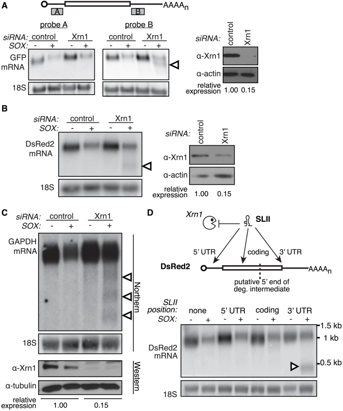
(A–B) 293T cells were treated with control or Xrn1 siRNAs, then transfected with the indicated reporters +/− SOX. The level of Xrn1 protein depletion is shown on the right, with actin as a loading control. (A) Diagram depicts location of probes on the GFP message. Northern blots using probes against either the 5′ (probe A; left panel) or 3′ (probe B; right panel) UTR of the GFP mRNA reporter or 18S. (B) Northern blots using probes against the 3′ UTR of the DsRed2 mRNA reporter, or 18S (left panels). (C) 293T cells were treated with control or Xrn1 shRNAs, then transfected with GFP (−) or GFP-SOX (+). Cells were then selected for GFP fluorescence by FACS before RNA and protein collection. RNA was Northern blotted with probes to the 3′ end of the GAPDH coding region, or to 18S. The level of Xrn1 knockdown was assessed by Western blot, using tubulin as a loading control. In panels A, B and C, arrowheads denote degradation intermediates. (D) A flaviviral Xrn1-blocking element (SLII) was inserted at different positions within the DsRed2 mRNA. RNA from 293T cells transfected with the indicated reporter +/− SOX was Northern blotted using a probe directed against the 3′ UTR of the DsRed2 reporter, or 18S. The full-length mRNA is ∼1.2 kb, and the expected size of the fragment protected from Xrn1 degradation by SLII within the 5′ UTR is ∼1.1 kb, within the coding region is ∼900 bp, and within the 3′ UTR is ∼500 bp. Arrowhead denotes protected fragment. We further confirmed that Xrn1 is involved in SOX-induced RNA degradation in an siRNA-independent manner by use of a flaviviral structural element (SLII) that can block 5′-3′ RNA degradation by Xrn1 [14]. In SOX expressing cells, insertion of the SLII element within the 3′ UTR of the DsRed2 reporter resulted in the appearance of a protected fragment of a size consistent with the portion of the mRNA downstream of the SLII (Figure 1D). In contrast, RNA fragments were not observed upon insertion of the SLII in the DsRed2 coding region upstream of the predicted 5′ end of the intermediate or in the 5′ UTR. This agrees with the observation that the 5′ portion of the mRNA is removed via an Xrn1-independent mechanism. Results consistent with this interpretation were also obtained for the GFP and β-globin reporters (Figure S1C–E). Collectively, our data suggest that SOX-mediated RNA depletion is a two-step process: an initiating event that removes a portion of the 5′ end of the mRNA, which is then followed by exonucleolytic degradation of the remaining fragment by the host Xrn1 enzyme.
SOX-induced mRNA degradation bypasses the requirement for deadenylation and decapping
In mammalian cells, sequential removal of the protective 3′ poly(A) tail and 5′ cap is generally required for Xrn1 to gain access to the RNA, as it can only degrade RNAs with a free 5′ monophosphate. Moreover, deadenylation is thought to be the rate-limiting step regulating mRNA degradation. Several results, however, indicate that SOX promotes mRNA degradation by Xrn1 while bypassing the requirement for deadenylation. Whereas the effects of a single copy of the Xrn1-blocking SLII element were only apparent in cells transfected with SOX (as these undergo enhanced mRNA turnover) (Figure 1D and S1D-E), insertion of two copies of the SLII (GFP-2xSLII, Figure S2A) led to a stronger Xrn1 block that can be visualized during basal GFP mRNA turnover even in cells lacking SOX (Figure 2A). In the absence of SOX, in vitro removal of the poly(A) tails by incubation with oligo(dT) and RNase H had little effect on the mobility of the degradation fragment resulting from the GFP-2xSLII construct, confirming that this fragment was generated after deadenylation. In contrast, the fragment produced in SOX-expressing cells was significantly larger prior to RNase H treatment, indicating that the cleaved fragment retained its poly(A) tail (Figure 2A).
Fig. 2. The 3′ degradation intermediate retains its poly(A) tail. 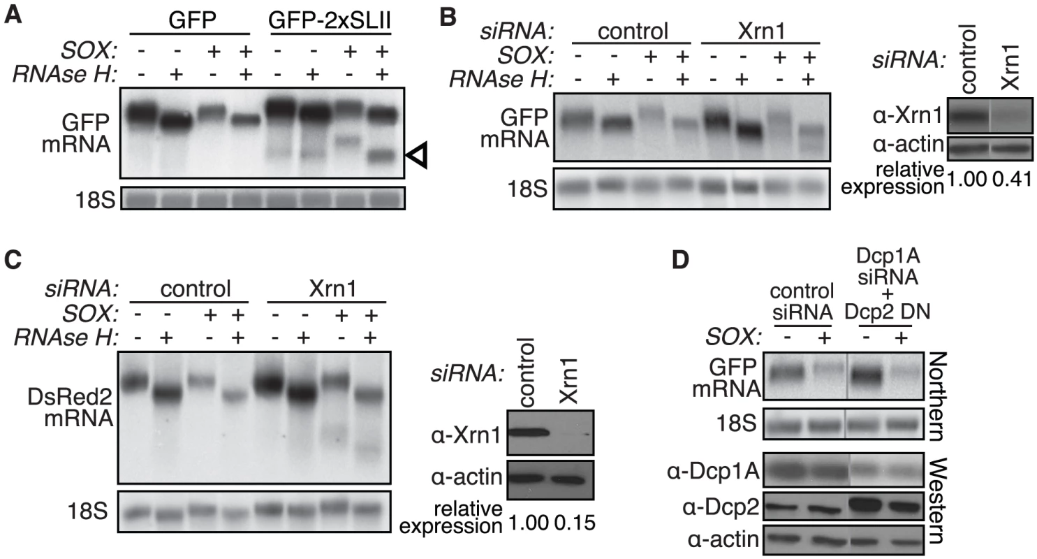
(A) 293T cells were transfected with GFP or a modified GFP reporter containing two copies of the flaviviral Xrn1-blocking element SLII within the GFP coding region, +/− SOX. A fraction of the RNA was treated with oligo(dT) and RNAse H to remove the poly(A) tail prior to Northern blotting with a GFP 3′ UTR or an 18S probe. Arrowhead denotes protected fragments. (B–C) 293T cells were transfected with control or Xrn1 siRNAs, followed by GFP (B) or DsRed2 (C) reporters +/− SOX. A fraction of the RNA was treated with oligo(dT) and RNAse H to remove the poly(A) tail prior to Northern blotting with a GFP 3′ UTR or an 18S probe. Xrn1 protein levels were assessed by Western blot (right panels in B and C). (D) 293T cells were treated with control siRNAs or siRNAs against the decapping complex protein Dcp1A. They were then transfected with GFP +/− SOX and/or the Dcp2 dominant negative mutant E148Q (Dcp2 DN). RNA was Northern blotted using a GFP 3′ UTR or an 18S probe. Western blots show the level of Dcp1A knockdown and Dcp2 DN overexpression. Actin serves as a loading control and grey lines indicate where intervening lanes have been cropped out. See also Figure S2. Similarly, we used RNAse H assays to evaluate the polyadenylation status of the SOX degradation intermediate present in Xrn1-depleted cells. Poly(A) tail removal decreased the mobility of both the full-length GFP and DsRed2 mRNAs, as well as the Xrn1-targeted degradation intermediates (Figure 2B and 2C). In addition, we were able to purify the GFP intermediate after poly(A) selection of the mRNA over oligo(dT) coupled beads (data not shown). Together, these data indicate that SOX can bypass the main regulatory step of normal mRNA degradation and render RNAs directly accessible to Xrn1.
Given that deadenylation generally precedes mRNA cap removal, we predicted that mRNA degradation in SOX-expressing cells would occur in a decapping-independent manner. Consistent with our hypothesis, we observed SOX-induced mRNA turnover in cells overexpressing a dominant negative mutant of the Dcp2 decapping enzyme, Dcp2 E148Q (J. Lykke-Andersen, personal communication; [34]), that had also been subjected to siRNA-mediated depletion of the decapping co-activator Dcp1A (Figure 2D and S2C). Although at least one additional decapping enzyme exists in mammalian cells [35], overexpression of the Dcp2 mutant was sufficient to reduce basal mRNA turnover of the GFP-2xSLII reporter, consistent with an inhibition of decapping activity (Figure S2B). We also depleted levels of the hRrp41 subunit of the 3′-5′ exosome and saw no difference between control and exosome siRNA-treated cells in our assay, in agreement with previous observations [25] (Figure S2D). Neither 5′ nor 3′ degradation fragments were detected upon hRrp41 knockdown (Figure S2E). A caveat with siRNA knockdown experiments in general is that negative results could be due to insufficient depletion of the protein. However, taken together our data suggest that the viral SOX protein interfaces specifically with the Xrn1 enzyme to accomplish host shutoff. In addition, these results further argue that removal of the 5′ portion of the RNA occurs independently of the canonical 5′-3′ decay pathway.
A specific element directs the initial cleavage
One way that mRNAs can be made accessible to Xrn1 without prior deadenylation or decapping is by internal endonucleolytic cleavage, a mechanism used by host quality control pathways to rapidly eliminate flawed mRNAs. Indeed, the defined length of the intermediates that we observe following Xrn1 depletion is strikingly reminiscent of the Smg6-cleaved intermediates seen during nonsense mediated decay (NMD) [36], [37]. A defined-length intermediate also suggests that cleavage is directed to a specific location or sequence within the mRNA. The different size of the intermediate observed in GFP and DsRed2 (compare Figures 1A and 1B) further indicated that a specific element, rather than positional information, is likely to direct the truncation. To test whether there is a specific sequence that leads to an endonucleolytic cut in the presence of SOX, we constructed a modified GFP reporter that contained an internal in-frame repeat of a 201 nt region encompassing the predicted cleavage site (Figure 3A, GFPrep). If this region contained an element targeted by an endonuclease, we expected that in the presence of SOX, GFPrep would give rise to two intermediates, assuming that the two cleavage events were equally likely and mutually exclusive. Indeed, we found that the GFPrep reporter generated two intermediates of sizes corresponding to two independent cleavage events (Figure 3B, arrowheads). These data suggest that an element within the initial 201 nucleotides of the GFP coding region directs an endonucleolytic cleavage in SOX-expressing cells.
Fig. 3. A specific element in the GFP coding region is sufficient for production of degradation intermediates by SOX. 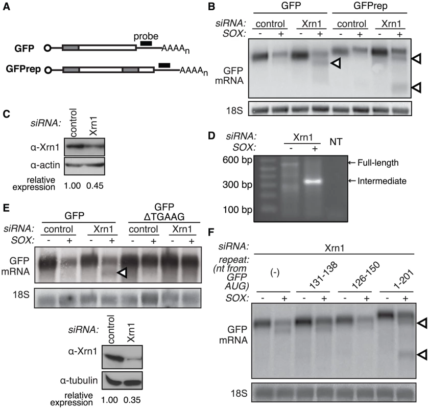
(A) The modified GFP mRNA (GFPrep) contains an internal repeat of the first 201 basepairs of the GFP coding region (grey shaded area). (B) 293T cells were treated with control or Xrn1 siRNAs, then transfected with GFP or GFPrep +/− SOX. RNA was Northern blotted with a GFP 3′ UTR or an 18S probe. Arrowheads denote degradation intermediates. (C) Western blot shows the level of Xrn1 depletion in the experiment depicted in panel B, with actin as a loading control. (D) 5′ RACE was carried out on total RNA from 293T cells transfected with Xrn1 siRNAs and a GFP expression vector in the presence or absence of SOX. An internal GFP primer and a primer to the adapter ligated to the 5′ end of the RNAs were used to amplify GFP in each sample. Full length refers to the band corresponding to the intact GFP transcript, intermediate refers to the ∼300 bp band preferentially amplified in the lysate from SOX-expressing cells, and NT denotes the no template control. (E) A conserved TGAAG site located just upstream of the cleavage site was deleted from GFP to create GFP-ΔTGAAG. 293T cells were treated with control or Xrn1 siRNAs, then transfected with GFP or GFP ΔTGAAG in the presence or absence of SOX. RNA was Northern blotted with a GFP 3′ UTR or 18S probe (upper panels). Arrowheads denote degradation intermediates. Western blot (lower panels) shows the level of Xrn1 depletion in experiments depicted in panel E and F, with tubulin as a loading control. (F) Modified GFP reporters bearing an internal duplication of sequences around the GFP cleavage site were transfected in 293T cells treated with Xrn1 siRNAs +/− SOX. The inserted nucleotides are numbered based on their position relative to the GFP start codon. GFPrep (nt 1–201) is included as a positive control. RNA was Northern blotted with a GFP 3′ UTR or 18S probe. Arrowheads denote degradation intermediates. To identify sequences involved in directing the internal cleavage, we used 5′ rapid amplification of cDNA ends (RACE) to map the 5′ end of the degradation intermediates found in GFP, DsRed2 and β-globin. In all cases our RACE results confirmed what we had previously seen with the Northern blot analysis, in that a single predominant intermediate was amplified in Xrn1-depleted cells in a SOX-dependent manner (Figure 3D and data not shown). The majority of the sequences within this band terminated at a single nucleotide or within few nucleotides of each other (Figure S3). Analysis of the sequences surrounding the cleavage site for each mRNA revealed a conserved stretch of five bases (TGAAG) just upstream of the cleavage site. Deletion of the TGAAG element from the GFP construct abolished production of the cleavage fragment, confirming its role in SOX targeting (Figure 3E). However, insertion of this sequence alone (nt 131–138) or of a 25-nucleotide stretch (nt 126–150) surrounding the GFP cleavage site was not sufficient to elicit generation of a second fragment (Figure 3F). We therefore hypothesize that the TGAAG sequence is an essential component of a larger element, perhaps structural, involved in directing endonucleolytic cleavage of mRNAs in SOX-expressing cells.
The catalytic residues of SOX are required for host shutoff
In vitro, several herpesviral homologs of SOX exhibit weak DNA endonuclease activity as well as RNase activity on linear RNA substrates [28], [29], [30], [38], [39], [40]. Thus, it is possible that SOX itself carries out the initial truncation of mRNAs in cells. Recently, the structure of SOX and its homolog BGLF5 in Epstein-Barr virus were solved, leading to the identification of catalytic residues responsible for the DNase activity of these proteins, as well as putative DNA binding residues [28], [30], [41]. Mutation of one of the catalytic residues in BGLF5 (D203S) was shown to abolish all in vitro nuclease activity without misfolding the protein [30]. To test whether the SOX catalytic core is required for host shutoff in vivo, we generated individual SOX mutants for each of the catalytic residues identified in the crystal structures: E184A, D221A or D221S (equivalent to D203S in BGLF5), E244A and K246I. Indeed, mutation of any one of the catalytic residues abolished the ability of SOX to deplete GFP mRNA in cells (Figure 4A). As a control, we mutated the putative DNA-binding residues (W135V, R139I, S144I, S146I, S219A, Q376G), which are also located in the active site cleft (Figure S4C), and found that only residues R139, S144 and Q376 were required for host shutoff (Figure 4B). This suggests that SOX may bind RNA and DNA using partially overlapping residues. In contrast with our host shutoff data, all the catalytic and putative DNA binding residues tested were required for in vitro DNase activity (Figure S4A and S4B). Thus, our mutational analysis suggests that the catalytic activity of SOX - presumably the RNase activity - is required for host shutoff.
Fig. 4. The catalytic residues of SOX are required for mRNA turnover and generation of the degradation intermediate. 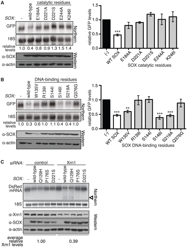
(A–B) Wild-type (WT) SOX or SOX putative catalytic (A) or DNA binding (B) residue mutants were transfected into 293T cells with a GFP reporter. RNA was Northern blotted using a 5′ GFP or 18S probe, and protein lysates were Western blotted for SOX and actin (as a loading control). Graphs depicting mean relative GFP levels ± s.e.m. from >3 experiments in cells transfected with the various mutants are also shown (right). **p<0.01, ***p<0.001, One-way ANOVA followed by Dunnett's test versus (−). (C) 293T cells were transfected with control or Xrn1 siRNAs, followed by a DsRed2 reporter +/− WT SOX or the indicated SOX mutant. Northern blots were performed using a probe directed against the 3′ UTR of the DsRed2 mRNA or 18S (upper panels), and Western blots show expression of the SOX mutants and the level of Xrn1 depletion (lower panels). Arrowhead denotes degradation intermediates. We next tested directly whether the RNA turnover activity of SOX was responsible for the generation of the degradation intermediate. We compared production of the degradation intermediate when expressing a SOX mutant lacking only DNase activity associated with viral genome processing (Q129H) [33], only the mRNA turnover activity (P176S) [33], or a catalytic mutant lacking both activities (D221S) (Figure 4C). The degradation intermediate was not produced in either of the two mutants lacking RNA turnover activity, but was readily detectable in cells expressing Q129H, which selectively lacks DNase function. These results indicate that generation of the intermediate by SOX is closely linked to its ability to degrade mRNAs, consistent with the two-step model for SOX-mediated mRNA degradation.
SOX depletes polysomes and co-sediments with 40S subunits
We next sought to determine how SOX targets cytoplasmic mRNAs for the initial cleavage event. In vitro, SOX exhibits relatively weak affinity for RNA (Kd of 87 µM), suggesting that in cells it is recruited to mRNAs via associations with host factors [28]. In cellular quality control pathways such as NMD, translation is required for error recognition and the primary endonucleolytic cleavage [42], [43]. In addition, it has recently been reported that the vast majority of mRNAs in the cytoplasm are polysome-associated [44], suggesting that targeting mRNAs engaged in translation would be an efficient mechanism to clear host messages during host shutoff.
To examine the effects of host shutoff on translating mRNAs, we performed polysome profiling of a B cell line (BCBL-1) derived from a patient with primary effusion lymphoma, which harbors KSHV in a latent state. We used a line of BCBL-1 cells bearing an inducible version of the KSHV major lytic transactivator RTA (TREx BCBL-1-RTA) [45] to allow efficient lytic reactivation following RTA induction. Upon chemical stimulation of lytic KSHV replication in these cells (when SOX is expressed from the viral genome), we observed a significant decrease in the polysome population and a corresponding increase in 80S monosomes, consistent with degradation of actively translating mRNAs (Figure 5A). It should be noted that the level of polysome depletion during viral infection is likely underestimated, as ∼20% of induced cells generally fail to enter the lytic cycle. The depletion of polyribosomes was not due to chemical treatment alone, because treatment of the KSHV negative BJAB B cell line did not result in a similar decrease in polysomes (data not shown). We next looked for the presence of SOX in gradient fractions from the corresponding polysome profiles of lytically reactivated BCBL-1 cells. As controls, we also blotted for PAIP2A, a protein not found in polysomes [46], as well as the ribosomal protein RPS3 (Figure 5B). SOX appeared to sediment primarily with the ribonucleoprotein (RNP), 40S, and monosome fractions, and exhibited partial overlap with RPS3 (Figure 5B). Puromycin treatment disrupted polysomes but failed to alter the SOX sedimentation profile, arguing against a specific association with the 80S and polysome fractions (data not shown). To more accurately determine its sedimentation profile, we increased the resolution of the lighter molecular weight complexes using lower density sucrose gradients. These experiments revealed that SOX indeed sediments in both the RNP and 40S fractions, similar to Xrn1 (Figure 5C). Its sedimentation profile also overlapped with the eIF3j and eIF2α components of the 40S pre-initiation complex, which is recruited to the 5′ cap prior to ribosomal scanning (Figure 5C). This is in contrast to the sedimentation of PABPC, which remains bound to the mRNA throughout the polysome fractions as well (Figure 5C; data not shown). Similar data were obtained upon transient expression of SOX in 293T cells (Figure S5A). We also tested the sedimentation profile of the SOX D221S catalytic mutant, and found it to mimic that of wild-type SOX (Figure S5B), indicating that the catalytic site of the protein is unlikely to be involved in its recruitment to target RNAs.
Fig. 5. SOX depletes polyribosomes and cosediments with 40S subunits. 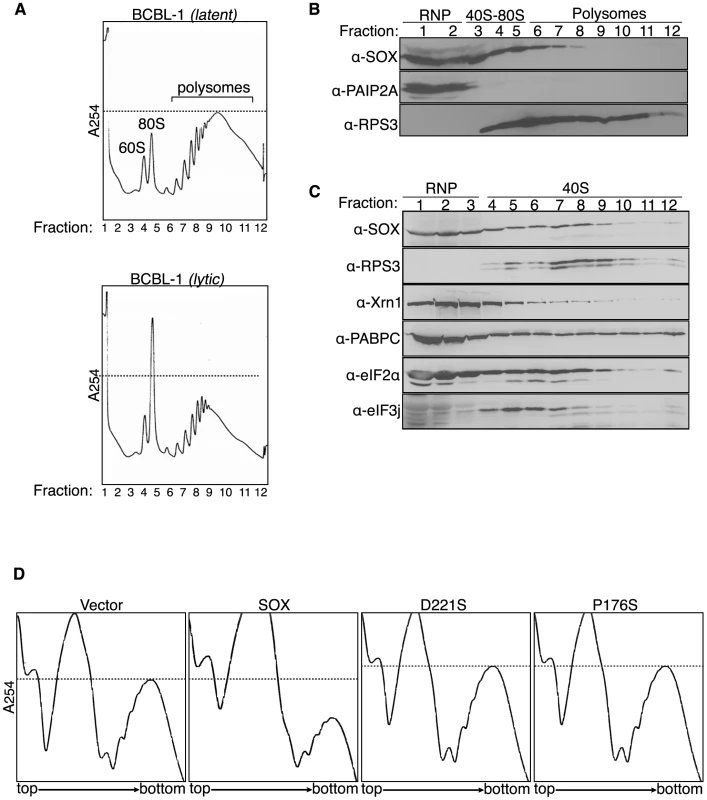
(A) TREx BCBL-1-RTA (BCBL-1) cells were mock treated (latent) or induced (lytic) with 1 µg/ml doxycycline, 500 ng/ml ionomycin, and 20 ng/ml 2-O-tetradecanoylphorbol-13-acetate for 24 h to stimulate KSHV replication, then subjected to sucrose gradient fractionation using a 15–60% sucrose gradient in order to monitor the abundance of translating polysomes. (B) Fractions collected from the induced BCBL-1 gradients shown in panel A were Western blotted with the indicated antibodies. (C) Lysate from induced BCBL-1 cells was fractionated using a 5–20% sucrose gradient and analyzed by Western blot with indicated antibodies. (D) 293T cells were transfected with the indicated construct and subjected to sucrose gradient fractionation to obtain polysome profiles. Dashed lines indicate polysome levels of either latent cells (A) or vector expressing cells (D). Given that SOX is the dominant effector of host shutoff during KSHV infection, we hypothesized that polysome depletion in BCBL-1 cells was a consequence of SOX activity. We therefore tested the effects of SOX on the endogenous translating mRNA pool through polysome profiling of 293T cells expressing either wild-type SOX, the SOX catalytic mutant D221S, or the single function SOX P176S mutant lacking host shutoff but not DNase activity. Indeed, expression of wild-type SOX alone caused a significant depletion of polysomes relative to vector-transfected cells, whereas neither of the host shutoff defective SOX mutants had this effect (Figure 5D). These results additionally confirm that the catalytic activity of SOX is required to initiate widespread turnover of endogenous host messages.
SOX specifically targets mRNAs at an early stage of translation
We had previously observed that a translationally incompetent version of the GFP mRNA, which terminates by ribozyme cleavage and lacks a poly(A) tail (GFP-HR) (Figure S6A), escaped SOX-mediated turnover [25]. Although this mRNA is inefficiently exported, subcellular fractionation experiments confirmed that even the exported cytoplasmic population of GFP-HR was not subject to SOX-induced turnover (Figure S6B). Thus, the failure of SOX to degrade this mRNA in the cytoplasm could be due to its translational incompetence.
To further explore a role for translation in SOX-induced mRNA turnover, we examined SOX turnover of RNAs transcribed by Pol I and Pol III. Pol I and Pol III transcription does not result in the addition of the cap and poly(A) tail, mRNA modifications critical to translation initiation specifically associated with Pol II transcription. Using a pure population of cells expressing GFP-SOX or GFP alone, we found that neither the endogenous Pol III-generated Y3 and 7SL cytoplasmic non-coding RNAs, nor the 18S rRNA transcribed by Pol I undergo turnover in the presence of SOX (Figure 6A). In contrast, we observed significant depletion of endogenous mRNAs transcribed by Pol II, including GAPDH, β-actin, and stearoyl-CoA desaturase (SCD) in SOX-expressing cells (Figure 6A). In principle, the inability of SOX to degrade the non-Pol II transcripts could be due to the absence of an ORF, or the presence of RNP complexes occluding SOX cleavage sites. To exclude these possibilities, we expressed the GFP reporter under the control of Pol I or Pol III promoters and found that in both cases, the GFP RNA was not targeted by SOX (Figure 6B). We confirmed using subcellular fractionation experiments that the inability of SOX to promote degradation of these RNAs was not due to a failure of the RNAs to be exported (Figure S6E and S6F). Collectively, these data indicate that RNAs must be translationally competent to be targeted by SOX.
Fig. 6. SOX targets mRNA at an early stage of translation. 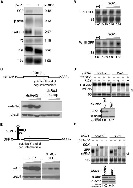
(A) 293T cells were transfected with control or Xrn1 shRNA-expressing constructs, and subsequently with either GFP or GFP-SOX. Cells were then sorted for GFP fluorescence to generate a pure population of SOX-expressing and control cells prior to RNA extraction. RNA was Northern blotted with the indicated probes against endogenous Pol I (18S), Pol II (SCD, β-actin, GAPDH), and Pol III (Y3, 7SL) RNAs. (B) 293T cells were transfected with either Pol I-driven GFP or Pol III-driven GFP reporters with or without increasing amounts of SOX (200–600 ng), then total RNA was Northern blotted using GFP or 18S probes. (C) 293T cells were transfected with increasing amounts (100–300 ng) of dsRed2 or a dsRed2–100stop construct containing a premature termination codon upstream of the predicted cleavage site (dashed line), then Western blotted for dsRed protein. (D) 293T cells were transfected with control or Xrn1 shRNAs and subsequently with either the dsRed2 or dsRed2-100stop (100stop) reporter in the presence or absence of SOX. RNA was Northern blotted with 3′ end dsRed2 or 18S probes. The arrowhead indicates the position of the SOX-induced cleavage product. (E) 293T cells were transfected with increasing amounts (100–300 ng) of GFP or a ΔEMCV-GFP construct containing the ΔEMCV IRES in the 5′ UTR, then Western blotted for GFP protein. (F) 293T cells were transfected with control or Xrn1 shRNAs and then with either the GFP or ΔEMCV-GFP reporter in the presence or absence of SOX. RNA was Northern blotted with 3′ GFP or 18S probes. The arrowhead indicates the position of the SOX-induced cleavage product. The Western blot shows level of Xrn1 depletion, and the actin loading control. To determine whether mRNA degradation by SOX required ribosomal passage over or near the putative initial cleavage site, we designed a reporter dsRed2 mRNA harboring a termination codon upstream of the putative SOX cleavage site (dsRed2-100stop). This prevented production of full-length dsRed2 protein (Figure 6C). Interestingly, in cells depleted of Xrn1, dsRed2-100stop was cleaved similarly to wild-type dsRed2 upon SOX expression (Figure 6D), indicating that ribosomal passage over the cleavage site is not necessary for its recognition.
To determine whether 60S joining is required for cleavage, we made use of a modified version of the encephalomyocarditis virus internal ribosomal entry site (IRES), termed ΔEMCV, which cannot promote cap-independent translation but is highly structured and decreases translation initiation in vitro [47]. Indeed, insertion of ΔEMCV into the GFP 5′ UTR 30 nt from the cap significantly reduced GFP protein accumulation (Figure 6E). However, in Xrn1-depleted cells, SOX still induced cleavage of this mRNA with similar efficiency as wild-type GFP, and at the same site (Figure 6F). In agreement with the ΔEMCV-GFP data, we observed that SOX could also promote turnover of a GFP mRNA with a 60 nt adenylate tract inserted in the 5′ UTR (5′A60-GFP) (Figure S6D), which similarly reduced GFP protein production (Figure S6C). A 50–70 nt adenylate-rich tract in the 5′ UTR of PABPC has likewise been shown to repress translation of its message in an autoregulatory manner as a consequence of PABPC protein binding to this region [48]. Consistent with the sedimentation profile of SOX, these data suggest that in SOX-expressing cells, mRNAs are targeted for cleavage at an early step during translation, perhaps prior to AUG recognition.
To monitor directly where cleavage occurs, we performed sucrose density gradient centrifugation on Xrn1-depleted cells expressing SOX and the GFP reporter. In agreement with our above findings, the cleaved intermediate preferentially accumulates in the 40S fraction (Figure 7A). The degradation intermediate is absent in all fractions from cells expressing the SOX catalytic mutant D221S (Figure 7B). These results lead us to favor a model in which mRNAs are recognized by SOX during formation of the 40S preinitiation complex, at which point they undergo SOX-induced endonucleolytic cleavage and subsequent destruction by Xrn1.
Fig. 7. The degradation intermediate sediments predominantly with the 40S fractions. 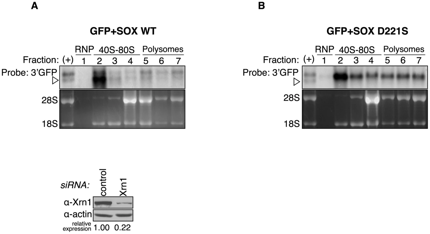
293T cells were transfected with Xrn1 shRNA, followed by expression of GFP and either wild-type (WT) SOX (A) or the catalytically dead SOX D221S (B). They were then fractionated over sucrose gradients and RNA from each fraction was Northern blotted with a 3′ GFP probe. Ribosomal RNA was visualized by ethidium bromide staining. In both panels the (+) lane shows the migration of full-length GFP and the degradation intermediate in unfractionated RNA from cells expressing wild-type SOX. It was used as a reference to identify the different RNA species in the fractionated RNA. Arrowheads point to degradation intermediates. Discussion
KSHV SOX and hXrn1 coordinate mRNA decay
This study shows that the global destruction of cellular mRNA during KSHV-induced host shutoff is enacted through the coordinated activities of both the viral SOX protein and cellular Xrn1. Our findings suggest that, in cells, SOX directs endonucleolytic cleavage of mRNAs at an early stage during translation, and subsequently employs host nucleases such as Xrn1 to execute degradation of the mRNA body. Thus, the virus is able to make use of the available host mRNA turnover machinery, yet bypass the rate limiting events that normally precede activation of Xrn1. Collectively, these data and our previous studies reconcile the seemingly disparate observations that the gammaherpesvirus SOX homologs promote potent host shutoff, yet exhibit relatively weak in vitro RNase activity. We hypothesize that while SOX can catalyze RNA cleavage, it does so in a site-specific manner in cells and requires one or more cellular factors to recruit and/or position it on the mRNA substrate, as well as host enzymes to complete the degradation process.
Evidence for SOX as an endonuclease
Endonucleolytic cleavages enable rapid mRNA turnover, because they generate unprotected RNA ends while bypassing the requirement for deadenylation and decapping. Cellular endonucleases are therefore generally subject to tight regulation. In eukaryotic cells, endonuclease-mediated decay is associated with mRNA quality control pathways, such as NMD, which targets messages containing premature stop codons [36], [37], and No-Go decay (NGD), which destroys mRNAs that experience stalls in translation [49]. In addition, recent studies have described previously unappreciated contributions of eukaryotic endonucleases to other basic processes such as miRNA-mediated mRNA decay [50], ER stress responses [51], [52], and exosome-mediated decay [53], [54], [55]. A few endonucleases that regulate decay of specific sets of otherwise normal mRNAs have also been described [10].
Several observations suggest that a primary role of SOX during host shutoff is to mediate endonucleolytic cleavage of target mRNAs, thereby enabling direct access by Xrn1. A key finding is that Xrn1-depleted cells undergoing host shutoff accumulate defined size degradation intermediates, similar to those derived from transcripts undergoing NMD or NGD under conditions of limiting Xrn1 [36], [37], [42], [49], [56]. Additionally, the observation that deadenylation and Dcp1/2-mediated decapping are not required to generate the degradation intermediate suggest that the 5′ and 3′ ends of the mRNA remain intact at the time of cleavage. We predict that SOX induces a sequence-specific rather than a position-dependent cut, as duplicating a 201 nt region surrounding the cut site yielded a second degradation intermediate, and addition of sequences upstream of the cleavage site did not alter the cleavage efficiency or location. We identified a conserved stretch of five nucleotides just upstream of the cut site in three different reporters. This consensus site was necessary, but not sufficient to elicit cleavage, suggesting that it represents a portion of a larger element required for SOX targeting. We hypothesize that the full element is a complex sequence or structure, perhaps degenerate, enabling it to be widespread among cellular mRNAs.
It is likely that additional enzymes besides Xrn1 participate in clearance of the fragments created by SOX, particularly in removal of the 5′ degradation intermediate. However, we have not observed a role for the canonical 3′-5′ exosome exonuclease complex. One possibility is that another host 3′-5′exonuclease instead mediates degradation of the upstream fragment, or plays a compensatory role upon exosome depletion. This might explain the limited protection of the 5′ fragment afforded by exosome knockdown in studies of another eukaryotic endonuclease [36]. Alternatively, more robust depletion of the exosome might be required to inhibit its activity in the context of cytoplasmic mRNA degradation.
SOX bears significant structural similarity to the PD-(D/E)XK type II restriction endonuclease superfamily [41], which includes several proteins that have been experimentally demonstrated to have endonuclease activity on RNA [57], [58]. The catalytic residues of SOX are essential for mRNA turnover in cells, further supporting a direct role for SOX in the primary cleavage event. Interestingly, in vitro RNase activity has recently been described for SOX and its EBV homolog BGLF5, although these studies concluded that it functioned as an exonuclease [28], [30]. However, RNases can harbor both endonuclease and exonuclease activity in the same active site [59], [60]. In addition, our data indicate that the endonucleolytic activity of SOX occurs in a site-specific manner, whereas the published assays used generic RNAs.
Mechanism of mRNA targeting
Our results support the involvement of an early stage of translation in the targeting of mRNA by SOX. This would allow the virus to selectively eliminate competing host mRNAs, while sparing regulatory RNAs that may be necessary for its own gene expression. Eukaryotic translation initiates with the recruitment of the 40S ribosomal subunit to the cap via interactions with translation initiation factors, followed by scanning to the AUG codon and recruitment of the 60S subunit. Two observations indicate that recognition and turnover in SOX-expressing cells are initiated prior to 60S joining. Specifically, insertion in the 5′ UTR of elements that presumably inhibit or prematurely terminate 40S scanning, and thus significantly reduce protein production, does not affect SOX activity. Moreover, the degradation intermediate accumulates preferentially in the 40S fraction. Our finding that SOX cosediments with the 40S initiation complex suggests that an association with translation initiation machinery directs it to mRNAs. Supporting this prediction is the failure of SOX to target translationally incompetent reporter and endogenous RNAs transcribed by RNA polymerase I or III.
We hypothesize that SOX targeting requires recognition of the mRNA 5′ end, likely via associated translation initiation factors, and that this recruitment somehow allows SOX access to the cleavage site(s) within the mRNA. Interestingly, our data show that cleavage can occur hundreds of nucleotides away from the site of translation initiation. One interesting parallel to these seemingly discrepant observations is that of the E. coli endonuclease RNase E, which can cleave anywhere along the length of the RNA (preferably within AU-rich sequences), but requires a monophosphate at the mRNA 5′ end. Thus, similar to our proposed model for SOX activity, RNase E can simultaneously recognize two non-adjacent regions of the primary RNA sequence [61]. These observations can be reconciled by the fact that RNAs adopt secondary and tertiary structures within a cell that could presumably enable such sequences to be juxtaposed. Alternatively, this distance could be bridged if there was an additional interaction of SOX with a host protein co-factor bound to the cleavage element.
Implications for virus-induced host shutoff
While this is the first example of a viral protein working in concert with host RNA turnover components to broadly target cellular messages, it is likely that several other viruses use similar mechanisms to enact host shutoff. Alphaherpesviruses and SARS coronavirus also encode host shutoff factors (vhs and nsp1, respectively) that promote mRNA degradation, but bear no homology to SOX [16], [17]. In vitro data indicate that they similarly induce endonucleolytic cleavage of RNAs, and it has been proposed that degradation of the mRNA body in cells may be assisted by host exonucleases, although this has yet to be shown [62], [63], [64], [65], [66]. In addition, all three viral proteins appear to use components of the translation machinery as a means to access mRNA [62], [65], [67], [68], [69]. Such parallels suggest that these viruses have adopted remarkably similar strategies to execute mRNA decay, resembling those of host pathways like NMD. In addition to the enhanced rate of decay afforded by an endonucleolytic mechanism, the advantage for the virus may be that this strategy of host shutoff generates intermediates that look indistinguishable from products of quality control pathways. Such intermediates would then be readily degraded by the core degradation machinery and might not be recognized as aberrant, perhaps avoiding activation of stress or innate immune responses. Thus, understanding how viral endonucleases interface with host pathways may provide insight into how manipulation of these pathways contributes to infectious disease, as well as into events that regulate cellular RNA decay.
Materials and Methods
Cell Extracts, Western blots, and Northern Blots
Protein lysates were either prepared in RIPA buffer [50 mM Tris-HCl (pH8.0), 150 mM NaCl, 1% (v/v) Nonidet P-40, 0.5% (w/v) sodium deoxycholate, 0.1% (w/v) sodium dodecyl sulfate (SDS)], or fractionated using the NE-PER kit (ThermoScientific). Western blots were performed with either mouse monoclonal anti-GFP (1∶2000, BD Biosciences), mouse monoclonal anti-dsRed (1∶500, Clontech), mouse monoclonal anti-HA (1∶2000, Invitrogen), mouse monoclonal anti-RPS3 (1∶1000, Abcam), mouse monoclonal anti-Flag (1∶1000, Sigma), rabbit polyclonal anti-PAIP2A (1 : 2000, kindly provided by N. Sonenberg [70]), rabbit polyclonal anti-hXrn1 (1∶5000, kindly provided by J. Lykke-Andersen), rabbit polyclonal anti-hDcp1a (1∶5000, kindly provided by J. Lykke-Andersen), rabbit polyclonal anti-hDcp2 (1∶400, kindly provided by M. Kiledjan), rabbit polyclonal anti-hRrp41 (1∶1000, see below), rabbit polyclonal anti-SOX J5803 (1∶5000, [33]), mouse monoclonal hnRNPC1/C2 (1∶2000, Abcam), mouse monoclonal anti-Hsp90 (1∶3000, Stressgen Bioreagents), mouse monoclonal anti-tubulin (1∶3000, Sigma Aldrich), goat polyclonal anti-actin (1∶5000, Santa Cruz Biotechnology), mouse monoclonal 10e10 anti-PABPC (1∶2000, Santa Cruz Biotechnology), rabbit polyclonal anti-eIF2α (1∶1000, Cell Signaling) or rabbit polyclonal anti-eIF3j (1∶1000, Cell Signaling). Rabbit polyclonal antibodies were raised against recombinant maltose binding protein (MPB)-fused hRrp41 purified from E. coli.
Total cellular RNA was isolated for Northern blotting using RNA-Bee (Tel-Test). Where indicated, the NE-PER kit (ThermoScientific) was used for cellular fractionation prior to RNA extraction. Northern blots were probed with 32P-labeled DNA probes made using either RediPrime II (GE Healthcare) or Decaprime II (Ambion) or, for β-globin, an SP6 transcribed 32P-labeled riboprobe. RNase H assays were carried out as previously described [25]. Results in each figure are a representative of at least 3 independent replicates of each experiment. Image J (http://rsbweb.nih.gov/ij/) was used for quantification of Northern and Western blots.
Polysome profiling
Profiles were obtained from uninduced or lytically reactivated TREx BCBL-1-RTA cells, or from 293T cells transfected with the indicated plasmids. Polysome profiles were carried out as described in Jackson and Larkins [71], except that cells were treated with 100 µg/ml CHX for 30 minutes prior to harvesting. BCBL-1 and 293T extracts were pelleted through 60% sucrose before layering or were directly layered on a 15–60% sucrose gradient containing 100 µg/ml CHX. Additional details, including the procedure for resolution of RNP/40S fractions are described in supplemental procedures (Text S1).
Additional experimental procedures, primer and siRNA/shRNA sequences used in the study are detailed in Supplemental Materials and Methods (Text S1).
Supporting Information
Zdroje
1. GrahamJRHendershottMCTerragniJCooperGM 2010 mRNA degradation plays a significant role in the program of gene expression regulated by phosphatidylinositol 3-kinase signaling. Mol Cell Biol 30 5295 5305
2. FanJYangXWangWWoodWH3rdBeckerKG 2002 Global analysis of stress-regulated mRNA turnover by using cDNA arrays. Proc Natl Acad Sci U S A 99 10611 10616
3. CheadleCFanJCho-ChungYSWernerTRayJ 2005 Control of gene expression during T cell activation: alternate regulation of mRNA transcription and mRNA stability. BMC Genomics 6 75
4. BelascoJG 2010 All things must pass: contrasts and commonalities in eukaryotic and bacterial mRNA decay. Nat Rev Mol Cell Biol 11 467 478
5. GarneauNLWiluszJWiluszCJ 2007 The highways and byways of mRNA decay. Nat Rev Mol Cell Biol 8 113 126
6. DeckerCJParkerR 1993 A turnover pathway for both stable and unstable mRNAs in yeast: evidence for a requirement for deadenylation. Genes Dev 7 1632 1643
7. MuhlradDDeckerCJParkerR 1994 Deadenylation of the unstable mRNA encoded by the yeast MFA2 gene leads to decapping followed by 5′–>3′ digestion of the transcript. Genes Dev 8 855 866
8. DunckleyTParkerR 1999 The DCP2 protein is required for mRNA decapping in Saccharomyces cerevisiae and contains a functional MutT motif. EMBO J 18 5411 5422
9. WangZJiaoXCarr-SchmidAKiledjianM 2002 The hDcp2 protein is a mammalian mRNA decapping enzyme. Proc Natl Acad Sci U S A 99 12663 12668
10. TomeckiRDziembowskiA 2010 Novel endoribonucleases as central players in various pathways of eukaryotic RNA metabolism. RNA 16 1692 1724
11. GagliaMMGlaunsingerB 2010 Viruses and the cellular RNA decay machinery. Wiley Interdisciplinary Reviews: RNA 1 47 59
12. SokoloskiKJDicksonAMChaskeyELGarneauNLWiluszCJ 2010 Sindbis virus usurps the cellular HuR protein to stabilize its transcripts and promote productive infections in mammalian and mosquito cells. Cell Host Microbe 8 196 207
13. UmbachJLCullenBR 2009 The role of RNAi and microRNAs in animal virus replication and antiviral immunity. Genes Dev 23 1151 1164
14. PijlmanGPFunkAKondratievaNLeungJTorresS 2008 A highly structured, nuclease-resistant, noncoding RNA produced by flaviviruses is required for pathogenicity. Cell Host Microbe 4 579 591
15. RoweMGlaunsingerBvan LeeuwenDZuoJSweetmanD 2007 Host shutoff during productive Epstein-Barr virus infection is mediated by BGLF5 and may contribute to immune evasion. Proc Natl Acad Sci U S A 104 3366 3371
16. KwongADKruperJAFrenkelN 1988 Herpes simplex virus virion host shutoff function. J Virol 62 912 921
17. KamitaniWNarayananKHuangCLokugamageKIkegamiT 2006 Severe acute respiratory syndrome coronavirus nsp1 protein suppresses host gene expression by promoting host mRNA degradation. Proc Natl Acad Sci U S A 103 12885 12890
18. GlaunsingerBGanemD 2004 Lytic KSHV infection inhibits host gene expression by accelerating global mRNA turnover. Mol Cell 13 713 723
19. ChangYCesarmanEPessinMSLeeFCulpepperJ 1994 Identification of herpesvirus-like DNA sequences in AIDS-associated Kaposi's sarcoma. Science 266 1865 1869
20. SoulierJGrolletLOksenhendlerECacoubPCazals-HatemD 1995 Kaposi's sarcoma-associated herpesvirus-like DNA sequences in multicentric Castleman's disease. Blood 86 1276 1280
21. CesarmanEChangYMoorePSSaidJWKnowlesDM 1995 Kaposi's sarcoma-associated herpesvirus-like DNA sequences in AIDS-related body-cavity-based lymphomas. N Engl J Med 332 1186 1191
22. GlaunsingerBGanemD 2004 Highly selective escape from KSHV-mediated host mRNA shutoff and its implications for viral pathogenesis. J Exp Med 200 391 398
23. CovarrubiasSRichnerJMClydeKLeeYJGlaunsingerBA 2009 Host shutoff is a conserved phenotype of gammaherpesvirus infection and is orchestrated exclusively from the cytoplasm. J Virol 83 9554 9566
24. ClydeKGlaunsingerB 2011 Deep sequencing reveals direct targets of gammaherpesvirus-induced mRNA decay and suggests that multiple mechanisms govern cellular transcript escape. PLoS One 6 e19655
25. LeeYJGlaunsingerBA 2009 Aberrant herpesvirus-induced polyadenylation correlates with cellular messenger RNA destruction. PLoS Biol 7 e1000107
26. KumarGRShumLGlaunsingerBA 2011 Importin {alpha}-Mediated Nuclear Import of Cytoplasmic Poly(A) Binding Protein Occurs as a Direct Consequence of Cytoplasmic mRNA Depletion. Mol Cell Biol 31 3113 3125
27. KumarGRGlaunsingerBA 2010 Nuclear import of cytoplasmic poly(A) binding protein restricts gene expression via hyperadenylation and nuclear retention of mRNA. Mol Cell Biol 30 4996 5008
28. BagnerisCBriggsLCSavvaREbrahimiBBarrettTE 2011 Crystal structure of a KSHV-SOX-DNA complex: insights into the molecular mechanisms underlying DNase activity and host shutoff. Nucleic Acids Res 39 5744 5756
29. KnopfCWWeisshartK 1990 Comparison of exonucleolytic activities of herpes simplex virus type-1 DNA polymerase and DNase. Eur J Biochem 191 263 273
30. BuissonMGeouiTFlotDTarbouriechNRessingME 2009 A bridge crosses the active-site canyon of the Epstein-Barr virus nuclease with DNase and RNase activities. J Mol Biol 391 717 728
31. HoffmannPJChengYC 1978 The deoxyribonuclease induced after infection of KB cells by herpes simplex virus type 1 or type 2. I. Purification and characterization of the enzyme. J Biol Chem 253 3557 3562
32. BaylisSAPurifoyDJLittlerE 1989 The characterization of the EBV alkaline deoxyribonuclease cloned and expressed in E. coli. Nucleic Acids Res 17 7609 7622
33. GlaunsingerBChavezLGanemD 2005 The exonuclease and host shutoff functions of the SOX protein of Kaposi's sarcoma-associated herpesvirus are genetically separable. J Virol 79 7396 7401
34. OzgurSChekulaevaMStoecklinG 2010 Human Pat1b connects deadenylation with mRNA decapping and controls the assembly of processing bodies. Mol Cell Biol 30 4308 4323
35. SongMGLiYKiledjianM 2010 Multiple mRNA decapping enzymes in mammalian cells. Mol Cell 40 423 432
36. EberleABLykke-AndersenSMuhlemannOJensenTH 2009 SMG6 promotes endonucleolytic cleavage of nonsense mRNA in human cells. Nat Struct Mol Biol 16 49 55
37. HuntzingerEKashimaIFauserMSauliereJIzaurraldeE 2008 SMG6 is the catalytic endonuclease that cleaves mRNAs containing nonsense codons in metazoan. RNA 14 2609 2617
38. HoffmannPJChengYC 1979 DNase induced after infection of KB cells by herpes simplex virus type 1 or type 2. II. Characterization of an associated endonuclease activity. J Virol 32 449 457
39. StolzenbergMCOokaT 1990 Purification and properties of Epstein-Barr virus DNase expressed in Escherichia coli. J Virol 64 96 104
40. SheafferAKWeinheimerSPTenneyDJ 1997 The human cytomegalovirus UL98 gene encodes the conserved herpesvirus alkaline nuclease. J Gen Virol 78 Pt 11 2953 2961
41. DahlrothSLGurmuDHaasJErlandsenHNordlundP 2009 Crystal structure of the shutoff and exonuclease protein from the oncogenic Kaposi's sarcoma-associated herpesvirus. FEBS J 276 6636 6645
42. GatfieldDIzaurraldeE 2004 Nonsense-mediated messenger RNA decay is initiated by endonucleolytic cleavage in Drosophila. Nature 429 575 578
43. IshigakiYLiXSerinGMaquatLE 2001 Evidence for a pioneer round of mRNA translation: mRNAs subject to nonsense-mediated decay in mammalian cells are bound by CBP80 and CBP20. Cell 106 607 617
44. HendricksonDGHoganDJMcCulloughHLMyersJWHerschlagD 2009 Concordant regulation of translation and mRNA abundance for hundreds of targets of a human microRNA. PLoS Biol 7 e1000238
45. NakamuraHLuMGwackYSouvlisJZeichnerSL 2003 Global changes in Kaposi's sarcoma-associated virus gene expression patterns following expression of a tetracycline-inducible Rta transactivator. J Virol 77 4205 4220
46. RiveraCILloydRE 2008 Modulation of enteroviral proteinase cleavage of poly(A)-binding protein (PABP) by conformation and PABP-associated factors. Virology 375 59 72
47. ChenCYSarnowP 1995 Initiation of protein synthesis by the eukaryotic translational apparatus on circular RNAs. Science 268 415 417
48. de Melo NetoOPStandartNMartins de SaC 1995 Autoregulation of poly(A)-binding protein synthesis in vitro. Nucleic Acids Res 23 2198 2205
49. DomaMKParkerR 2006 Endonucleolytic cleavage of eukaryotic mRNAs with stalls in translation elongation. Nature 440 561 564
50. MeisterGLandthalerMPatkaniowskaADorsettYTengG 2004 Human Argonaute2 mediates RNA cleavage targeted by miRNAs and siRNAs. Mol Cell 15 185 197
51. HollienJWeissmanJS 2006 Decay of endoplasmic reticulum-localized mRNAs during the unfolded protein response. Science 313 104 107
52. HanDLernerAGVande WalleLUptonJPXuW 2009 IRE1alpha kinase activation modes control alternate endoribonuclease outputs to determine divergent cell fates. Cell 138 562 575
53. LebretonATomeckiRDziembowskiASeraphinB 2008 Endonucleolytic RNA cleavage by a eukaryotic exosome. Nature 456 993 996
54. SchneiderCLeungEBrownJTollerveyD 2009 The N-terminal PIN domain of the exosome subunit Rrp44 harbors endonuclease activity and tethers Rrp44 to the yeast core exosome. Nucleic Acids Res 37 1127 1140
55. SchaefferDTsanovaBBarbasAReisFPDastidarEG 2009 The exosome contains domains with specific endoribonuclease, exoribonuclease and cytoplasmic mRNA decay activities. Nat Struct Mol Biol 16 56 62
56. ChenCYXuNShyuAB 1995 mRNA decay mediated by two distinct AU-rich elements from c-fos and granulocyte-macrophage colony-stimulating factor transcripts: different deadenylation kinetics and uncoupling from translation. Mol Cell Biol 15 5777 5788
57. MorinBCoutardBLelkeMFerronFKerberR 2010 The N-terminal domain of the arenavirus L protein is an RNA endonuclease essential in mRNA transcription. PLoS Pathog 6 e1001038 doi:1001010.1001371/journal.ppat.1001038
58. YuanPBartlamMLouZChenSZhouJ 2009 Crystal structure of an avian influenza polymerase PA(N) reveals an endonuclease active site. Nature 458 909 913
59. YangXCSullivanKDMarzluffWFDominskiZ 2009 Studies of the 5′ exonuclease and endonuclease activities of CPSF-73 in histone pre-mRNA processing. Mol Cell Biol 29 31 42
60. MathyNBenardLPellegriniODaouRWenT 2007 5′-to-3′ exoribonuclease activity in bacteria: role of RNase J1 in rRNA maturation and 5′ stability of mRNA. Cell 129 681 692
61. CarpousisAJLuisiBFMcDowallKJ 2009 Endonucleolytic initiation of mRNA decay in Escherichia coli. Prog Mol Biol Transl Sci 85 91 135
62. KamitaniWHuangCNarayananKLokugamageKGMakinoS 2009 A two-pronged strategy to suppress host protein synthesis by SARS coronavirus Nsp1 protein. Nat Struct Mol Biol 16 1134 1140
63. TaddeoBRoizmanB 2006 The virion host shutoff protein (UL41) of herpes simplex virus 1 is an endoribonuclease with a substrate specificity similar to that of RNase A. J Virol 80 9341 9345
64. TaddeoBZhangWRoizmanB 2006 The U(L)41 protein of herpes simplex virus 1 degrades RNA by endonucleolytic cleavage in absence of other cellular or viral proteins. Proc Natl Acad Sci U S A 103 2827 2832
65. ElgadiMMHayesCESmileyJR 1999 The herpes simplex virus vhs protein induces endoribonucleolytic cleavage of target RNAs in cell extracts. J Virol 73 7153 7164
66. ZelusBDStewartRSRossJ 1996 The virion host shutoff protein of herpes simplex virus type 1: messenger ribonucleolytic activity in vitro. J Virol 70 2411 2419
67. FengPEverlyDNJrReadGS 2005 mRNA decay during herpes simplex virus (HSV) infections: protein-protein interactions involving the HSV virion host shutoff protein and translation factors eIF4H and eIF4A. J Virol 79 9651 9664
68. FengPEverlyDNJrReadGS 2001 mRNA decay during herpesvirus infections: interaction between a putative viral nuclease and a cellular translation factor. J Virol 75 10272 10280
69. TaddeoBZhangWRoizmanB 2009 The virion-packaged endoribonuclease of herpes simplex virus 1 cleaves mRNA in polyribosomes. Proc Natl Acad Sci U S A 106 12139 12144
70. KhaleghpourKSvitkinYVCraigAWDeMariaCTDeoRC 2001 Translational repression by a novel partner of human poly(A) binding protein, Paip2. Mol Cell 7 205 216
71. JacksonAOLarkinsBA 1976 Influence of Ionic Strength, pH, and Chelation of Divalent Metals on Isolation of Polyribosomes from Tobacco Leaves. Plant Physiol 57 5 10
Štítky
Hygiena a epidemiologie Infekční lékařství Laboratoř
Článek Quorum Sensing in Fungi: Q&AČlánek Blood Feeding and Insulin-like Peptide 3 Stimulate Proliferation of Hemocytes in the MosquitoČlánek The DEAD-box RNA Helicase DDX6 is Required for Efficient Encapsidation of a Retroviral GenomeČlánek A Phenome-Based Functional Analysis of Transcription Factors in the Cereal Head Blight Fungus,Článek A Wide Extent of Inter-Strain Diversity in Virulent and Vaccine Strains of AlphaherpesvirusesČlánek The Anti-Sigma Factor TcdC Modulates Hypervirulence in an Epidemic BI/NAP1/027 Clinical Isolate ofČlánek Critical Roles for LIGHT and Its Receptors in Generating T Cell-Mediated Immunity during InfectionČlánek Frequent and Recent Human Acquisition of Simian Foamy Viruses Through Apes' Bites in Central Africa
Článek vyšel v časopisePLOS Pathogens
Nejčtenější tento týden
2011 Číslo 10- Jak souvisí postcovidový syndrom s poškozením mozku?
- Měli bychom postcovidový syndrom léčit antidepresivy?
- Farmakovigilanční studie perorálních antivirotik indikovaných v léčbě COVID-19
- 10 bodů k očkování proti COVID-19: stanovisko České společnosti alergologie a klinické imunologie ČLS JEP
-
Všechny články tohoto čísla
- Quorum Sensing in Fungi: Q&A
- Discovery of an Ebolavirus-Like Filovirus in Europe
- Toll-like Receptor 7 Controls the Anti-Retroviral Germinal Center Response
- Tubule-Guided Cell-to-Cell Movement of a Plant Virus Requires Class XI Myosin Motors
- Herpesvirus Telomerase RNA (vTR) with a Mutated Template Sequence Abrogates Herpesvirus-Induced Lymphomagenesis
- Mitochondrial Peroxiredoxin Plays a Crucial Peroxidase-Unrelated Role during Infection: Insight into Its Novel Chaperone Activity
- Sustained CD8+ T Cell Memory Inflation after Infection with a Single-Cycle Cytomegalovirus
- Novel Mouse Xenograft Models Reveal a Critical Role of CD4 T Cells in the Proliferation of EBV-Infected T and NK Cells
- Toll-8/Tollo Negatively Regulates Antimicrobial Response in the Respiratory Epithelium
- Exhausted Cytotoxic Control of Epstein-Barr Virus in Human Lupus
- Structural and Functional Analysis of Laninamivir and its Octanoate Prodrug Reveals Group Specific Mechanisms for Influenza NA Inhibition
- Infection Drives IL-17-Mediated Neutrophilic Allergic Airways Disease
- Blood Feeding and Insulin-like Peptide 3 Stimulate Proliferation of Hemocytes in the Mosquito
- HIV-1 Replication in the Central Nervous System Occurs in Two Distinct Cell Types
- Deep Molecular Characterization of HIV-1 Dynamics under Suppressive HAART
- Fitness Landscape of Antibiotic Tolerance in Biofilms
- The DEAD-box RNA Helicase DDX6 is Required for Efficient Encapsidation of a Retroviral Genome
- Preventing Sepsis through the Inhibition of Its Agglutination in Blood
- A Phenome-Based Functional Analysis of Transcription Factors in the Cereal Head Blight Fungus,
- IFITM3 Inhibits Influenza A Virus Infection by Preventing Cytosolic Entry
- Targeting Cattle-Borne Zoonoses and Cattle Pathogens Using a Novel Trypanosomatid-Based Delivery System
- A Wide Extent of Inter-Strain Diversity in Virulent and Vaccine Strains of Alphaherpesviruses
- Coordinated Destruction of Cellular Messages in Translation Complexes by the Gammaherpesvirus Host Shutoff Factor and the Mammalian Exonuclease Xrn1
- Signal Transduction through CsrRS Confers an Invasive Phenotype in Group A
- Biochemical and Structural Insights into the Mechanisms of SARS Coronavirus RNA Ribose 2′-O-Methylation by nsp16/nsp10 Protein Complex
- Histone Deacetylase 8 Is Required for Centrosome Cohesion and Influenza A Virus Entry
- Severe Acute Respiratory Syndrome Coronavirus Envelope Protein Regulates Cell Stress Response and Apoptosis
- Co-opts the FGF2 Signaling Pathway to Enhance Infection
- IRAK-2 Regulates IL-1-Mediated Pathogenic Th17 Cell Development in Helminthic Infection
- Trafficking of Hepatitis C Virus Core Protein during Virus Particle Assembly
- The Anti-interferon Activity of Conserved Viral dUTPase ORF54 is Essential for an Effective MHV-68 Infection
- A Viral Nuclear Noncoding RNA Binds Re-localized Poly(A) Binding Protein and Is Required for Late KSHV Gene Expression
- Suppression of Methylation-Mediated Transcriptional Gene Silencing by βC1-SAHH Protein Interaction during Geminivirus-Betasatellite Infection
- ISG15 Is Critical in the Control of Chikungunya Virus Infection Independent of UbE1L Mediated Conjugation
- Non-Hematopoietic Cells in Lymph Nodes Drive Memory CD8 T Cell Inflation during Murine Cytomegalovirus Infection
- RNA Polymerase II Stalling Promotes Nucleosome Occlusion and pTEFb Recruitment to Drive Immortalization by Epstein-Barr Virus
- Noninfectious Retrovirus Particles Drive the / Dependent Neutralizing Antibody Response
- Endophytic Life Strategies Decoded by Genome and Transcriptome Analyses of the Mutualistic Root Symbiont
- An Integrated Approach to Elucidate the Intra-Viral and Viral-Cellular Protein Interaction Networks of a Gamma-Herpesvirus
- as an Animal Model for the Study of Biofilm Infections
- Homeostatic Proliferation Fails to Efficiently Reactivate HIV-1 Latently Infected Central Memory CD4+ T Cells
- The Anti-Sigma Factor TcdC Modulates Hypervirulence in an Epidemic BI/NAP1/027 Clinical Isolate of
- Enhances Protective and Detrimental HLA Class I-Mediated Immunity in Chronic Viral Infection
- The Mouse IAPE Endogenous Retrovirus Can Infect Cells through Any of the Five GPI-Anchored EphrinA Proteins
- The Urgent Need for Robust Coral Disease Diagnostics
- HacA-Independent Functions of the ER Stress Sensor IreA Synergize with the Canonical UPR to Influence Virulence Traits in
- A Novel Core Genome-Encoded Superantigen Contributes to Lethality of Community-Associated MRSA Necrotizing Pneumonia
- Critical Roles for LIGHT and Its Receptors in Generating T Cell-Mediated Immunity during Infection
- The SARS-Coronavirus-Host Interactome: Identification of Cyclophilins as Target for Pan-Coronavirus Inhibitors
- Frequent and Recent Human Acquisition of Simian Foamy Viruses Through Apes' Bites in Central Africa
- Mechanisms of Trafficking to the Brain
- Defining Emerging Roles for NF-κB in Antivirus Responses: Revisiting the Enhanceosome Paradigm
- The Role of Sialyl Glycan Recognition in Host Tissue Tropism of the Avian Parasite
- Evolutionarily Divergent, Unstable Filamentous Actin Is Essential for Gliding Motility in Apicomplexan Parasites
- The Herpes Simplex Virus-1 Transactivator Infected Cell Protein-4 Drives VEGF-A Dependent Neovascularization
- Distinct Single Amino Acid Replacements in the Control of Virulence Regulator Protein Differentially Impact Streptococcal Pathogenesis
- Soluble Rhesus Lymphocryptovirus gp350 Protects against Infection and Reduces Viral Loads in Animals that Become Infected with Virus after Challenge
- A Genetic Screen Reveals Arabidopsis Stomatal and/or Apoplastic Defenses against pv. DC3000
- Hepatitis C Virus Reveals a Novel Early Control in Acute Immune Response
- Fumarate Reductase Activity Maintains an Energized Membrane in Anaerobic
- PLOS Pathogens
- Archiv čísel
- Aktuální číslo
- Informace o časopisu
Nejčtenější v tomto čísle- Severe Acute Respiratory Syndrome Coronavirus Envelope Protein Regulates Cell Stress Response and Apoptosis
- The SARS-Coronavirus-Host Interactome: Identification of Cyclophilins as Target for Pan-Coronavirus Inhibitors
- Biochemical and Structural Insights into the Mechanisms of SARS Coronavirus RNA Ribose 2′-O-Methylation by nsp16/nsp10 Protein Complex
- Evolutionarily Divergent, Unstable Filamentous Actin Is Essential for Gliding Motility in Apicomplexan Parasites
Kurzy
Zvyšte si kvalifikaci online z pohodlí domova
Současné možnosti léčby obezity
nový kurzAutoři: MUDr. Martin Hrubý
Všechny kurzyPřihlášení#ADS_BOTTOM_SCRIPTS#Zapomenuté hesloZadejte e-mailovou adresu, se kterou jste vytvářel(a) účet, budou Vám na ni zaslány informace k nastavení nového hesla.
- Vzdělávání


