-
Články
Top novinky
Reklama- Vzdělávání
- Časopisy
Top články
Nové číslo
- Témata
Top novinky
Reklama- Videa
- Podcasty
Nové podcasty
Reklama- Kariéra
Doporučené pozice
Reklama- Praxe
Top novinky
ReklamaA Typhimurium-Typhi Genomic Chimera: A Model to Study Vi Polysaccharide Capsule Function In Vivo
The Vi capsular polysaccharide is a virulence-associated factor expressed by Salmonella enterica serotype Typhi but absent from virtually all other Salmonella serotypes. In order to study this determinant in vivo, we characterised a Vi-positive S. Typhimurium (C5.507 Vi+), harbouring the Salmonella pathogenicity island (SPI)-7, which encodes the Vi locus. S. Typhimurium C5.507 Vi+ colonised and persisted in mice at similar levels compared to the parent strain, S. Typhimurium C5. However, the innate immune response to infection with C5.507 Vi+ and SGB1, an isogenic derivative not expressing Vi, differed markedly. Infection with C5.507 Vi+ resulted in a significant reduction in cellular trafficking of innate immune cells, including PMN and NK cells, compared to SGB1 Vi− infected animals. C5.507 Vi+ infection stimulated reduced numbers of TNF-α, MIP-2 and perforin producing cells compared to SGB1 Vi−. The modulating effect associated with Vi was not observed in MyD88−/− and was reduced in TLR4−/− mice. The presence of the Vi capsule also correlated with induction of the anti-inflammatory cytokine IL-10 in vivo, a factor that impacted on chemotaxis and the activation of immune cells in vitro.
Published in the journal: . PLoS Pathog 7(7): e32767. doi:10.1371/journal.ppat.1002131
Category: Research Article
doi: https://doi.org/10.1371/journal.ppat.1002131Summary
The Vi capsular polysaccharide is a virulence-associated factor expressed by Salmonella enterica serotype Typhi but absent from virtually all other Salmonella serotypes. In order to study this determinant in vivo, we characterised a Vi-positive S. Typhimurium (C5.507 Vi+), harbouring the Salmonella pathogenicity island (SPI)-7, which encodes the Vi locus. S. Typhimurium C5.507 Vi+ colonised and persisted in mice at similar levels compared to the parent strain, S. Typhimurium C5. However, the innate immune response to infection with C5.507 Vi+ and SGB1, an isogenic derivative not expressing Vi, differed markedly. Infection with C5.507 Vi+ resulted in a significant reduction in cellular trafficking of innate immune cells, including PMN and NK cells, compared to SGB1 Vi− infected animals. C5.507 Vi+ infection stimulated reduced numbers of TNF-α, MIP-2 and perforin producing cells compared to SGB1 Vi−. The modulating effect associated with Vi was not observed in MyD88−/− and was reduced in TLR4−/− mice. The presence of the Vi capsule also correlated with induction of the anti-inflammatory cytokine IL-10 in vivo, a factor that impacted on chemotaxis and the activation of immune cells in vitro.
Introduction
The genus Salmonella comprises serotypes with a range of host adaptation, and spectrum of disease syndromes ranging from self-limiting gastroenteritis, bacteraemia and typhoid fever. The outcome of the host-pathogen interaction is dependent on the combination of the host species, host immune status and the repertoire of virulence factors encoded in the genome of the Salmonella pathogen. Typhoid fever is a systemic disease caused by Salmonella enterica serovar Typhi (S. Typhi), a serotype that is highly host-adapted to the human host. Typhoid disease is characterised by a slow onset, protracted fever and a relatively high frequency of chronic carriage [1]. Although fever is ultimately an important feature of typhoid, progression of the disease is relatively slow and septic shock is uncommon. Although pyrogenic cytokines are elevated in typhoid patients [2], [3], they are nonetheless low relative to patients with sepsis [4], [5].
Typhoid fever has been extensively studied using the surrogate pathogen S. Typhimurium infections in genetically susceptible mouse. This model has been used successfully to study many aspects of typhoid fever where S. Typhi and S. Typhimurium employ common virulence mechanisms. A significant antigenic difference between S. Typhi and S. Typhimurium is the expression of the Vi polysaccharide capsule by Typhi. The Vi locus is encoded on the 134 kb Salmonella pathogenicity island (SPI) 7 that is not present in non-typhoid Salmonella serotypes such as S. Typhimurium. The Vi locus, known as viaB, encodes genes involved in Vi regulation (tviA), biosynthesis (tviBCDE) and export (vexABCDE) [6]. S. Typhi that express Vi are more virulent than equivalent Vi-negative S. Typhi in volunteers and Vi is expressed by virtually all clinical isolates of S. Typhi [7]. TNF-α production by J774 macrophage-like cells and transcription of GRO-a and IL-17 genes in the intestine of streptomycin pre-treated mice, bovine ileal loops and human colonic explants was decreased as a result of expression of the Vi polysaccharide by S. Typhimurium [8], [9]. Furthermore, TNF-α and i-NOS expression in the liver of mice was similarly decreased in response to expression of Vi [10].
Here we characterise the expression of the Vi polysaccharide capsule by a S. Typhimurium/S. Typhi genomic chimera in vitro, and the early innate immune response to infection in the murine typhoid model. We test the hypothesis that S. Typhimurium containing the entire SPI-7 region and expressing the Vi polysaccharide capsule modulates the murine immune response during the systemic phase of infection resulting in altered immune cell populations in the spleen and mesenteric lymph nodes and the intracellular cytokine response. Our results further define the genetic basis of S. Typhi pathogenesis and host adaptation, and propose an improved murine typhoid model for developing intervention strategies to combat typhoid fever, including Vi polysaccharide based vaccines.
Results
S. Typhimurium C5507 Vi+ harbours SPI-7 of S. Typhi and expresses Vi capsular polysaccharide
S. Typhimurium C5.507 Vi+ was constructed by hfr conjugation between S. Typhi Ty2 and S. Typhimurium C5 during which a previously undefined region of the chromosome of S. Typhi Ty2 was transferred to S. Typhimurium (Personal Communication, M.Y. Poppoff). An exconjugant designated C5.507 Vi+, was agglutinated with anti-Vi antiserum and antiserum raised to the somatic antigens O4, O5 and O12. This suggested that genes required for Vi biosynthesis are present in C5.507 Vi+, but that the S. Typhi-derived genome did not include genes encoding the determinants of S. Typhi O antigens (O9, O12). To define the extent of S. Typhi genome we used an Illumina Genome Analyzer (Illumina, GA) to determine 36 bp single end nucleotide reads from 300 bp fragments of C5.507 Vi+ genome. We then determined S. Typhi and S. Typhimurium-specific single nucleotide polymorphisms (SNPs) by mapping reads to the complete genome sequences of S. Typhimurium LT2 and S. Typhi CT18 (Figure 1). As these were single end reads we were unable to assemble the genome, but nonetheless the S. Typhimurium and S. Typhi SNP density defined the origin of the chimeric genome sequence (Figure 1). Most of the uniquely mapped reads of C5.507 Vi+ contained a low SNP frequency when mapped to the LT2 genome and a relatively high SNP frequency when mapped to S. Typhi CT18 consistent with a relatively small region of the S. Typhi genome recombining into a predominantly S. Typhimurium C5 background. Two regions of elevated SNP frequency were identified, when Illumina reads were mapped to the LT2 genome. The first mapped to the Fels-2 prophage element of LT2, showing that a related but distinct phage is present in the C5.507 Vi+ genome reflecting the distinct phage repertoire of LT2 and C5. A second region of high SNP density mapped to the SPI-7 region of S. Typhi. This spanned the region from the 5′ end of the gltP gene to the intergenic region of STY4805 and STY4806, a total of 298 kb of the S. Typhi genome including the entire SPI-7 genomic island. This indicated that nearly 7% of the S. Typhi genome was present in the C5.507 Vi+.
Fig. 1. SNP density of S. Typhimurium C5.507 Vi+ mapped to S. Typhimurium LT2 and S. Typhi CT18. 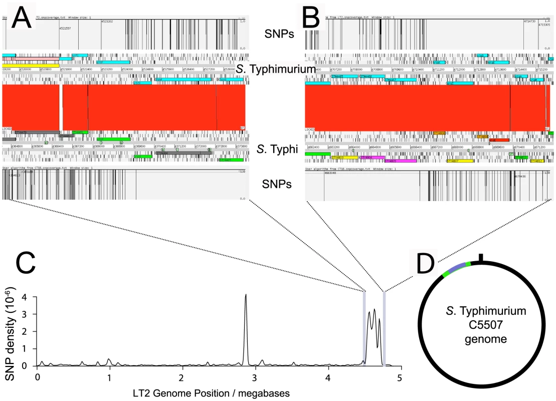
The boundaries of genomic sequence of S. Typhimurium and S. Typhi origin were determined by calculating the SNP density identified by mapping Illumina GA short reads from S. Typhimurium C5507 to the S. Typhimurium LT2 and S. Typhi Ty2 reference genome sequence. Artemis comparison tool (ACT) view of the left (A) and right (B) boundaries are shown with the position of SNPs in S. Typhimurium C5507 sequence reads mapped to either the S. Typhimurium LT2 or S. Typhi Ty2 genome. The SNP density (C) determined by mapping Illumina GA short reads from S. Typhimurium C5507 to the S. Typhimurium LT2 reference genome. The peak at 2.8 megabases is due to related but distinct prophage elements in S. Typhimurium C5507 genome relative to LT2 and the peak at 4.5 to 4.8 Mb is the S. Typhi genome sequence flanking SPI-7. Sequence reads from SPI-7 were not mapped to LT2 since this region is absent from S. Typhimurium. The deduced mosaic structure of the S. Typhimurium C5507 genome (D) with genomic sequence of S. Typhimurium C5 origin (black line) and S. Typhi Ty2 genome (green line) and SPI-7 (blue line) indicated. The viaB locus harbours genes necessary for the biosynthesis, secretion and anchoring of the Vi polysaccharide antigen on the bacterial cell surface [6]. Surface structures resembling a capsule were visualised by transmission electron microscopy (TEM) of the control S. Typhi BRD948 and S. Typhimurium C5.507 Vi+ cultured in rich medium containing 0.09 M NaCl. This structure was absent from S. Typhimurium C5.507 Vi+ in which the tviB gene, that encodes an essential component of the biosynthesis pathway, had been deleted (SGB1) (Figures 2A–C). Furthermore, SGB1 did not agglutinate with anti-Vi antiserum. The presence of Vi on the surface of S. Typhi and S. Typhimurium was visualised and semi-quantified by immunogold labelling with anti-Vi coated gold beads (Figures 2D–E and Figure 3). S. Typhimurium SGB1 cells were not associated with gold beads, while in contrast C5.507 Vi+ were significantly associated with anti-Vi+ coated gold beads. In S. Typhi the viaB locus is positively regulated by the two-component regulator OmpR/EnvZ in response to osmotic tension. In elevated NaCl concentration the viaB locus is reported to be down-regulated. [11], [12], [13]. We quantified immuno-gold labelling with anti-Vi antibody following culture at 0.09 M and 0.3 M NaCl. Culture of S. Typhimurium C5.507 Vi+ in media containing 0.09 M NaCl resulted in ∼2-fold increase in labelling than that observed following culture in 0.3 M NaCl. Furthermore, a derivative in which the ompR gene was inactivated by deletion was not labelled, even when cultured in low osmolarity medium (LB+0.09 M NaCl). The quantification of labelling with anti-Vi+ coated gold beads correlated with the lack of agglutination with anti-Vi serum. Together these data indicate that the entire SPI-7 region of S. Typhi is integrated into the S. Typhimurium genome in C5.507 Vi+ and the pattern of expression of Vi antigen is similar to that in S. Typhi.
Fig. 2. Transmission electron microscopy (TEM) images of S. Typhi and S. Typhimurium showing expression of Vi polysaccharide. 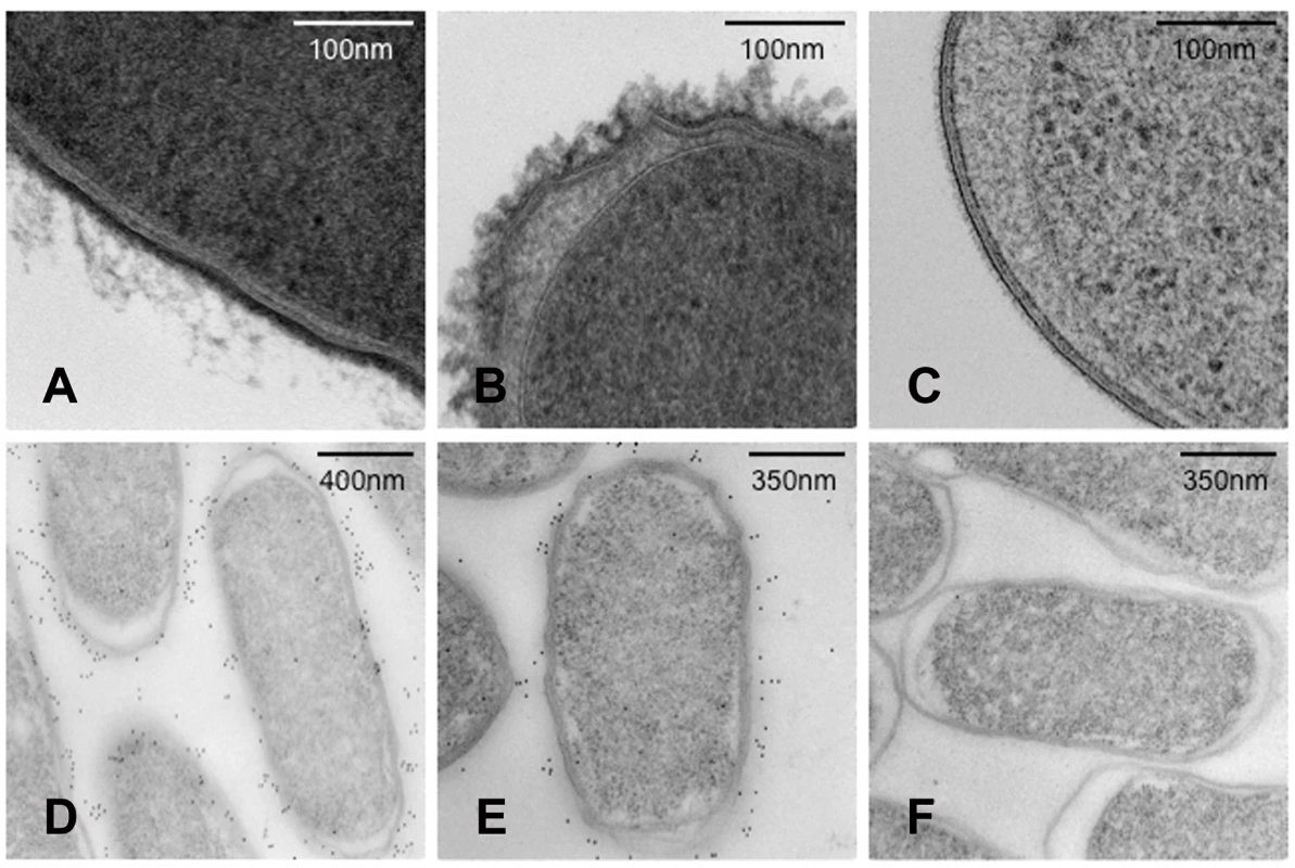
S. Typhi (A), S. Typhimurium C5507 (B), S. Typhimurium SGB1 (C5507 DtviB::kanr) (C) visualised using TEM. S. Typhi (D), S. Typhimurium C5507 (E), S. Typhimurium SGB1 (C5507 DtviB::kanr) (F) visualised using TEM in conjunction with immunogold labelling using anti-Vi antibody. Fig. 3. Enumeration of anti-Vi antibody immuno-gold labelling of S. Typhi and S. Typhimurium. 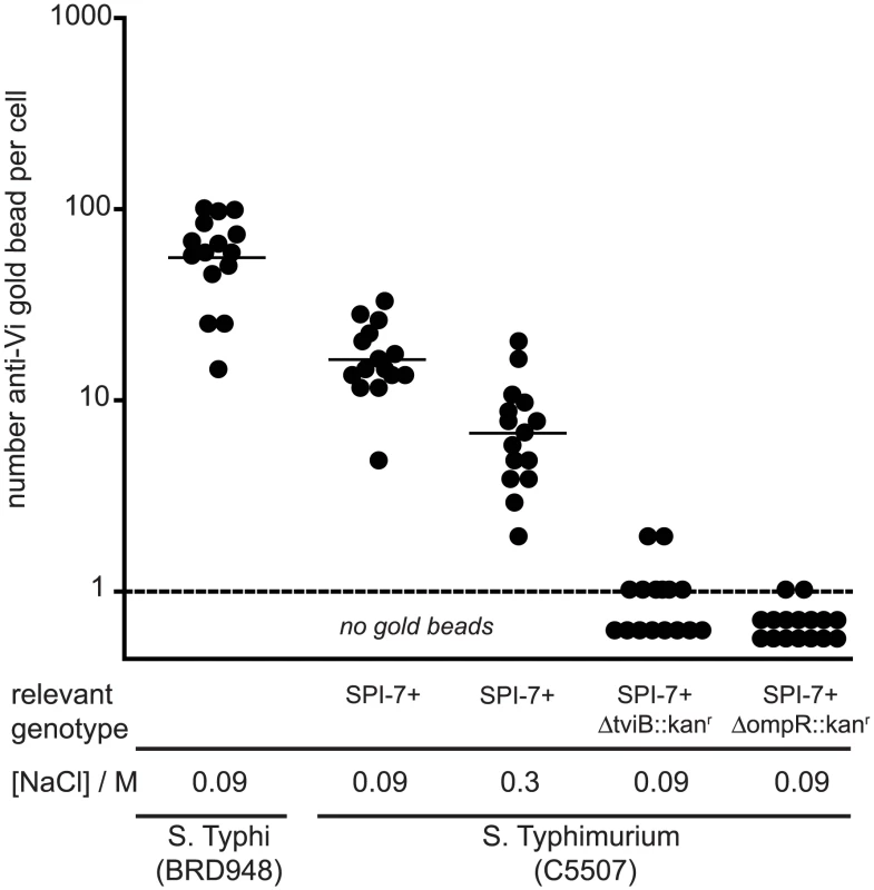
The number (filled circle) and the mean (horizontal bar) of anti-Vi antibody coated gold particles associated with S. Typhi BRD948, and S. Typhimurium C5507 either with no additional mutations or in which the tviB or ompR gene were deleted. Bacteria were cultured in medium containing NaCl of either 0.09 M (low osmolarity) or 0.3 M (high osmolarity). Expression of Vi polysaccharide by S. Typhimurium does not impact colonisation of C57BL/6 mice following oral inoculation
S. Typhi are host-adapted to higher primates and attenuated in mice following inoculation by the oral or parenteral routes. Consequently, we determined if S. Typhimurium C5.507 Vi+ could colonise the genetically susceptible C57BL/6 mouse. Mice were inoculated by oral gavage with approximately 1×10⁸ CFU S. Typhimurium C5, C5.507 Vi+ or SGB1 (ΔtviB). No significant difference in the colonisation of MLN, ceacum, ileum, spleen or liver was observed for these derivatives five days post inoculation (Figure 4A). To further determine the effects of the Vi capsule on chronic colonization as well as shedding within the faeces, we inoculated 129/sv mice by oral gavage with a mixture containing approximately 1×10⁹ CFU C5.507 Vi+ or SGB1. C5.507 Vi+ and SGB1 Vi− were shed in the stool at similar levels on day 1, 4, 7 and 10 post inoculation (Figure 4B).
Fig. 4. Colonisation of C57BL/6 mice with S. Typhimurium C5, S. Typhimurium C5507, and S. Typhimurium SGB1 (C5507 DtviB::kanr). 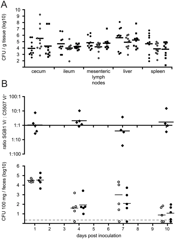
(A) Groups of ten C57BL/6 genetically susceptible mice were inoculated orally with 1×10⁸ cfu of S. Typhimurium C5 (closed circles), S. Typhimurium C5507 (open circles), or S. Typhimurium SGB1 (C5507 DtviB::kanr) (closed squares). Horizontal bar indicates the geometric mean. Mice were culled on day 5 post-inoculation and the cfu in mesenteric lymph nodes, cecum, ileum, spleen and liver homogenates determined. (B) A group of five 129/sv genetically resistant mice were inoculated orally with an equal mixture of 1×10⁹ CFU S. Typhimurium C5507 Vi+, and S. Typhimurium SGB1 Vi− (C5507 DtviB::kanr). The mean log10 ratio of these two strains in fresh fecal pellets on days 1, 4, 7 and 10 post inoculation are plotted (top), the CFU per 100 mg of S. Typhimurium C5507 Vi+ (open circles) and S. Typhimurium SGB1 Vi− (filled circles) are plotted (below). Infection with S. Typhimurium C5.507 Vi+ results in altered innate immune cell population in spleen compared to a Vi-negative derivative
To determine the impact of Vi expression on early innate immune responses to S. Typhimurium infection, spleens and MLN from naïve mice or from mice given a single i.v. or oral dose of C5.507 Vi+ or SGB1 tviB (Vi−) were examined by flow cytometry 24 hours post-inoculation. This time point was chosen since we were interested in determining the early innate immune response and because Vi is expressed on the surface of C5.507 Vi+ during this period but is down regulated by four days post inoculation [14]. Interestingly, mice inoculated with the SGB1 Vi−, had a small but significant increase in the levels of bacterial spleen colonisation (p = 0.025, unpaired, two tail, Mann Whitney) at 24 h compared to mice infected with C5.507 Vi+ (Figure 5).
Fig. 5. Colonisation of C57BL/6 mouse spleen by S. Typhimurium C5507, and S. Typhimurium SGB1 (C5507 DtviB::kanr). 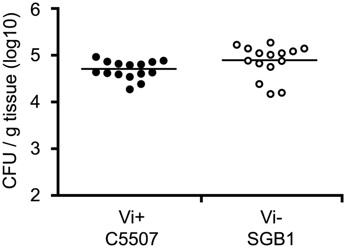
Groups of fifteen C57BL/6 mice were inoculated orally with 1×10⁵ cfu of S. Typhimurium C5507 (closed circles) or S. Typhimurium SGB1 (C5507 DtviB::kanr) (open circles). Mice were culled on 24 hours post-inoculation and the cfu in spleen homogenates determined. Examination of the spleen population of CD11c+ (dendritic cells, DC), F4/80+ (macrophage, MΦ), DX5+/CD3− (natural killer, NK) cells and Ly6G+ (polymorphonuclear, PMN) in C5.507 Vi+ or SGB1 (Vi−) infected mice revealed differences in immune cell populations during early infection that correlated with the presence of a functional Vi locus (Figure 6A and Table 1). Infection with C5.507 Vi+ resulted in moderate but significant increases (p<0.05) in both percentage and total cell numbers of PMN in spleens at 24 h when compared to naïve animals, although other immune cells monitored were largely unchanged. In contrast, spleens from mice at 24 h after inoculation with SGB1 Vi− had dramatically increased (p<0.001) percentage of PMN and NK cells, a significant increase (p<0.001) in the total number of DC and MΦ, although as a percentage they were not different form the population of these cells in spleen from naïve animals. We also observed an increase in the percentage of NK and PMN cells in mice infected with SGB1 Vi− compared to C5.507 Vi+ and an increase in the total number of both DC and MΦ populations.
Fig. 6. Expression of the Vi capsule induces differential innate immune responses shortly after infection. 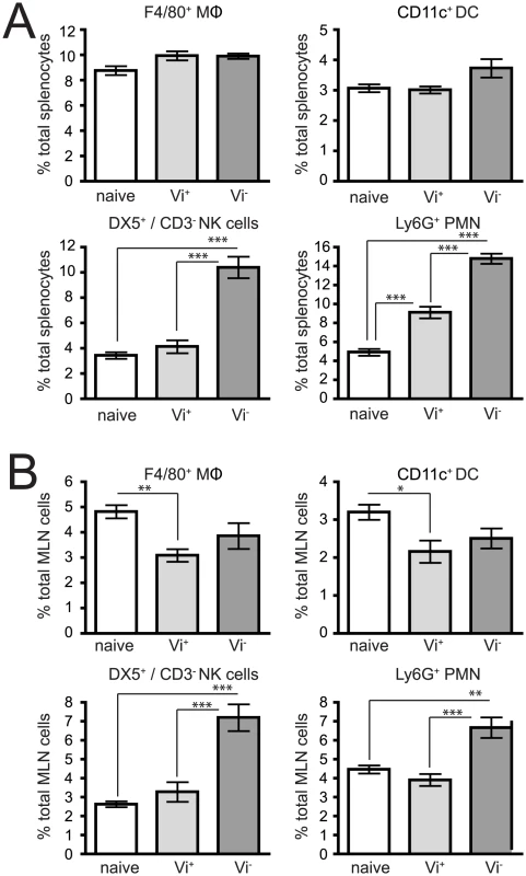
Cells were isolated from the spleens after i.v. infection (A) or MLN after oral infection (B) of C57BL/6 mice 24 h after infection with C5507 (Vi+) or SGB1 (Vi−) S. Typhimurium and stained with flurochrome-labelled mAb and analysed by flow cytometry in which 20,000–200,000 events were recorded. Columns represent the percentage ± SEM. Significant differences in values of * p<0.05; **, p<0.01; ***, p<0.001, as determined by one-way ANOVA followed by Bonferroni's multiple comparison test. Tab. 1. Innate cell numbers after infection with Vi+ or Vi− S. Typhimurium. 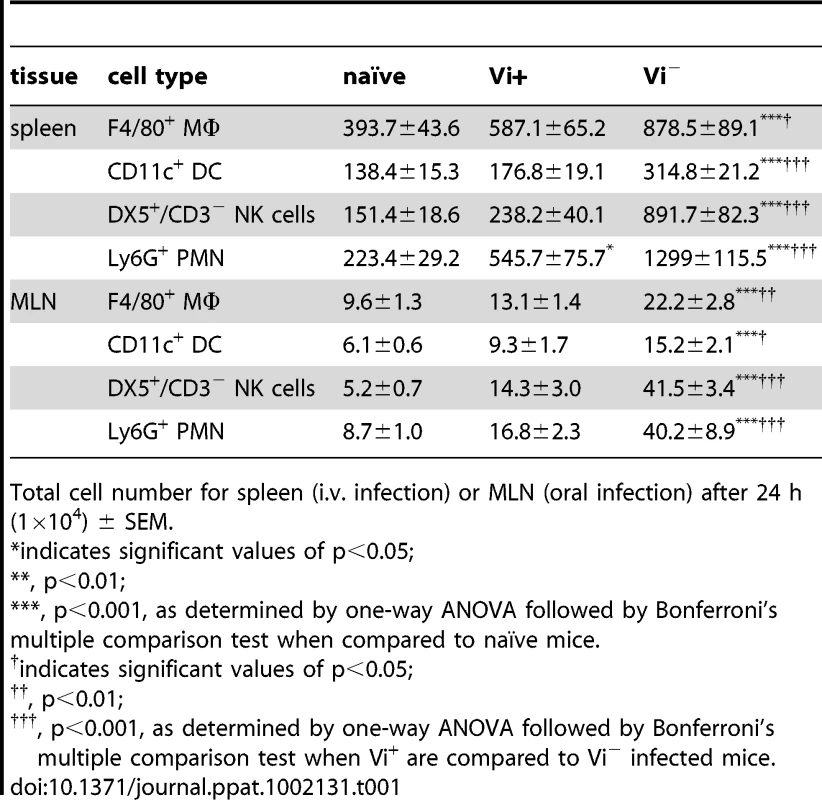
Total cell number for spleen (i.v. infection) or MLN (oral infection) after 24 h (1×10⁴) ± SEM. During natural infection following oral ingestion S. Typhimurium invades the enterocytes and M cells of the terminal ileum and then enters the lymph system that drains via the mesenteric lymph nodes (MLN). We therefore determined the impact of Vi expression on the innate immune cell populations of the MLN 24 hours after oral infection (Figure 6B and Table 1). Similar but not identical patterns of splenocyte immune cell populations were observed. For example, mice inoculated with SGB1 Vi− had a dramatically increased (p<0.001) percentages of PMN and NK cells in the MLN compared with MLN from C5.507 Vi+ infected or naïve mice. However, in contrast to splenocyte population in i.v. inoculated mice in which we observed a significant increase in the percentage of PMN with C5.507 Vi+, PMN in MLN following infection with C5.507 was not significantly different from naïve animals.
Vi expression modulates cytokine and chemokine responses
The intracellular cytokine response to Vi+ and Vi− S. Typhimurium were determined following ex vivo stimulation of splenocyte and MLN cells with phorbol 12-myristate 13-acetate (PMA). Flow cytometric analysis of intracellular cytokine expression in both splenocytes (after i.v. infection) and MLN cells (after oral infection) from Salmonella-infected mice were determined (Figure 7 and Table 2). Infection with SGB1 Vi− was associated with a significant increase (p<0.05) in the percentage and total cell number of MIP-2, TNF-α, IFN-γ and perforin producing splenocytes cells when compared to similarly stimulated naïve cells. In the case of MIP-2, IFN-γ and perforin producing cells from mice infected with C5.507 Vi+ the levels were not significantly different than those from naïve animals. Additionally, although, we did observe a significant increase in the proportion of TNF-α positive cells compared to naïve animals this was significantly lower (p<0.05) than observed in cells from mice infected with SGB1 Vi−. There was no observed difference in IL-6 expression by splenocytes from naïve mice or mice infected with C5507 Vi+ or SGB1 Vi−.
Fig. 7. Vi expression also impacts on the cytokine profile of cells after S. Typhimurium infection. 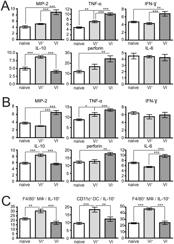
Isolated splenocytes after i.v. infection (A) or MLN after oral infection (B) from naïve, or 24 h infected C5507 (Vi+) and SGB1 (Vi−) mice were stimulated for 6 h with BD Leukocyte Activation Cocktail with BD GolgiPlug (BD Biosciences) in vitro, permeabilised and stained with anti-cytokine flurochrome-labelled mAb. (C) Cells were stained with mAb to specific surface markers F4/80+, CD11c+ and DX5+/CD3− the identity of innate populations, permeabilised and stained with anti-IL-10. Data represent percent of cytokine positive cells out of total spleen populations ± SEM. Significant differences in values of * p<0.05; **, p<0.01; ***, p<0.001, as determined by one-way ANOVA followed by Bonferroni's multiple comparison test. Tab. 2. Total number of cytokine producing cells after infection with Vi+ or Vi− S. Typhimurium. 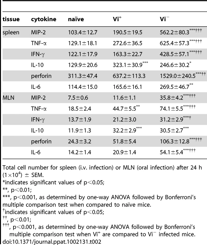
Total cell number for spleen (i.v. infection) or MLN (oral infection) after 24 h (1×10⁴) ± SEM. Since the natural route of infection for Salmonella is via the oral route we also determined the impact of infection with S. Typhimurium C5507 Vi+ and SGB1 Vi− on intracellular production of MIP-2, TNF-α, IFN-γ and perforin by immune cells of the MLNs 24 h post inoculation. Similar observations were made to those found in splenocytes following i.v. inoculation but differences in IFN-γ+ cell populations and IL-6 producing cells were observed. IFN-γ+ splenocytes were elevated, but not IFN-γ+ MLN cells, and significantly greater percentages and numbers of IL-6 producing MLN cells, but no significant differences in the IL-6 splenocyte population in SGB1 Vi− infected mice when compared to naïve animals. With respect to SGB1 Vi− infected mice, we also detected a significant increase (p<0.05) in the percentage of TNF-α producing splenocytes and MLN cells.
We also determined the cellular source of these cytokines within the lymphoid tissues after infection (Table 3). Splenocytes isolated from SGB1 Vi− infected mice were found to have significantly more MIP-2+ and IFN-γ+ (p<0.001) producing cells compared to both naive and C5.507 Vi+ infected mice. TNF-α was mainly expressed by macrophage and NK cells and to a lesser extent PMN. IL-6 expression was only significantly different in NK cells in SGB1 Vi− infected compared toC5.507 Vi+ infected or uninfected mice. Also the numbers of macrophage detectably producing IL-6 from Vi+ infected mice was significantly (p<0.05) lower than both naïve and Vi− infected mice. The numbers of perforin+ NK cells was significant higher (p<0.001) in those mice infected with SGB1 Vi− compared to naïve and C5.507 Vi+ infected mice. A similar cytokine profile was also observed within the MLN from orally infected mice. Notably, the cellular sources of the significant increase in IL-6+ MLN cells from SGB1 Vi− infected mice included DC, MΦ and NK cells (data not shown).
Tab. 3. Vi impacts on the cytokine profile of innate cells shortly after infection. 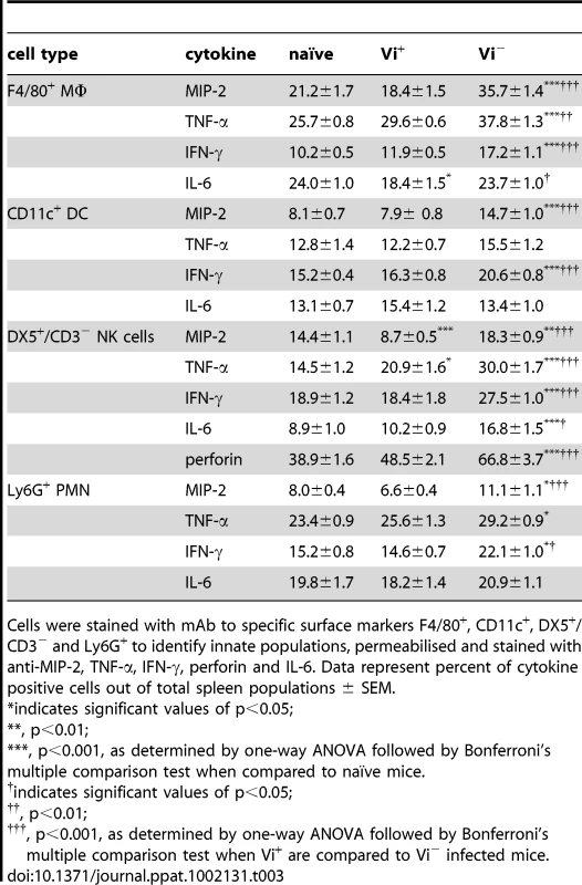
Cells were stained with mAb to specific surface markers F4/80+, CD11c+, DX5+/CD3− and Ly6G+ to identify innate populations, permeabilised and stained with anti-MIP-2, TNF-α, IFN-γ, perforin and IL-6. Data represent percent of cytokine positive cells out of total spleen populations ± SEM. Strikingly, infection with C5507 Vi+ was also associated with a significant increase in the percentage and total number of cells producing the anti-inflammatory cytokine IL-10 when compared to both stimulated naïve and SGB1 Vi− infected cells. Indeed, no increase in the number of cells producing IL-10 above that in naïve resulted from infection with SGB1 Vi−. We observed that re-stimulated MΦ, DC and NK cells, but not PMN, from C5.507 Vi+ infected mice expressed significantly more (p<0.01) IL-10 when compared to similarly stimulated naïve and Vi− infected splenocytes (Figure 7C).
Vi expression modulates in vivo innate immune responses in a TLR-dependent manner
To determine if the observed differences in the innate immune response in mice infected with Vi+ S. Typhimurium were due to detection of PAMPs by TLRs, we infected both MyD88−/− and TLR4−/− mice with SGB1 Vi− or C5507Vi+. Both bacterial colonisation and splenocyte PMN and NK cell populations were examined 24 h post-infection in wild type and KO mice (Figure 8 and Table 4).
Fig. 8. Vi modulates innate immune responses through TLR. 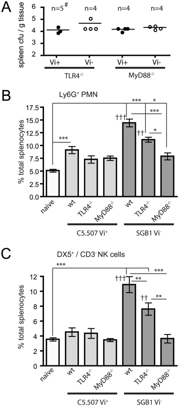
Spleens were removed from naïve, WT or KO animals (both MyD88−/− and TLR4−/−) infected with C5.507 (Vi+) or SGB1 (Vi−) 24 h after infection. The cfu per g of tissue (A) and the percentage of splenocytes that were Ly6G+ (B) or DX5+/CD3− (C) were determined. Columns represent the percentage of cells ± SEM. Significant differences are indicated * p<0.05; **, p<0.01; ***, p<0.001, and for comparison of Vi+ compared to Vi− infected mice, †† p<0.01; †††, p<0.001, as determined by one-way ANOVA followed by Bonferroni's multiple comparison test. # Colonisation data for two of the mice in the TLR4−/− mouse group inoculated with C5.507 were not determined. Tab. 4. Total number of PMN and NK cells after infection with Vi+ or Vi− S. Typhimurium in TLR4−/− and MyD88−/− mice. 
Total cell number of PMN and NK cells in the spleen 24 h post (×104) ± SEM. As observed previously, spleens from wild type mice infected with C5.507 Vi+ had significantly increased percentage and number of PMN compared to naïve mice (p<0.01). In contrast, in both TLR4−/− and MyD88−/− mice infected with C5.507 Vi+ the numbers of PMN was not significantly different (p>0.05) in percentage or total cell number compared with naïve mice. Indeed, overall, in TLR4−/− and MyD88−/− infected mice the PMN and NK cell response to C5.507 Vi+ infection was indistinguishable from naïve mice. In contrast, although the response to SGB1 Vi− infection in TLR4−/− and MyD88−/− was reduced compared to that in WT mice, there was still a significant response compared to naïve mice.
Infection of wild type mice with SGB1 Vi− as before resulted in a significant increase (p<0.001) in the PMN population compared to both naïve and C5.507 Vi+ infected mice. Furthermore, unlike infections with C5.507 Vi+, both TLR4−/− and MyD88−/− mice infected with SGB1 Vi− also had significantly more (p<0.05) PMN within their spleen compared with naïve mice. Indeed, when wild type TLR4−/− and MyD88−/− mice infected with SGB1 Vi− were compared, both TLR4−/− and MyD88−/− mice had significantly less (p<0.001) PMN than wild type infected mice. Furthermore MyD88−/− mice had a significant reduction (p<0.05) compared to TLR4−/− SGB1 Vi− infected mice. Importantly, there were also significantly fewer (p<0.01) PMN in C5.507 Vi+ infected TLR4−/− mice compared to SGB1 Vi− infected mice, but not within the MyD88−/− groups.
The splenic NK cell population showed very similar responses to Vi+ and Vi− S. Typhimurium PMN population in wild type and TLR4−/− and MyD88−/− mice (Figure 8B). Specifically, only Vi− infected wild type and TLR4−/− infected mice had significantly greater percentages and numbers of NK cells compared to naïve and C5.507 Vi+ infected mice. Additionally, both TLR4−/− and MyD88−/− SGB1 Vi− infected mice had significantly lower levels of NK cells when compared to similarly infected wild type mice. MyD88−/− infected mice also had significantly less NK cells when compared to TLR4−/− SGB1 Vi− infected mice. We again observed that C5.507 Vi+ infected TLR4−/− mice had significantly less (p<0.01) NK cells than similar SGB1 Vi− infected mice.
Vi associated IL-10 expression regulates activation and chemotaxis of splenocytes in vitro
Splenocytes purified from naïve C57BL/6 mice were cultured with C5.507 Vi+ or SGB1 Vi− bacteria for 24 h and the supernatant assayed for the presence of IL-10. The culture supernatant from cells stimulated with C5.507 Vi+ contained higher (p<0.001) IL-10 levels than SGB1 Vi− stimulated splenocytes (Figure 9A). To address directly if enhanced expression of IL-10 associated with C5.507 Vi+ infection was at least in part responsible for the observed immune suppression in infected mice, splenocytes from naïve mice were stimulated with Vi+ or Vi− S. Typhimurium in the presence of anti-IL-10 or isotype control antibody. Decreased chemotaxis (p<0.01) was observed in SGB1 Vi− stimulated isotype treated cultures compared to both isotype control antibody and anti-IL-10 SGB1 Vi− stimulated splenocytes. Notably, when C5.507 Vi+ stimulated cultures were grown in the presence of anti-IL-10 we no longer observed a significant reduction (p>0.05) in the movement of cells (Figure 9B). C5.507 Vi+ co-culture with isotype antibody significantly reduced (p<0.01) both the percentage and mean fluorescent intensity (MFI), expression levels, of the early activation marker CD69 on both PMN and NK cells. Again, presence of anti-IL-10 in C5.507 Vi+ stimulated cultures led to an increase in CD69 expression that was not significantly different (p>0.05) from SGB1 Vi− stimulated splenocytes (Figure 9C). The addition of anti-IL-10 did not appear to have any significant effect on migration, although we did observe a significantly greater percentage of CD69+ PMN in anti-IL-10 SGB1 Vi− treated cultures compared to isotype controls (Figure 9B and C). Further, addition of rIL-10 to naïve splenocytes gave a similar chemotaxis and immune activation profile (p>0.05) to that observed in C5.507 Vi+ stimulated splenocytes containing control antibody (Figure 9B and C).
Fig. 9. Vi induction of IL-10 impacts on chemotaxis and activation of splenocytes in vitro. 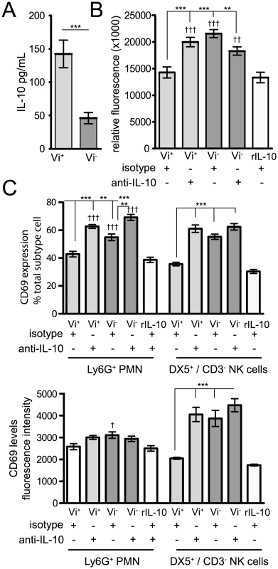
(A) Spleen cells were isolated from naïve mice and stimulated for 24 h with 1∶1 ratio of Vi+ or Vi− Salmonella strains and supernatants were analysed for levels of IL-10. Mean ± SEM are presented from six individual animals. Significance was determined using Mann-Whitney test, *** p<0.001. (B) Splenocytes were also stimulated with the various S. Typhimurium strains in the presence of anti-IL-10, isotype control antibody or rIL-10. Cells were then stained with surface antibodies and the percentage and expression/mean fluorescent intensity (MFI) of the activation marker CD69 on NK cells and PMN was analysed by flow cytometry. Columns represent the percentage of MFI ± SEM and significant differences were determined as in Figure 6. (C) Chemotactic migration of splenocytes towards supernatants from Salmonella stimulated cultures (in the presence of anti-IL-10, control antibody or rIL-10). After 4 h incubation, the level of cell migration was determined by reading the relative fluorescent units (RFU) at 480/520 nm. Data represents mean ± SEM minus negative (media alone) controls. Significant differences in values of * p<0.05; **, p<0.01; ***, p<0.001, as determined by Kruskal-Wallis followed by Dunn's multiple comparison test or † indicating significant values when compared to r-IL-10. Discussion
In this study we addressed the hypothesis that early innate immune responses to Salmonella can be modulated at systemic sites by the expression of Vi. We exploit a S. Typhimurium/S. Typhi chimera (C5.507) harbouring ∼300 kb of the S. Typhi genome including the entire SPI-7 island containing the viaB locus. In our assays the colonisation level of mice following oral inoculation with this chimera strain was indistinguishable from the parent strain lacking the S. Typhi genomic region. However, we cannot discount small differences in colonisation that may be detectable in more sensitive experiments. Importantly the viaB locus in this strain is encoded as a single copy and in the natural genomic context. Consequently, the Vi polysaccharide capsule is expressed on the surface of C5.507 Vi+ at similar levels to that in S. Typhi and expression is controlled by osmotic stress in an OmpR dependent manner as in S. Typhi [11]. The pathogen chimera approach could be used to study other S. Typhi specific determinants or to overcome technical constraints associated with other host-adapted pathogens.
An immune-modulator role for Vi has been proposed previously based on observations of the effect of the expression of this polysaccharide on cytokine responses in T84, THP-1 and HEK293 cells in tissue culture [15], bone-marrow derived macrophage [10], and in an in vivo colitis models [9]. In these studies determination of relative transcription of IL-6, TNF-α, or IL-17 genes was generally used as a measure of the immune response to infection. Here we use an alternative approach to directly measure both the population of immune cells (macrophage, dendritic cells, NK cells and polymorphonuclear cells), present in the spleen of mice infected with S. Typhimurium C5.507 Vi+, and directly determine the intracellular level of a series of key inflammatory or anti-inflammatory cytokines; MIP-2, TNF-α, IFN-γ, IL-6 and IL-10. When we compared the splenocyte population 24 h after infection we observed significantly fewer PMN in mice inoculated with C5.507 Vi+ compared to SGB1 Vi−. This could be explained by the observation that a greater number of TNF-α and MIP-2 producing MΦ, NK cells and DC were found in those mice infected with the Vi− compared to the Vi+ S. Typhimurium. Through neutrophil recruitment and activation, TNF-α and MIP-2 are known to be important mediators of intestinal inflammation and associated pathology, and the difference in the induction of these mediators by S. Typhi and S. Typhimurium may explains some of the differences in disease outcome associated with these pathogens [9]. We also report that Vi expression is associated with decreased TNF-α and MIP-2 expression in the spleen of infected mice following inoculation by the intravenous route. Vi mediated decrease in IL-6 production by bone marrow derived macrophages in vitro has been described previously [10]. However, we only observed reduced IL-6 expression in cells (MΦ, DC and NK cells) of the mesenteric lymph node following oral inoculation but no detectable difference in intracellular IL-6 levels in mice infected via the intravenous route regardless of Vi expression. Notably, our data shows that reductions in neutrophil chemo attractants directly impacts on their trafficking in vivo. NK cells were also influenced by the expression of Vi following either intravenous or oral infection with C5.507 Vi+ as NK cell populations were not significantly different from those in naïve mice. In contrast, mice infected with SGB1 Vi− showed a marked increase in the NK cell population. We also observed an increase in the proportion of perforin positive NK cells. Perforin is stored in NK and CD8+ T cells as granules and is key to their ability to destroy infected host cells. The reduction in NK cell influx and associated decrease in perforin in response to C5.507 Vi+ infection may also explain in part how expression of this polysaccharide contributes to the ability of the S. Typhi to disseminate to systemic sites, colonise and replicate.
The effect of Vi expression by S. Typhimurium on DC and MΦ populations was not as great as that observed for PMN and NK cell populations. Nonetheless, we did observe significantly lower numbers of these innate cells in mice infected with Vi+ compared to Vi− S. Typhimurium. The apparent impact of Vi expression on the infux of DC and MΦ may also be attributed to MIP-2 since this chemokine can also modulate the trafficking of DC [16]. As well as being a chemo attractant in its own right, TNF-α induces the synthesis of a number of chemokines, including IP-10, RANTES, KC, in a cell-type and tissue-specific manner [17]. Impairment of this response may also account for the reduction in splenic MΦ and NK cells observed in C5.507 Vi+ infected mice. Together, these data support a role for Vi in modulating the recruitment of immune cells to sites beyond the intestinal mucosa, including the mesenteric lymph nodes and spleen.
The mechanism by which Vi modulates innate responses is currently not known, although it has been postulated that Vi may mask LPS therefore preventing its detection by TLR4 [10]. We report that spleen from TLR4−/− and MyD88−/− mice infected with SGB1 Vi− have reduced immune cell splenocyte populations compared to wild type control mice. However, there were significantly greater immune cell populations in TLR4−/− and MyD88−/− mice than in naïve mice suggesting that proinflammatory signals other than those dependent on these were in operation. These data suggest Vi may play a role as a physical barrier separating Salmonella PAMPs from TLR4 activation. However, data from MyD88−/− mice suggests that while TLR4 signalling plays some role in the immuno-modulatory aspect of Vi, other pathways may also be involved.
Much of what is known of the immune-modulator effects of Vi is from observations of induction of pro-inflammatory cytokines and chemokines. We additionally examined the production of an anti-inflammatory cytokine, IL-10, and report a significant increase in the number of IL-10-expressing cells in mice infected with Vi+ compared to Vi− S. Typhimurium. In part, IL-10 acts on macrophage and myeloid dendritic cells to inhibit the development of a TH-1 response (reviewed in [18]). IL-10 is produced by many different cell types of the innate and adaptive immune systems, and the temporal and spatial expression is likely important to moderating immune response [19]. Importantly, IL-10 is induced in macrophage and myeloid dendritic cells by a variety of pathogen derived products, including LPS following detection by TLR-4 [20]. We did not observe induction of IL-10 in splenocytes from mice infected with SGB1 Vi− relative to naïve mice at the time point studied, but induction was specifically observed in mice infected with C5.507 expressing Vi. Indeed, when we determined the innate populations producing IL-10 we observed that MΦ, DC and NK cells were the cell types responsible for this increased IL-10 phenotype. Importantly, we also observed that inhibition of IL-10 from C5.507 Vi+ stimulated splenocytes in vitro directly impacted both chemotaxis and activation status of immune cells. Notably, previous studies have shown that IL-10 can inhibit chemokine expression and reduce iNOS production from PMN [21], [22]. Therefore, early production of IL-10 after infection with Vi+ Salmonella may be important in diminishing neutrophil influx and activation and thereby increasing the ability of this pathogen to efficiently colonise and infect distal sites within the host. At later infection time-points Vi may be an important mechanism associated with dampening the TH-1 response that is normally associated with resolving infections by invasive bacterial pathogens. Indeed, higher levels of IL-10, as well as IL-4, have been detected in PBMC culture of typhoid fever patients when compared to healthy control subjects.
S. Typhimurium C5507 Vi+ provides a model pathogen to study the impact of Vi and potentially other S. Typhi specific virulence genes encoded on SPI-7, in the well characterized murine typhoid model. Recently, two excellent murine models for typhoid fever have been described [23], [24]. The utility of S. Typhimurium C5507 Vi+ as a model system has previously been demonstrated in studies to determine the protection afforded by a conjugated Vi subunit vaccine [25]. Furthermore, S. Typhimurium C5507 Vi+ has been used to studies expression of Vi polysaccharide capsule from heterologous promoters including PssaG, to improve the anti-Vi immune response to live oral vaccines against typhoid (unpublished observations). However, observations from the use of a surrogate infection models should be considered with care. Many differences between the chimera infection of mice remain compared with the natural S. Typhi infections of humans. One of these is that rate at which the infection proceeds and the dynamics of colonisation and clearance. These differences are likely accounted for in the genetic differences outside of the chimera region and also differences in the host species.
Together these data describe the impact of Vi expression on the outcome and innate immune response to S. Typhimurium infections in mice. Modulation of the innate immune response by the Vi polysaccharide capsule decreased innate cell recruitment and provided insights into how S. Typhi effects its interaction with the host to provide the desired outcome from the host-pathogen interaction. This is consistent with a pathogenic strategy central to which is a slowly progressing systemic disease that is rarely associated with death from sepsis or cytokine storm, but that ultimately provides access for this pathogen to the gall bladder and potentially other immune privileged niches, required for chronic carriage.
Materials and Methods
Bacterial strains and culture conditions
S. Typhimurium C5507 Vi+ was a gift from M. Popoff (Institute Pasteur, France) and was generated by conjugation of S. enterica Typhi Ty2 with S. enterica Typhimurium C5. A serotype Typhimurium strain in which the phoN gene was replaced by the aph gene was constructed by transferring the aph gene from AJB715 into strain SL1344 by P22 transduction. This derivative was designated AMJ204. SGB1 Vi − strain was generated by precise deletion of the tviB gene and replacement of this with the aph gene that confers resistance to kanamycin using the Datsenko and Wanner red recombinase allelic exchange methodology [26]. Oligonucleotide primers 5′ GCCAGAACCAGTTTGGTCCGTAGTTCTTCGTAAGCCGTCATGATTGTGTAGGCTGGAGCTGCTTCG 3′ and 5′ AATTAACTTTGTAAATATAAAATTTTAGTAAAGGATTAATAAGAGCATATGAATATCCTCCTTAG 3′ were used to amplify the aph gene from pKD4 template. Strain SGB1 expressed comparable amounts of flagellin on culure in LB broth in vitro as determined by crude flagella preparation and separation of flagellin monomer by SDS PAGE (data not shown). Bacteria were cultured aerobically at 37°C in Luria-Bertani (LB) broth or LB with 15% agar supplemented with antibiotics at appropriate concentrations; ampicillin (Amp), 100 mg/L (LB+Amp); chloramphenicol (Chl), 30 mg/L (LB+Chl); and kanamycin (Kan), 30 mg/L (LB+Kan).
Sequence analysis
To identify the boundaries of S. Typhi and S. Typhimurium-derived genomic sequence, genomic DNA was prepared from S. Typhimurium C5507 and sequenced using the Illumina/Solexa Genome Analyser. Over 6.5 million single end reads of 36 bp were generated, giving a theoretical 48-fold coverage of the S. Typhimurium genome. Reads were mapped to the reference genomes S. Typhimurium LT2 (EMBL:AE006468) and S. Typhi CT18 (EMBL:AL513382) using Maq (maq.sourceforge.net), single nucleotide polymorphisms between C5507 and the reference genomes were identified. The position and density of these substitutions compared to the two reference genomes was visualised using Artemis and ACT (Artemis Comparison Tool) [27].
Transmission Electron Microscopy (TEM)
Bacteria cultured for 24 hr at 37°c were sampled selecting a single colony from each strain, mixing with 20 µl sterile distilled water and rapidly freezing in a Bal-Tec HPM010 high pressure freezer. Samples for immunogold-labelling were freeze-substituted in a Leica EM AFS at −90°C in methanol containing 0.2% uranyl actetate and 0.1% glutaraldehyde followed by low temperature embedding in Lowicryl HM20 resin. Samples for ultrastructural analysis were freeze-substituted with acetone containing 0.1% tannic acid, 0.5% glutaraldehyde sequentially with acetone containing 1% osmium tetroxide and 0.1% uranyl actetate followed by room temperature embedding in TAAB 812 resin. 50 µm ultrathin sections were cut on a Leica EM UC6 and contrasted with lead citrate and uranyl acetate For immunogold-localisation ultrathin sections were labelled with anti Vi antibody and probed with protein A gold as previously described [28]. Images were taken on an FEI Tecnai Spirit 120 kV TEM with a Tietz F415 CCD camera. For quantification of labelling fifteen random clearly defined bacteria were selected and the number of associated 10 nm gold particles counted.
Experimental infections of mice
Female wild-type (WT) C57BL/6 and 129/sv mice (6–8 week old) and knock-out (KO) C57BL/6 mice, including MyD88−/− [29] and TLR4−/− [30] were kind gifts and were bred at The Sanger Institute Research Support Facility (RSF). All animals were given food and water ad libitum. Mice were sacrificed by cervical dislocation or exsanguination. The S. Typhimurium strains were grown with shaking in LB broth (with appropriate antibiotics) for 20 h. Bacteria were then harvested by centrifugation, washed, and suspended in PBS (pH 7.4) to approximately 109 CFU per ml. Groups of 5–10 mice were inoculated orally by gavage or intravenously with 0.2 ml of bacterial suspension. Viable counts were determined in the inoculum by serial dilutions onto LB agar. After 24 h or five days the mice were culled and their MLN, terminal end ileum, cecum, spleen, and liver were aseptically removed and weighed. Organs were homogenised in sterile water (5 ml) using a Seward Stomacher 80 (Seward, London UK) for 2 minutes at high speed. Serial dilutions of each organ were plated onto LB agar. Colonies were enumerated after overnight incubation at 37°C.
Ethics statement
All animal procedures were performed in accordance with the United Kingdom Home Office Inspectorate under the Animals (Scientific Procedures) Act 1986. Ethical approval for these procedures were granted by the Wellcome Trust Sanger Institutes Ethical Review Committee.
Flow cytometry and ELISA
Single cell suspensions from the spleens and MLN of individual mice were prepared to obtain a final concentration of 5×10⁵ cells/well in blocking buffer (1× PBS/1% BSA/0.05% sodium azide/1% rat, hamster and mouse serum). 0.05 ml of each mAb dye mix, 0.005 ml of the amine-reactive viability dye ViViD (Invitrogen) to determine dead cells, with incubation in the dark at 4°C for 30 minutes. The mAb used for flow cytometry were (BD Biosciences unless stated otherwise); CD11c, clone HL3 with PE-Cy7 conjugate (BD Biosciences), Ly6G, clone RB6-8C5 with PE conjugate (BD Biosciences), CD49b, clone DX5 with FITC or PE conjugate (BD Biosciences), F4/80, clone BM8 with TRI-COLOR conjugate (Invitrogen), CD3, clone 145-2C11 with APC, CD69, clone H1.2F3 with PE-Cy7, Alexa Fluor 700, conjugate (BD Biosciences), CXCL2/MIP-2, clone 40605 with biotin conjugate (AbD Serotec), IL-10, clone JES5-16E3 with APC conjugate (BD Biosciences), TNF-α, clone MP6-XT22 with PE-Cy7 conjugate (BD Biosciences), IL-6, clone MP5-20F3 with PE conjugate (BD Biosciences), IFN-γ, clone XMG1.2 with PE-Cy7 conjugate (BD Biosciences), perforin, clone eBioOMAK-D with FITC conjugate (eBioscience), streptavidin, with PE-Texas Red conjugate (BD Biosciences). Cells were washed twice with blocking buffer and finally resuspended in 0.2 ml 1% paraformaldehyde. To perform flow cytometric analyses and measure relative fluorescence intensities a FACSAria cytometer and BD Diva software (Becton Dickinson) were used. For each mouse 20,000–200,000 events were recorded. The percentage of cells labelled with each mAb was calculated in comparison with cells stained with isotype control antibody. Background staining was controlled by labelled isotype controls and fluorescence-minus-one (FMO). The results represent the percentage of positively stained cells in the total cell population exceeding the background staining signal. For determination of intracellular cytokine production by splenocytes, cells were incubated for 6 h at 37°C with BD Activation Cocktail (BD Biosciences) containing Phorbol 12-Myristate 13-Acetate (PMA), plus GolgiPlug or GolgiPlug alone (BD Biosciences). Cells were then washed with staining buffer and stained at 4°C with surface mAbs. Cells were then fixed and saponin-permeabilised (Perm/Fix solution, BD Biosciences) and incubated with cytokine mAb as listed above or isotypic controls. After 30 min cells were twice washed in permealisation buffer (BD Biosciences) and then analysed by flow cytometry as described above. For determination of IL-10 levels from in vitro stimulated splenocyte supernatants the IL-10 Instant ELISA (Bender MedSystems) system was used according to manufacturer's instructions.
In vitro Salmonella stimulations
Spleen suspensions were prepared as described above and 2×10⁵ cells added to a 96-well round bottom plate and were left for 1 h at 37°C 5% CO2. Cells were then stimulated with appropriate 1∶1 ratio of Salmonella strains supplemented with 20 µg/mL anti-IL-10 or appropriate isotype control (PeproTech). In others, cells were supplemented with 100 ng/mL recombinant (r)-IL-10 (PeproTech). Splenocytes were also stimulated with LPS (100 ng/mL) or RPMI+ medium as positive and negative controls respectively. Cells were then incubated for approximately 24 h before being centrifuged at 800× g for 5 min. Some supernatant was removed and stored at −80°C for subsequent cytokine analysis. Remaining cells from stimulation were then stained for flow cytometry with surface (DX5+/CD3− or Ly6G+) and the activation marker CD69 as described above.
Chemotaxis assay
Migration of Salmonella stimulated splenocytes was analysed in 96-well QCM Chemotaxis Cell Migration Assay (Millipore, UK) with 5-µm pore polycarbonate filters. Briefly, 2×10⁵ cells in 0.1 ml of RPMI+ were placed into the migration chambers. Supernatant from stimulated splenocytes were added to the lower chamber. Migration was performed for 4 h at 37°C with 5% CO2. Cells/media from the top chamber of the insert was discarded and remaining cells removed using Cell Detachment Solution (Millipore, UK). Lysis Buffer/Dye Solution (Millipore, UK) were added to each well and incubated for 15 min. 0.15 ml of this mixture was then transferred to new 96-well plate and read with a fluorescence plate reader using 480/520 nm filter set.
Statistical analysis
Experimental results were plotted and analysed for statistical significance with Prism4 software (GraphPad, San Diego, CA). A p-value of <0.05 was used as significant in all cases.
Zdroje
1. SantosRLZhangSTsolisRMKingsleyRAAdamsLG 2001 Animal models of Salmonella infections: enteritis versus typhoid fever. Microbes Infect 3 1335 1344
2. ButlerTHoMAcharyaGTiwariMGallatiH 1993 Interleukin-6, gamma interferon, and tumor necrosis factor receptors in typhoid fever related to outcome of antimicrobial therapy. Antimicrob Agents Chemother 37 2418 2421
3. KeuterMDharmanaEGasemMHvan der Ven-JongekrijgJDjokomoeljantoR 1994 Patterns of proinflammatory cytokines and inhibitors during typhoid fever. J Infect Dis 169 1306 1311
4. GirardinEGrauGEDayerJMRoux-LombardPLambertPH 1988 Tumor necrosis factor and interleukin-1 in the serum of children with severe infectious purpura. N Engl J Med 319 397 400
5. WaageABrandtzaegPHalstensenAKierulfPEspevikT 1989 The complex pattern of cytokines in serum from patients with meningococcal septic shock. Association between interleukin 6, interleukin 1, and fatal outcome. J Exp Med 169 333 338
6. PickardDWainJBakerSLineAChohanS 2003 Composition, acquisition, and distribution of the Vi exopolysaccharide-encoding Salmonella enterica pathogenicity island SPI-7. J Bacteriol 185 5055 5065
7. WainJHouseDZafarABakerSNairS 2005 Vi antigen expression in Salmonella enterica serovar Typhi clinical isolates from Pakistan. J Clin Microbiol 43 1158 1165
8. RaffatelluMChessaDWilsonRPTukelCAkcelikM 2006 Capsule-mediated immune evasion: a new hypothesis explaining aspects of typhoid fever pathogenesis. Infect Immun 74 19 27
9. RaffatelluMSantosRLChessaDWilsonRPWinterSE 2007 The capsule encoding the viaB locus reduces interleukin-17 expression and mucosal innate responses in the bovine intestinal mucosa during infection with Salmonella enterica serotype Typhi. Infect Immun 75 4342 4350
10. WilsonRPRaffatelluMChessaDWinterSETukelC 2008 The Vi-capsule prevents Toll-like receptor 4 recognition of Salmonella. Cell Microbiol 10 876 890
11. PickardDLiJRobertsMMaskellDHoneD 1994 Characterization of defined ompR mutants of Salmonella typhi: ompR is involved in the regulation of Vi polysaccharide expression. Infect Immun 62 3984 3993
12. ArricauNHermantDWaxinHEcobichonCDuffeyPS 1998 The RcsB-RcsC regulatory system of Salmonella typhi differentially modulates the expression of invasion proteins, flagellin and Vi antigen in response to osmolarity. Mol Microbiol 29 835 850
13. VirlogeuxIWaxinHEcobichonCPopoffMY 1995 Role of the viaB locus in synthesis, transport and expression of Salmonella typhi Vi antigen. Microbiology 141 Pt 12 3039 3047
14. JanisCGrantAJMcKinleyTJMorganFJJohnVF 2011 In vivo regulation of the Vi antigen in Salmonella and induction of immune responses with an in vivo inducible promoter. Infect Immun 79 2481 2488
15. RaffatelluMChessaDWilsonRPDusoldRRubinoS 2005 The Vi capsular antigen of Salmonella enterica serotype Typhi reduces Toll-like receptor-dependent interleukin-8 expression in the intestinal mucosa. Infect Immun 73 3367 3374
16. CaoXZhangWWanTHeLChenT 2000 Molecular cloning and characterization of a novel CXC chemokine macrophage inflammatory protein-2 gamma chemoattractant for human neutrophils and dendritic cells. J Immunol 165 2588 2595
17. AlgoodHMLinPLYankuraDJonesAChanJ 2004 TNF influences chemokine expression of macrophages in vitro and that of CD11b+ cells in vivo during Mycobacterium tuberculosis infection. J Immunol 172 6846 6857
18. MooreKWde Waal MalefytRCoffmanRLO'GarraA 2001 Interleukin-10 and the interleukin-10 receptor. Annu Rev Immunol 19 683 765
19. SaraivaMO'GarraA 2010 The regulation of IL-10 production by immune cells. Nat Rev Immunol 10 170 181
20. BoonstraARajsbaumRHolmanMMarquesRAsselin-PaturelC 2006 Macrophages and myeloid dendritic cells, but not plasmacytoid dendritic cells, produce IL-10 in response to MyD88 - and TRIF-dependent TLR signals, and TLR-independent signals. J Immunol 177 7551 7558
21. SunLGuoRFNewsteadMWStandifordTJMacariolaDR 2009 Effect of IL-10 on neutrophil recruitment and survival after Pseudomonas aeruginosa challenge. Am J Respir Cell Mol Biol 41 76 84
22. KobbePSchmidtJStoffelsBChanthaphavongRSBauerAJ 2009 IL-10 administration attenuates pulmonary neutrophil infiltration and alters pulmonary iNOS activation following hemorrhagic shock. Inflamm Res 58 170 174
23. SongJWillingerTRongvauxAEynonEEStevensS 2010 A mouse model for the human pathogen Salmonella typhi. Cell Host Microbe 8 369 376
24. LibbySJBrehmMAGreinerDLShultzLDMcClellandM 2010 Humanized nonobese diabetic-scid IL2rgammanull mice are susceptible to lethal Salmonella Typhi infection. Proc Natl Acad Sci U S A 107 15589 15594
25. HaleCBoweFPickardDClareSHaeuwJF 2006 Evaluation of a novel Vi conjugate vaccine in a murine model of salmonellosis. Vaccine 24 4312 4320
26. DatsenkoKAWannerBL 2000 One-step inactivation of chromosomal genes in Escherichia coli K-12 using PCR products. Proc Natl Acad Sci U S A 97 6640 6645
27. CarverTBerrimanMTiveyAPatelCBohmeU 2008 Artemis and ACT: viewing, annotating and comparing sequences stored in a relational database. Bioinformatics 24 2672 2676
28. GouldingDThompsonHEmersonJFairweatherNFDouganG 2009 Distinctive profiles of infection and pathology in hamsters infected with Clostridium difficile strains 630 and B1. Infect Immun 77 5478 5485
29. AdachiOKawaiTTakedaKMatsumotoMTsutsuiH 1998 Targeted disruption of the MyD88 gene results in loss of IL-1 - and IL-18-mediated function. Immunity 9 143 150
30. HoshinoKTakeuchiOKawaiTSanjoHOgawaT 1999 Cutting edge: Toll-like receptor 4 (TLR4)-deficient mice are hyporesponsive to lipopolysaccharide: evidence for TLR4 as the Lps gene product. J Immunol 162 3749 3752
Štítky
Hygiena a epidemiologie Infekční lékařství Laboratoř
Článek SUMO Pathway Dependent Recruitment of Cellular Repressors to Herpes Simplex Virus Type 1 GenomesČlánek A Structural Model for Binding of the Serine-Rich Repeat Adhesin GspB to Host Carbohydrate ReceptorsČlánek Dynamic Evolution of Pathogenicity Revealed by Sequencing and Comparative Genomics of 19 IsolatesČlánek Widespread Endogenization of Genome Sequences of Non-Retroviral RNA Viruses into Plant GenomesČlánek The Cost of Virulence: Retarded Growth of Typhimurium Cells Expressing Type III Secretion System 1Článek A Role for the Chemokine RANTES in Regulating CD8 T Cell Responses during Chronic Viral Infection
Článek vyšel v časopisePLOS Pathogens
Nejčtenější tento týden
2011 Číslo 7- Jak souvisí postcovidový syndrom s poškozením mozku?
- Měli bychom postcovidový syndrom léčit antidepresivy?
- Farmakovigilanční studie perorálních antivirotik indikovaných v léčbě COVID-19
- 10 bodů k očkování proti COVID-19: stanovisko České společnosti alergologie a klinické imunologie ČLS JEP
-
Všechny články tohoto čísla
- What Do We Really Know about How CD4 T Cells Control ?
- “Persisters”: Survival at the Cellular Level
- E6 and E7 from Beta Hpv38 Cooperate with Ultraviolet Light in the Development of Actinic Keratosis-Like Lesions and Squamous Cell Carcinoma in Mice
- Selection of Resistant Bacteria at Very Low Antibiotic Concentrations
- The Extracytoplasmic Domain of the Ser/Thr Kinase PknB Binds Specific Muropeptides and Is Required for PknB Localization
- CD39/Adenosine Pathway Is Involved in AIDS Progression
- Hypoxia and a Fungal Alcohol Dehydrogenase Influence the Pathogenesis of Invasive Pulmonary Aspergillosis
- One Is Enough: Effective Population Size Is Dose-Dependent for a Plant RNA Virus
- Effects of Interferon-α/β on HBV Replication Determined by Viral Load
- A Typhimurium-Typhi Genomic Chimera: A Model to Study Vi Polysaccharide Capsule Function In Vivo
- Dual Chaperone Role of the C-Terminal Propeptide in Folding and Oligomerization of the Pore-Forming Toxin Aerolysin
- Rotavirus Stimulates Release of Serotonin (5-HT) from Human Enterochromaffin Cells and Activates Brain Structures Involved in Nausea and Vomiting
- Dissociation of Infectivity from Seeding Ability in Prions with Alternate Docking Mechanism
- The Impact of Recombination on dN/dS within Recently Emerged Bacterial Clones
- The Regulation of Sulfur Metabolism in
- Illumination of Parainfluenza Virus Infection and Transmission in Living Animals Reveals a Tissue-Specific Dichotomy
- A Permeable Cuticle Is Associated with the Release of Reactive Oxygen Species and Induction of Innate Immunity
- A Concerted Action of Hepatitis C Virus P7 and Nonstructural Protein 2 Regulates Core Localization at the Endoplasmic Reticulum and Virus Assembly
- SUMO Pathway Dependent Recruitment of Cellular Repressors to Herpes Simplex Virus Type 1 Genomes
- Re-localization of Cellular Protein SRp20 during Poliovirus Infection: Bridging a Viral IRES to the Host Cell Translation Apparatus
- Divergent Effects of Human Cytomegalovirus and Herpes Simplex Virus-1 on Cellular Metabolism
- A Structural Model for Binding of the Serine-Rich Repeat Adhesin GspB to Host Carbohydrate Receptors
- Transformation of Natural Genetic Variation into Genomes
- EBV Latency Types Adopt Alternative Chromatin Conformations
- Global mRNA Degradation during Lytic Gammaherpesvirus Infection Contributes to Establishment of Viral Latency
- Dynamic Evolution of Pathogenicity Revealed by Sequencing and Comparative Genomics of 19 Isolates
- Microbial Virulence as an Emergent Property: Consequences and Opportunities
- Widespread Endogenization of Genome Sequences of Non-Retroviral RNA Viruses into Plant Genomes
- Structural Basis of Chemokine Sequestration by CrmD, a Poxvirus-Encoded Tumor Necrosis Factor Receptor
- Cross-Species Transmission of a Novel Adenovirus Associated with a Fulminant Pneumonia Outbreak in a New World Monkey Colony
- An Interaction between KSHV ORF57 and UIF Provides mRNA-Adaptor Redundancy in Herpesvirus Intronless mRNA Export
- Elevated 17β-Estradiol Protects Females from Influenza A Virus Pathogenesis by Suppressing Inflammatory Responses
- The Role of IL-15 Deficiency in the Pathogenesis of Virus-Induced Asthma Exacerbations
- Fluorescence Lifetime Imaging Unravels Metabolism and Its Crosstalk with the Host Cell
- Programmed Death (PD)-1-Deficient Mice Are Extremely Sensitive to Murine Hepatitis Virus Strain-3 (MHV-3) Infection
- Hemoglobin Promotes Nasal Colonization
- Crystallography of a Lewis-Binding Norovirus, Elucidation of Strain-Specificity to the Polymorphic Human Histo-Blood Group Antigens
- The Cost of Virulence: Retarded Growth of Typhimurium Cells Expressing Type III Secretion System 1
- A Genome-Wide Approach to Discovery of Small RNAs Involved in Regulation of Virulence in
- Requires Glycerol for Maximum Fitness During The Tick Phase of the Enzootic Cycle
- C Metabolic Flux Analysis Identifies an Unusual Route for Pyruvate Dissimilation in Mycobacteria which Requires Isocitrate Lyase and Carbon Dioxide Fixation
- A Role for the Chemokine RANTES in Regulating CD8 T Cell Responses during Chronic Viral Infection
- Glycosaminoglycans and Sialylated Glycans Sequentially Facilitate Merkel Cell Polyomavirus Infectious Entry
- Regulation of Stomatal Tropism and Infection by Light in : Evidence for Coordinated Host/Pathogen Responses to Photoperiod?
- Multiple Translocation of the Effector Gene among Chromosomes of the Rice Blast Fungus and Related Species
- Comparative Genomics Yields Insights into Niche Adaptation of Plant Vascular Wilt Pathogens
- Unique Cell Adhesion and Invasion Properties of O:3, the Most Frequent Cause of Human Yersiniosis
- C-Terminal Region of EBNA-2 Determines the Superior Transforming Ability of Type 1 Epstein-Barr Virus by Enhanced Gene Regulation of LMP-1 and CXCR7
- Novel Chikungunya Vaccine Candidate with an IRES-Based Attenuation and Host Range Alteration Mechanism
- PLOS Pathogens
- Archiv čísel
- Aktuální číslo
- Informace o časopisu
Nejčtenější v tomto čísle- Requires Glycerol for Maximum Fitness During The Tick Phase of the Enzootic Cycle
- Comparative Genomics Yields Insights into Niche Adaptation of Plant Vascular Wilt Pathogens
- The Role of IL-15 Deficiency in the Pathogenesis of Virus-Induced Asthma Exacerbations
- “Persisters”: Survival at the Cellular Level
Kurzy
Zvyšte si kvalifikaci online z pohodlí domova
Současné možnosti léčby obezity
nový kurzAutoři: MUDr. Martin Hrubý
Všechny kurzyPřihlášení#ADS_BOTTOM_SCRIPTS#Zapomenuté hesloZadejte e-mailovou adresu, se kterou jste vytvářel(a) účet, budou Vám na ni zaslány informace k nastavení nového hesla.
- Vzdělávání


