-
Články
Top novinky
Reklama- Vzdělávání
- Časopisy
Top články
Nové číslo
- Témata
Top novinky
Reklama- Videa
- Podcasty
Nové podcasty
Reklama- Kariéra
Doporučené pozice
Reklama- Praxe
Top novinky
ReklamaRole of Individual Subunits of the CSN Complex in Regulation of Deneddylation and Stability of Cullin Proteins
The Cop9 signalosome (CSN) is an evolutionarily conserved multifunctional complex that controls ubiquitin-dependent protein degradation in eukaryotes. We found seven CSN subunits in Neurospora crassa in a previous study, but only one subunit, CSN-2, was functionally characterized. In this study, we created knockout mutants for the remaining individual CSN subunits in N. crassa. By phenotypic observation, we found that loss of CSN-1, CSN-2, CSN-4, CSN-5, CSN-6, or CSN-7 resulted in severe defects in growth, conidiation, and circadian rhythm; the defect severity was gene-dependent. Unexpectedly, CSN-3 knockout mutants displayed the same phenotype as wild-type N. crassa. Consistent with these phenotypic observations, deneddylation of cullin proteins in csn-1, csn-2, csn-4, csn-5, csn-6, or csn-7 mutants was dramatically impaired, while deletion of csn-3 did not cause any alteration in the neddylation/deneddylation state of cullins. We further demonstrated that CSN-1, CSN-2, CSN-4, CSN-5, CSN-6, and CSN-7, but not CSN-3, were essential for maintaining the stability of Cul1 in SCF complexes and Cul3 and BTB proteins in Cul3-BTB E3s, while five of the CSN subunits, but not CSN-3 and CSN-5, were also required for maintaining the stability of SKP-1 in SCF complexes. All seven CSN subunits were necessary for maintaining the stability of Cul4-DDB1 complexes. In addition, CSN-3 was also required for maintaining the stability of the CSN-2 subunit and FWD-1 in the SCFFWD-1 complex. Together, these results not only provide functional insights into the different roles of individual subunits in the CSN complex, but also establish a functional framework for understanding the multiple functions of the CSN complex in biological processes.
Published in the journal: . PLoS Genet 6(12): e32767. doi:10.1371/journal.pgen.1001232
Category: Research Article
doi: https://doi.org/10.1371/journal.pgen.1001232Summary
The Cop9 signalosome (CSN) is an evolutionarily conserved multifunctional complex that controls ubiquitin-dependent protein degradation in eukaryotes. We found seven CSN subunits in Neurospora crassa in a previous study, but only one subunit, CSN-2, was functionally characterized. In this study, we created knockout mutants for the remaining individual CSN subunits in N. crassa. By phenotypic observation, we found that loss of CSN-1, CSN-2, CSN-4, CSN-5, CSN-6, or CSN-7 resulted in severe defects in growth, conidiation, and circadian rhythm; the defect severity was gene-dependent. Unexpectedly, CSN-3 knockout mutants displayed the same phenotype as wild-type N. crassa. Consistent with these phenotypic observations, deneddylation of cullin proteins in csn-1, csn-2, csn-4, csn-5, csn-6, or csn-7 mutants was dramatically impaired, while deletion of csn-3 did not cause any alteration in the neddylation/deneddylation state of cullins. We further demonstrated that CSN-1, CSN-2, CSN-4, CSN-5, CSN-6, and CSN-7, but not CSN-3, were essential for maintaining the stability of Cul1 in SCF complexes and Cul3 and BTB proteins in Cul3-BTB E3s, while five of the CSN subunits, but not CSN-3 and CSN-5, were also required for maintaining the stability of SKP-1 in SCF complexes. All seven CSN subunits were necessary for maintaining the stability of Cul4-DDB1 complexes. In addition, CSN-3 was also required for maintaining the stability of the CSN-2 subunit and FWD-1 in the SCFFWD-1 complex. Together, these results not only provide functional insights into the different roles of individual subunits in the CSN complex, but also establish a functional framework for understanding the multiple functions of the CSN complex in biological processes.
Introduction
The Cop9 signalosome (CSN) is a multiprotein complex that was initially discovered in Arabidopsis thaliana as an important regulator of photomorphogenesis, and was later found to participate in a wide range of processes in eukaryotes [1]. The CSN usually contains eight subunits (CSN1–CSN8) in higher eukaryotes, and each CSN subunit has evolutionarily conserved counterparts in the 26S proteasome lid complex and eukaryotic translation initiation factor 3 (eIF3) [2], [3]. All known CSNs regulate ubiquitin-dependent protein degradation [4].
The ubiquitin–proteasome system is the major pathway responsible for the degradation of intracellular proteins. In this pathway, proteins targeted for rapid degradation are conjugated to ubiquitin, a small conserved protein with 76 amino acids [5]. The attachment of ubiquitin to its target proteins is mediated by a cascade of enzymatic reactions involving the ubiquitin-activating enzyme (E1), ubiquitin-conjugating enzyme (E2), and ubiquitin ligase (E3). After recruiting the specific substrate, the ubiquitin ligase (E3) complex bridges the targeted protein and E2-ubiquitin to form a polyubiquitinated protein, which is subsequently degraded by the 26S proteasome [6]. Cullin-RING ubiquitin ligases (CRLs) are the major group of E3s. A typical CRL complex consists of a cullin subunit (Cul1, Cul3, or Cul4), a RING protein (Hrt1/Roc1/Rbx1), an adaptor protein (Skp1 in SCF complexes, DDB1 in Cul4-based E3), and a substrate-recognition subunit such as F-box proteins (FBPs) in SCF complexes [7], BTB proteins in Cul3-type E3 complexes [8], and DCAFs in Cul4-DDB1 E3 complexes [9]–[11]. In eukaryotic systems, CRLs play essential roles in many processes, including cell division, cell proliferation, cell differentiation, and circadian clock function [12]. The CRLs are activated by the neddylation process, in which Nedd8, a ubiquitin-like protein, is attached to a conserved lysine site on cullin proteins. The neddylated cullin may accelerate assembly of the CRL E3 complex, which promotes the ubiquitination of its substrate. The CSN negatively regulates the activity of CRLs by deneddylation, in which Nedd8 is cleaved from cullin proteins [4], [13]. Disruption of CSN subunits generally causes hyperneddylation of Cul1 and other cullins in many organisms [4], [12]–[16]. Genetic evidence indicates that CSN promotes CRL-mediated degradation of substrates in vivo [4], [17], [18]. Therefore, CSN has been proposed to mediate the assembly/disassembly of CRLs [17], and recent studies demonstrate that a major function of the CSN complex is to control the stability of CRL ubiquitin ligases in vivo [19]–.
Eight subunits of CSN are present in higher eukaryotes, as well as in Dictyostelium discoideum and Aspergillus nidulans. Interestingly, three CSN subunits are missing in Saccharomyces cerevisiae (CSN4, CSN6, and CSN8) [22] and two are missing in Schizosaccharomyces pombe (CSN6 and CSN8) [23], while both Caenorhabditis elegans [24] and N. crassa [20] lack CSN8. The absence of one or more CSN complex subunits in lower eukaryotes suggests that the composition of the CSN complex is species specific. The role of each CSN subunit may differ in some species, and different CSN subunits may not contribute equally to the function of the CSN complex. For example, in S. pombe, mutants with different CSN subunits deleted display distinct phenotypes [14]. In Drosophila melanogaster, both csn4 and csn5 mutants have defects in oogenesis and embryo patterning, as well as larval lethality. However, csn4-null flies exhibit molting defects, while csn5-null flies develop melanotic tumors [25]. In addition, csn4 and csn5 mutants show different gene expression patterns [26].
Compared to yeast and higher organisms, the functions and roles of the individual CSN subunits are poorly understood in filamentous fungi. To date, only four out of the eight subunits in A. nidulans and one out of the seven subunits in N. crassa have been investigated [20], [27]. Furthermore, the functions of individual CSN subunits within the CSN complex are largely unknown in eukaryotes. Therefore, to further understand the functions and roles of the CSN complex with regard to the regulation of CRLs, we performed a systematic functional analysis of each subunit in the N. crassa CSN complex.
Results
N. crassa CSN complex composed of seven subunits
Recently, the purification of Myc-His-CSN-2 protein expressed in a csn-2KO mutant led to the identification of the N. crassa CSN complex, which contains seven subunits (CSN-1 to CSN-7a) (Table S1) [20]. Like most other fungi, the N. crassa genome does not encode a csn-8–like gene. Protein sequence alignment indicated that the N. crassa CSN subunits were more closely related to the CSN subunits of animals and A. thaliana than to those of yeast. Bioinformatics analyses further showed that N. crassa CSN-3 was the least-conserved subunit in the CSN complex, with a lower e-value PCI (proteasome, COP9 signalosome, eukaryotic initiation factor 3) domain. Similarly, in A. nidulans, CsnC (CSN-3) and CsnH (CSN-8) are also less-conserved subunits in the CSN complex [28]. Interestingly, we also found that there is another csn-7–like gene (csn-7b, NCU02813) in the N. crassa genome that encodes a protein with a highly conserved PCI domain. However, this hypothetical protein was not detected in purification products of the CSN complex [20]. Thus, the N. crassa CSN complex consists of seven subunits: five PCI domain proteins (CSN-1, CSN-2, CSN-3, CSN-4, and CSN-7) and two MPN (Mpr-Pad1-N-terminal) domain proteins (CSN-5 and CSN-6) (Table S1).
Deletion of individual CSN subunits led to different defects in growth and conidiation
Of the seven CSN subunits in N. crassa, only CSN-2 has been functionally characterized [20]. To systematically analyze the function of each CSN subunit, we generated deletion mutants for each of the remaining six csn genes by gene replacement with a hygromycin resistance gene (hph). As with the csn-2KO mutant, we obtained homokaryotic deletion strains of each single csn gene, indicating that none of the seven csn genes was essential for the cell viability of N. crassa. However, attempts to generate homokaryotic mutants for the csn-7–like gene csn-7b (NCU02813) were unsuccessful, suggesting that this gene is essential for cell viability. The failure to generate csn-7b homokaryotic deletion mutants suggests a functional difference between csn-7b and the other csn genes. This result, together with the absence of CSN-7b in Myc-His-CSN-2 purification products, further suggests that the product of the csn-7b gene is not a component of the N. crassa CSN complex.
We recently showed that the CSN-2 subunit plays important roles in N. crassa growth and development [20]. The csn-1KO, csn-4KO, and csn-7KO strains produced fewer conidia and aerial hyphae on slants than the csn-2KO mutant (Figure 1A), while the csn-5KO and csn-6KO strains exhibited similar phenotypes to the csn-2 mutant (Figure 1B). These results suggest that these subunits are important for N. crassa development. In addition, the growth rates of the csn-1KO, csn-2KO, csn-4KO, csn-5KO, csn-6KO, and csn-7KO strains were markedly slower than that of the wild-type strain at normal temperature (Figure 1D). The severity of the growth defects was gene dependent. Compared with that of the csn-2KO mutant, the growth rates of csn-1KO, csn-4KO, csn-5KO, csn-6KO, and csn-7KO were slower (Figure 1D). This indicates that these subunits are important for N. crassa growth. Unexpectedly, the csn-3 mutant exhibited hyphal formation and conidiation that was the same as the wild-type strain (Figure 1C). Furthermore, growth of the csn-3KO strain was slightly faster than that of the wild-type strain (Figure 1D), suggesting that CSN-3 is not a key regulator of growth and development in N. crassa. Taken together, these observations demonstrate that the seven subunits of the CSN complex play different roles in the growth and development of N. crassa.
Fig. 1. Effect of disrupting individual csn subunits on growth and developmental phenotypes. 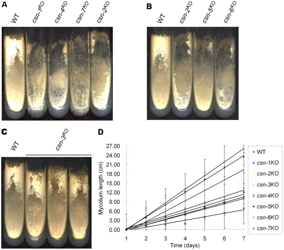
(A) Wild-type and csn-1, csn-4, and csn-7 mutant strains growing on minimal slants. csn-1, csn-4, and csn-7 mutant strains produced significantly less conidia and aerial hyphae than the wild-type strain and were weaker than the csn-2 mutant. (B) csn-2KO, csn-5KO, and csn-6KO had similar phenotypes on minimal slants. (C) Disruption of csn-3 had little effect on growth and development on minimal slants. (D) Growth rate of the wild-type strain and seven csn subunit mutants, measured at 25°C using the race tube assay in constant darkness after one day of light treatment. Different CSN subunits differently regulate conidiation rhythms
CSN-2 was found to be a regulator of the N. crassa circadian clock [20]. To find out whether other CSN subunits have a similar role, we examined the conidiation rhythm of csn knockout mutants by race tube assays. After entrainment by light, the wild-type strain exhibited a robust circadian conidiation rhythm with a period of about 22 hours at 25°C in constant darkness (Figure 2A), while conidiation was very irregular in the csn-1KO, csn-2KO, csn-4KO, csn-5KO, csn-6KO, and csn-7KO strains (Figure 2A), suggesting that these subunits are essential for circadian rhythms. In contrast, the csn-3 mutant grew faster on race tubes than the wild-type strain, and exhibited a robust and precise period of conidiation identical to that of the wild-type strain (Figure 2B), indicating that the CSN-3 subunit was not essential for normal conidiation rhythms in N. crassa. Taken together, these observations demonstrate that, of the seven CSN subunits, all but CSN-3 play an important role in circadian rhythm.
Fig. 2. Effect of disrupting different csn subunit genes on conidiation rhythms. 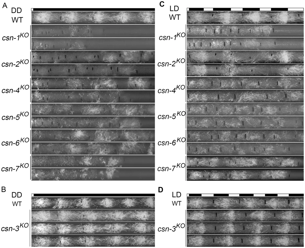
In DD (A, B) and in LD (C, D). At least four replicates were tested under each condition, and two representatives are shown. The black lines indicate the growth fronts every 24 h. The CSN complex regulates photomorphogenesis in plants [1], and CSN-2 is required for light entrainment of conidiation rhythms in N. crassa [20]. To test whether the rest of the CSN subunits have a similar function, we examined the conidiation rhythms of each csn mutant in light–darkness cycles (12 h light/12 h darkness). As shown in Figure 2C, the conidiation rhythms of the csn-1KO, csn-2KO, csn-4KO, csn-5KO, csn-6KO, and csn-7KO strains were not entrained by light–darkness cycles, indicating that these six CSN subunits play key roles in light regulation of the circadian clock. As expected, like the wild-type strain, the csn-3KO mutant could be entrained by light–darkness cycles (Figure 2D), demonstrating that CSN-3 is not required for the light-response process in N. crassa. These results further confirm that each CSN subunit contributes unequally to light-dependent processes in N. crassa.
Because the temperature-regulated conidiation process is affected in the csn-2 mutant [20], we examined the responses of the other CSN subunit mutants to temperature entrainment using race tube assays. In 12 h 25°C/12 h 20°C temperature cycles, the conidiation rhythm of the wild-type strain was synchronized, with conidial peaks in the cold phase (Figure 3A). As expected, temperature cycles failed to entrain the conidiation rhythm in the csn-1KO, csn-4KO, csn-5KO, csn-6KO, and csn-7KO strains, as was the case with the csn-2 mutant (Figure 3A) [20], suggesting that these CSN subunits play important roles in the temperature response of N. crassa. In contrast, the conidiation rhythm of the csn-3KO mutant (Figure 3B) could be synchronized by temperature cycles, similar to the wild-type strain, indicating that the CSN-3 subunit is not required for the temperature-response process. Very similar results were seen with 12 h 28°C/12 h 25°C temperature cycles (Figure 3C and 3D). These results suggest that the CSN complex is involved in the regulation of temperature response, and that all of the subunits except for CSN-3 may play key roles.
Fig. 3. Disruption of different csn genes resulted in different responses to temperature cycles in DD. 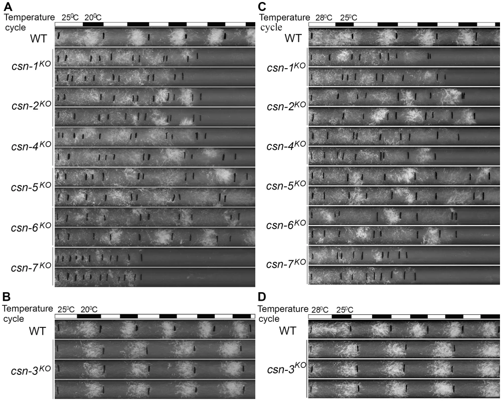
At 25/20°C (A, B) and at 28/25°C (C, D). At least four replicates were carried out under each condition and two representatives are shown. The black lines indicate the growth fronts every 24 h. Different csn mutants differently regulate the neddylation/deneddylation state of cullin proteins
CSN negatively regulates the activities of cullin-based E3 ubiquitin ligases by cleaving the Nedd8 modification from cullins [4], [13]. The distinct phenotypes of each csn mutant suggest that they may contribute differently to this particular CSN function. To test this possibility, we examined the neddylation status of three N. crassa cullin proteins: Cul1, Cul3, and Cul4. The neddylation status of these cullin proteins reflects the functional activity of the CSN complex in each strain.
To monitor the neddylation state of cullins in vivo, we introduced a construct expressing Myc-tagged Cul1, Cul3, or Cul4 into the wild-type strain and into each csn knockout strain, respectively. Expression of Myc-Cul1, Myc-Cul3, or Myc-Cul4 proteins with the predicted molecular weight was confirmed by western blot analysis. Neddylated Myc-tagged cullins were distinguished from unneddylated Myc-tagged cullins based on their slower migration in an SDS-PAGE gel compared to the unneddylated forms. In the wild-type strain, neddylated Myc-Cul1 (upper bands in Figure 4) represented less than 10% of the total of Myc-Cul1 (Figure 4A). We demonstrated previously that disruption of the csn-2 gene causes the hyperneddylation of Cul1 [20] (Figure 4A), confirming that normal functioning of the CSN complex is severely impaired in csn-2 mutants. Similarly, csn-1KO, csn-4KO, csn-5KO, csn-6KO, and csn-7KO mutants displayed increased Cul1 neddylation levels (Figure 4A). The Csn5 subunit in S. pombe and D. melanogaster underlies the Nedd8 isopeptidase activity of the CSN [15]. Our results suggest that the N. crassa CSN-5 subunit performed a similar Nedd8 isopeptidase function, and that the other five subunits were also functional subunits for Cul1 deneddylation in N. crassa. However, the neddylated/deneddylated Cul1 levels in the csn-3KO mutant were similar to the pattern in the wild-type strain (Figure 4A), suggesting that CSN-3 is not required for Cul1 deneddylation activity in N. crassa. The same Cul1 neddylated/deneddylated patterns were also observed in a csn-3 deletion mutant (non-band background) derived from FGSC11275 (Fungal Genetics Stock Center) using the Cul1 detection approach described above (unpublished), further confirming that N. crassa CSN-3 is not required for the Cul1 deneddylation activity of the CSN complex.
Fig. 4. Different cullin neddylation states associated with loss of individual csn genes. 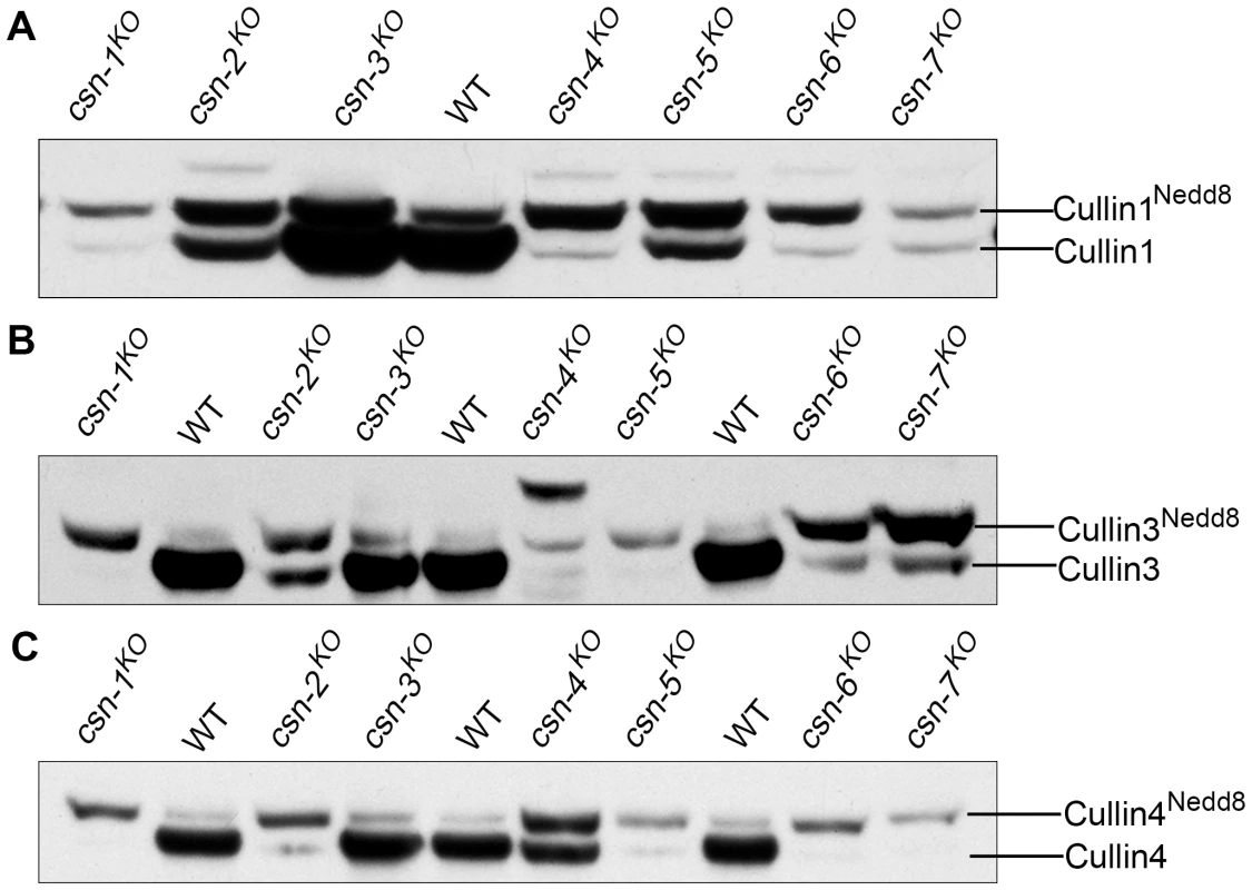
Western blot analyses with c-Myc antibody of expression profiles of Myc-Cul1 (A), Myc-Cul3 (B), and Myc-Cul4 (C) in the wild-type strain and csn mutants. We further examined the neddylation states of two other N. crassa cullin proteins, Cul3 and Cul4, in the wild-type strain and csn mutants. As shown in Figure 4B, the csn-3KO mutant had a Myc-Cul3 neddylated/deneddylated pattern that was similar to that in the wild-type strain, whereas in the other csn knockout mutants, Myc-Cul3 was hyperneddylated. The neddylated/deneddylated pattern of Myc-Cul4 was similar to the patterns of Cul1 and Cul3 in each strain (Figure 4C). Taken together, these results indicate that CSN-3 is not critical for CSN deneddylation activity, suggesting that CSN-3 is not a key subunit in the CSN complex in N. crassa. This molecular evidence, together with the genetic data, strongly suggests that CSN-1, CSN-2, CSN-4, CSN-5, CSN-6, and CSN-7 form the functional core of the CSN complex for cleavage of Nedd8 from cullins in N. crassa.
Different CSN subunits differently contribute to the stability of the SCFFWD-1 complex
Recent studies demonstrated that a major function of the CSN complex is to control the stability of E3 ubiquitin ligases in vivo [19]–[21]. To determine whether the CSN subunits contribute unequally to the stability control of CRLs in N. crassa, we examined the stabilities of Cul1, SKP-1, and FWD-1, the major components of the SCFFWD-1 complex, in each of the csn strains. As shown in Figure 5A, Myc-Cul1 was very stable in the wild-type strain and the csn-3KO mutant, with a half-life of more than 9 h in the presence of cycloheximide (CHX). In contrast, Myc-Cul1 in the csn-1, csn-2, csn-4, csn-5, csn-6, and csn-7 mutants was unstable, with a half-life of less than 3 h (Figure 5A and 5D). These results demonstrate that the functional core subunits of CSN are responsible not only for Cul1 deneddylation, but also for maintenance of Cul1 stability. In N. crassa, SKP-1 is an adaptor protein in the SCF complex that becomes very unstable in csn-2 mutants [20]. As shown in Figure 5B, Myc-SKP-1 remained very stable in the csn-3KO mutant and the wild-type strain, with a half-life of approximately 12 h. However, Myc-SKP-1 was unstable in the csn-1, csn-4, csn-6, and csn-7 mutants, with a half-life of approximately 1.5–3 h (Figure 5B and 5E). Interestingly, as in the wild-type strain and csn-3KO mutant, the stability of Myc-SKP-1 was not affected in the csn-5KO mutant (Figure 5B and 5E), indicating that the key deneddylation isopeptidase subunit of the CSN functional core was dispensable for maintaining SKP-1 stability in the SCF complex.
Fig. 5. Differing stabilities of components of the SCF complex, Cullin1, and SKP-1 in csn mutants. 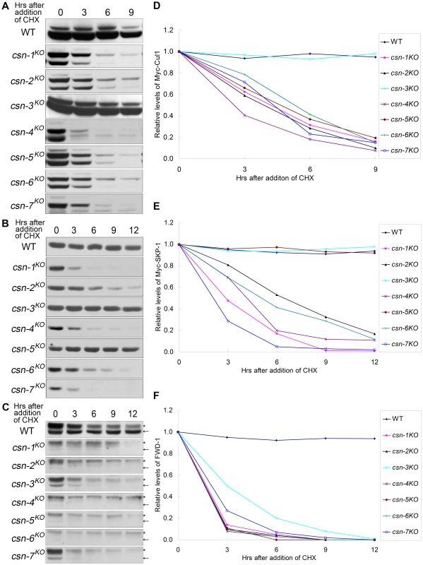
(A, B, C) Western blot analyses showing degradation of Myc-Cul1, Myc-SKP-1, and FWD-1 in csn mutants after the addition of CHX (10 µg/ml). Densitometric analyses from four independent experiments showing the degradation of Cullin 1 (D), SKP-1 (E), and FWD-1 (F). Arrows point out FWD-1 protein bands. Asterisks indicate nonspecific bands detected by our FWD-1 antibody. The F-box domain-containing protein FWD-1 is a component of the SCFFWD-1 complex; it specifically recognizes phosphorylated FRQ protein and targets it for ubiquitination, which is a key process for circadian clock control in N. crassa [20], [29]. As shown in Figure 5C, FWD-1 levels were drastically reduced in the core csn subunit knockout mutants and in the csn-3 mutant, with a half-life of less than 3 h. In contrast, the FWD-1 protein remained very stable in the wild-type strain, with a half-life of more than 12 h (Figure 5C and 5F). Together, these results indicate that the CSN is important for maintaining the stability of F-box domain–containing proteins, such as FWD-1, in N. crassa. Although CSN-3 in the CSN complex was not required for deneddylation of Cul1 or for maintaining the stability of Cul1 and SKP-1, it was required for preventing the degradation of FWD-1. These observations suggest that CSN-3 is also required to maintain normal functioning of the intact CSN complex. Taken together, the differing stability of Cul1, SKP-1, and FWD-1 in the csn mutants indicates that each subunit of the CSN complex functions differently in maintaining the stability of SCF complexes in N. crassa.
Different contributions of CSN subunits to the stability of Cul3 and associated protein BTB1
In N. crassa, Cul3-binding proteins have not been reported previously; therefore, we searched for BTB domain protein coding genes in the N. crassa genome and found eight predicted proteins with highly conserved BTB domains. To test the interactions between Cul3 and the BTB domain proteins, we created Myc-tagged BTB domain proteins and co-expressed each of them in the wild-type strain with Flag-tagged Cul3. As shown in Figure 6A, BTB1 protein (NCU04838) strongly interacted with Cul3 in the immunoprecipitation reaction, indicating that they may form a Cul3-BTB ubiquitin ligase complex in N. crassa.
Fig. 6. Protein levels of Cul3-based E3 components reduced dramatically in the six core CSN subunit mutants, but not in csn-3KO mutant. 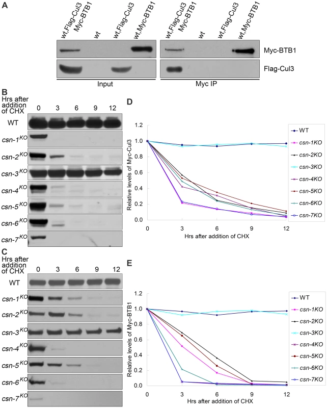
(A) Immunoprecipitation assay confirming the interaction between Flag-Cul3 and Myc-BTB1. Wild-type strain and wild-type strain expressing Flag-Cul3 were used as negative controls. Lysates were subjected to immunoprecipitation with c-Myc antibody, followed by western blot analyses with c-Myc and Flag antibodies. Western blot analyses showing degradation of Myc-Cul3 (B) and Myc-BTB1 (C) in csn mutants after the addition of CHX. Densitometric analyses from four independent experiments showing degradation of Cul3 (D) and BTB1 (E). We next examined whether the stability of Myc-Cul3 was affected in each csn mutant. As shown in Figure 6B, Myc-Cul3 was very stable in the wild-type strain and the csn-3KO mutant, with a half-life of more than 12 h in the presence of CHX. In contrast, this protein was very unstable in the other csn mutants, with a half-life of approximately 1.5–3 h (Figure 6B and 6D). These results indicate that the CSN functional core subunits were necessary for maintaining the stability of Cul3 in N. crassa.
We then investigated the stability of BTB1 protein in the wild-type stain and csn mutants. Myc-BTB1 was very stable in the wild-type strain and the csn-3KO mutant, with a half-life of more than 12 h after CHX treatment; however, it was very unstable in other csn mutants, with a half-life of about 3 h (Figure 6C and 6E). Taken together, these results further demonstrate that the functional core subunits of CSN were important for Cul3 deneddylation and maintaining the stability of the entire Cul3-BTB E3 complex in N. crassa.
Cul4 and DDB1 proteins unstable in all csn mutants
N. crassa Cul4 was previously shown to interact with DDB1 [30]. Therefore, we tested the effect of loss of different CSN subunits on the regulation of Cul4-DDB1 E3s. We showed above that the neddylation/deneddylation pattern of Cul4 in the csn-3 knockout strain was similar to that of the wild-type strain, in which the half-life of Myc-Cul4 was 12 h in the presence of CHX (Figure 7A and 7C). Unexpectedly, Myc-Cul4 was very unstable in all of the csn mutants, with a half-life of approximately 3 h after CHX treatment (Figure 7A and 7C). These results suggest that although CSN-3 is not in the deneddylation core of the CSN complex, it was necessary for maintaining the stability of Cul4 in N. crassa. We next measured the stability of DDB1 in the wild-type strain and csn mutants. As expected, Myc-DDB1 was very unstable in the csn mutants, with a half-life of approximately 3 h after CHX treatment (Figure 7B and 7D), while it remained very stable in the wild-type strain, with a half-life of more than 12 h (Figure 7B and 7D). Therefore, unlike in Cul1 - or Cul3-based E3 complexes, all of the CSN subunits are required for maintaining the stability of Cul4-DDB1 ubiquitin ligases.
Fig. 7. Components of Cul4-DDB1 E3 ubiquitin ligase unstable in all csn mutants. 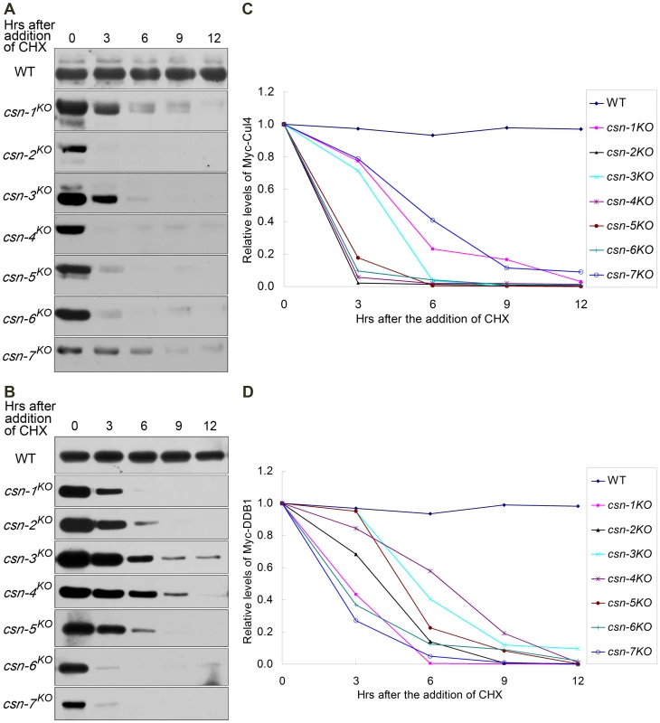
Western blot analyses showing degradation of Myc-Cul4 (A) and Myc-DDB1 (B) in csn mutants after the addition of CHX. Densitometric analyses from four independent experiments showing degradation of Cul4 (C) and DDB1 (D). DDB1 - and Cul4-associated factor DCAF11 unstable in all csn mutants
We previously showed that in N. crassa, DCAF11 is an adaptor protein in a Cul4-DDB1 E3 ligase complex by association with DDB1 protein [31]. In the present study, we performed an immunoprecipitation assay to detect interactions between Myc-DCAF11 and Flag-Cul4. As shown in Figure 8A, Myc-DCAF11 interacted with Flag-Cul4, confirming that it forms an E3 complex with Cul4 and DDB1 proteins. To examine whether the adaptor protein DCAF11 is unstable in the csn mutants, we compared DCAF11 degradation rates in the wild-type and seven csnKO strains. As shown in Figure 8B, DCAF11 was stable in the wild-type strain, as a majority of DCAF11 was still present after 12 h of CHX incubation. In contrast, DCAF11 became undetectable after only 3 h of CHX treatment in the core csn mutants and in the csn-3KO strain, indicating that it was very unstable (Figure 8B and 8G). The accelerated DCAF11 degradation rate in the csn mutants was likely due to its increased autoubiquitination, which is counteracted by normal CSN activity. If this is indeed the case, mutation of a conserved arginine in the WDXR motif of DCAF11 (Figure 8C) should disrupt binding of DCAF11 to the Cul4-DDB1 complex, thus preventing its autoubiquitination and degradation. As shown in Figure 8D, interactions between DDB1 and DCAF11 were disrupted by substitution of the conserved arginine with alanine in the DCAF11 WDXR motif. This point mutation also abolished interactions between DCAF11 and Cul4 (Figure 8E). Indeed, Myc-tagged DCAF11 with an arginine-to-alanine point mutation was very stable and accumulated to reach high steady-state levels in all csn mutants, including the csn-3KO strain (Figure 8F and 8H).
Fig. 8. DCAF11 levels drastically reduced in all csn mutants, possibly because of rapid autoubiquitination-mediated degradation. 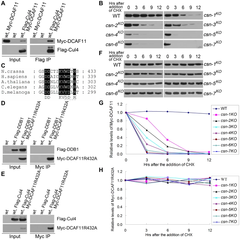
(A) Immunoprecipitation assay confirming the interaction between Flag-Cul4 and Myc-DCAF11. Wild-type strain and wild-type strain expressing Myc-DCAF11 were used as negative controls. (B) Western blot analyses showing the degradation of Myc-DCAF11 in csn mutants after the addition of CHX. (C) Amino acid alignment of conserved WDXR motifs of DCAF11 homologs. (D) Immunoprecipitation assay with c-Myc antibody showing that Myc-DCAF11R432A failed to interact with Flag-DDB1. (E) Myc immunoprecipitation assay showing that Myc-DCAF11R432A failed to interact with Flag-Cul4. (F) Degradation of Myc-DCAF11R432A expressed in the wild-type strain and all csn mutants after the addition of CHX. Densitometric analyses from four independent experiments showing degradation of DCAF11 (G) and DCAF11R432A (H). Taken together, these data indicate that CSN is important for maintaining the stability of DCAF11 in N. crassa, probably by preventing its autoubiquitination. DCAF11 levels were also low in the csn-3 mutant, indicating that the functional core complex alone was not sufficient to protect Cul4-DDB1 E3 ligase complexes from autoubiquitination and degradation. Because the adaptor proteins FWD-1 and DCAF11 are very unstable in the csn-3 strain, maintenance of functional E3 ligase complexes may rely mostly on newly synthesized FWD-1 and DCAF11 proteins. These observations suggest that the CSN-3 subunit is also required to maintain the function of intact CSN complexes in protecting cullin-RING E3 ligase adaptor proteins from autoubiquitination.
Purification and identification of components of the CSN complex in csn-3KO
We next investigated whether loss of the CSN-3 subunit affects the protein levels of other CSN subunits and proper assembly of the CSN complex. Recent studies demonstrated that downregulation of CSN1 and CSN3 causes a proportional reduction in all CSN subunits and a decrease in levels of the holocomplex [32]. We first introduced a Myc-His-tagged CSN-1–, CSN-2–, CSN-4–, CSN-5–, CSN-6–, or CSN-7–expressing construct into a csn-3 mutant and a wild-type strain, respectively. Western blot analyses showed that Myc-His-CSN-2 became very unstable in the csn-3KO mutant, with a half-life of less than 3 h in the presence of CHX (Figure 9A). In contrast, Myc-His-CSN-2 was very stable in the wild-type strains, with a half-life of more than 12 h (Figure 9A). Other five Myc-His-CSN proteins remained very stable in the wild-type strain and in the csn-3KO mutant, with a half-life of more than 12 h (Figure 9A). These results indicate that CSN-3 is required for maintaining the stability of the CSN-2 subunit in N. crassa.
Fig. 9. Comparison of CSN assembly in a csn-3 mutant and a wild-type strain. 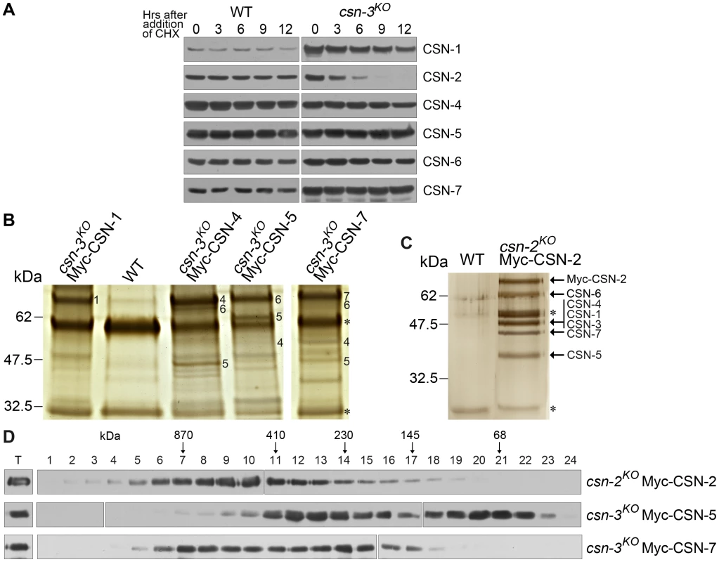
(A) Western blot analyses showing stability of Myc-His-tagged CSN-1, CSN-2, CSN-4, CSN-5, CSN-6, and CSN-7 in the wild-type strain and csn-3 mutant after the addition of CHX. (B) Silver-stained SDS-PAGE showing the two-step purification of Myc-His-CSN-1, CSN-4, CSN-5, or CSN-7 in the csn-3 mutant and the purification of Myc-His-CSN-2 in the csn-2 mutant (positive control). Wild-type strain was used as the negative control. CSN subunits identified by mass spectrometry analysis are indicated. Asterisks indicate the two IgG bands. 1 for CSN-1 protein, 4 for CSN-4, 5 for CSN-5, 6 for CSN-6 and 7 for CSN-7. (D) The Myc-His-CSN-2 in csn-2 mutant, Myc-His-CSN-5 or Myc-His-CSN-7 in csn-3 mutant properly incorporate into CSN complex, but recombinant CSN-7 or CSN-5 form subcomplexes or remained monomer state in the absence of CSN-3. Expression of Myc-His-tagged CSN did not affect the phenotype of the csn-3KO mutant, suggesting that the CSN fusion protein may function similar to the endogenous counterpart subunit. Thus, we used above transformants to purify the CSN complex in the absence of the CSN-3 subunit. Myc-His-tagged CSN-1, CSN-4, CSN-5, or CSN-7 protein was purified on a nickel column followed by immunoprecipitation using a c-Myc monoclonal antibody, respectively. As shown in Figure 9B, several major protein bands were detected in the Myc-His-CSN-4, Myc-His-CSN-5 or Myc-His-CSN-7 sample, but not in Myc-His-CSN-1 sample and the wild-type strain (a negative control). Liquid chromatography–mass spectrometry/mass spectrometry (LC-MS/MS) analysis of excised gel bands led to the identification of CSN-5 and CSN-6 in the Myc-His-CSN-4 purified products, CSN-4 and CSN-6 in the Myc-His-CSN-5 purified products, while CSN-4, CSN-5, and CSN-6 in the Myc-His-CSN-7 purified products, but no CSN-1 and CSN-2 were detected in any of these purifications (Figure 9B). However, there was substantially more CSN-4, CSN-5 or CSN-7 than other CSN subunits in the purification products from csn-3KO, as revealed by a silver-stained SDS-PAGE gel (Figure 9B); in contrast, the amount of Myc-His-CSN-2 was similar to that of other CSN subunits in a csn-2KO, QA-Myc-His-CSN-2 transformant (Figure 9C). These results suggest that under this purification condition, CSN-4, CSN-5 and CSN-6 formed a more stable subcomplex, while Myc-His-CSN-7 incorporated subcomplex with CSN-4, CSN-5, and CSN-6. To test whether Myc-His-tagged CSN subunit incorporates into larger molecular mass complex in the absence of the CSN-3 subunit, we performed gel filtration using above purified Myc-His-tagged CSN proteins. As shown in Figure 9D, CSN-7 and CSN-5 fusion proteins were eluted in larger molecular mass fractions and lower molecular mass fractions, while CSN-2 fusion protein remain in high molecular mass fractions. These results confirmed that both Myc-His-CSN-7 and Myc-His-CSN-5 participate in the formation of CSN and the lower molecular mass form. These data indicate that although proper assembly of the N. crassa CSN complex is not affected in the csn-3KO mutant, the amount of CSN complex is decreased and subcomplexes such as CSN4/5/6 and CSN4/5/6/7 are formed in the absence of the CSN-3 subunit, further suggesting that the phenotypes we observed in csn-3 mutants are due to functional CSN subcomplexes.
Discussion
CSN is an evolutionarily conserved multifunctional complex found in eukaryotes. In the present study, we used mutant analysis to confirm that the N. crassa CSN complex is composed of seven CSN subunits and to demonstrate that each CSN subunit makes a different contribution to CSN function. To the best of our knowledge, this is the first systematic functional analysis of all of the subunits of a complete CSN complex in filamentous fungi. Our results suggest that, of the seven CSN subunits in N. crassa, six act as a functional core in cullin deneddylation. Although CSN-3 is dispensable for cullin deneddylation, it plays an important role in maintaining the stability of FBPs such as FWD-1 in SCF complexes, as well as the stability of Cullin4-based ubiquitin ligases and the CSN-2 subunit. In addition, although the absence of CSN-3 did not affect proper assembly of the CSN complex, the amount of functional core CSN complex was reduced and subcomplexes such as CSN4/5/6 and CSN4/5/6/7 were formed.
Functional core subunits of CSN regulate deneddylation of cullins
In general, CSN complexes contain 7–8 subunits. Accurate assessment of the contribution of individual CSN subunits to the function of the complex as a whole in plants and animals has been difficult, because deletion of any CSN subunit can lead to lethality. Phenotypic features among plants with mutations in different CSN subunit genes are indistinguishable, and plant CSN mutants display almost overlapping misregulation of hormone and other response pathways [33]–[37], indicating that plant CSN subunits function coordinately to support critical deneddylation activity. Fortunately, CSN subunit genes are not essential genes in fungi, such as yeast and N. crassa, which are therefore excellent model systems in which to investigate the function of individual CSN subunits.
We successfully created knockout mutants of all of the individual csn subunits in N.crassa. Of the seven CSN subunits, deletion of CSN-1, CSN-2, CSN-4, CSN-5, CSN-6, or CSN-7 caused similar defects in growth, development, circadian clock, light response, and temperature-entrained conidiation, while deletion of CSN-3 did not cause the defects detected in the other CSN mutants. Similar results were found for other organisms in previous studies. For example, in S. pombe, mutations of different CSN subunits cause distinct phenotypes [14]. In D. melanogaster, csn4- and csn5-null flies display different phenotypes [25] and different gene expression patterns [26]. These results, together with the findings of the present study, indicate that different subunits may have different roles in the CSN complex. Consistent with phenotypic analysis results, our molecular analyses demonstrated that CSN-3 was not required for cullin deneddylation, while deletion of any of the other six csn genes caused hyperneddylation of all three cullins, indicating that in N. crassa, these six CSN subunits were essential for cleavage of Nedd8 from the cullins of CRLs in vivo. Nedd8 modification of cullin positively regulates the activities of CRLs. Recent genetic and biochemical analyses demonstrated that the CSN complex is required for removal of Nedd8 from cullin proteins [4], [13], [17], [18]. Thus, the differences in the growth and developmental phenotypes between csn-3 and the other csn mutants are due to the unequal contributions of different subunits to the CSN deneddylation function. Six of the conserved CSN subunits, CSN-1, CSN-2, CSN-4, CSN-5, CSN-6 and CSN-7, likely act as a functional core for CSN deneddylation activity. This idea is supported by the finding that A. thaliana plants with an N-terminal deletion in the CSN1 subunit (CSNCSN1–C231) exhibit a wild-type pattern of Cul1 neddylation, suggesting that these mutants have normal deneddylation activity [38]. Moreover, the core composition of the CSN complex may also explain why some lower eukaryotes, such as Candida albicans, Cyanidioschyzon merolae, and Saccharomyces cerevisiae, have fully functioning CSN complexes that lack individual subunits [2], [39]. Because CSN is a highly conserved complex that is necessary for regulating the function of CRLs in higher eukaryotes, we propose that a basic functional CSN complex core may exist in these organisms, similar to that found in lower eukaryotes.
CSN-3 required for maintaining the stability of FBPs and Cul4-DDB1 E3 ubiquitin ligases
CSN3 was first identified in A. thaliana as an essential regulator of light-mediated development [1], [40]. Although it is the least-conserved subunit, CSN3 is a component of CSN complexes from plants to mammals. In A. nidulans, deletion of genes encoding CSN subunits 1, 2, 4, or 5 resulted in identical blocks in fruit body formation [28]. However, the two nonconserved subunits C (CSN-3) and H (CSN-8) did not interact in a yeast two-hybrid experiment, suggesting that they may require other subunits or posttranslational modifications for stable interactions [28]. We found that deletion of csn-3 from the genome of N. crassa did not affect the neddylation of cullins and slightly increased the growth rate in the mutant strain compared to the wild-type strain. These findings suggest that the role of CSN-3 in the CSN complex is different from those of the other subunits. Hemizygous deletion of human chromosome 17, band p11.2 results in a multiple congenital anomalies/mental retardation syndrome called Smith–Magenis syndrome (SMS) [41]–[43]. The deleted region spans 1.5–2.0 Mb of DNA, which contains about 20 genes, including CSN3. To investigate the role of CSN3 in mammalian development and in Smit–Magenis syndrome, csn3 was disrupted in mice [44]. Interestingly, there are no visible defects in heterozygous csn3-disrupted mice, although the protein level was reduced. Embryonic development of homozygous Csn3−/− mice is arrested at an early developmental stage [44]. Although Csn2+/− heterozygous mice are phenotypically healthy, loss of Csn2 causes embryonic lethality [45]. These studies imply that CSN3, the least-conserved subunit in the CSN complex, may not be important to the function of the CSN complex compared to CSN2 in mice.
In the present study, we demonstrate that the six core subunits of CSN, but not CSN-3, were essential for maintaining the stability of Cul1 in SCF complexes and of Cul3 and BTB proteins in Cul3-BTB E3 ubiquitin ligasess, while five subunits, but not CSN-3 or CSN-5, were required for maintaining the stability of SKP-1 in the SCF complex. This molecular evidence further supports the idea that individual subunits of the CSN complex contribute differently to CSN functions. Consistent with the phenotype of the csn-3 mutant, CSN-3 does not appear to play an important role in maintaining the stability of these E3s in N. crassa. In mutants lacking different CSN subunits, autoubiquitination of FBPs is enhanced, resulting in increased instability. Several FBPs in fission yeast, N. crassa, and humans are under the protection of the CSN complex [19], [20], [46]. We further show that FWD-1 stability and levels were drastically reduced in the csn-3 mutant and other csn mutants, confirming that CSN-3 was required for preventing autoubiquitination of FBPs after destruction of their substrates. The modular architecture of SCF complex is apparently shared by several other cullin-RING E3 complexes, such as Cul3, that directly interact with a family of substrate receptors through their common BTB domain, which has a Skp1-like structural fold [12]. Our results show that CSN-3 was not required for maintaining the stability of Cul1, SKP-1, Cul3, or BTB protein in the SCF and SCF-like E3s. We further confirmed that CSN-3 was required for maintaining the stability of CSN-2 in the presence of CHX, and for normal levels of the CSN functional core complex. These data provide evidence that the functional core complex efficiently cleaved Nedd8 from cullins and protected scaffold components of the SCF and SCF-like E3s from autoubiquitination; however, it was not sufficient to protect the FBPs from autoubiquitination. Although FBP levels were also low in the csn-3 strain, levels of Cul1 and SKP-1 were not affected; some functional SCF complexes formed, most likely by incorporating newly synthesized FBPs, and mediated the degradation of their substrates. These data may explain the normal circadian rhythms that were observed in the csn-3 mutant, but not in other csn mutants.
In contrast, all seven subunits of the CSN, including CSN3, were required to maintain the stability of Cul4-DDB1 E3s in the presence of CHX. The difference in stability compared to SCF and SCF-like E3s is most likely due to architectural differences between Cul4-DDB1 E3s and SCF E3s; recent studies show that DDB1 displays a flexible linkage between the major protein binding domain (BPA-BPC) and the cullin binding domain (BPB) [10], while SCF and SCF-like E3s exhibit more rigid architectures [47]. In support of this possibility, we found that autoubiquitination of DCAF11 was enhanced in all seven csn mutants. However, disruption of the interactions between DCAF11 and DDB1 completely abrogated the autoubiquitination and degradation of DCAF11 in csn mutants. These data suggest that the instability of Cul4-DDB1 E3s in csn mutants could also be due to the strong interactions among their components, which are more powerful than those in the SCF and SCF-like E3s. In this regard, the function of CSN-3 is similar to that of the functional core subunits. These results provide new insight into how CSN functions to protect different CRLs. Based on these data, we speculate that the different subunits play different roles in maintaining the stability of different CRLs, and that the biochemical functions of the CSN subunits are distinct from one another. Although CSN-3 is the least-conserved subunit, it is found in all of the CSN complexes studied to date, from fungi to mammals [28], [40], [44]. Thus, we propose that CSN-3 may have a similar role in regulating the function of CRLs in higher eukaryotes.
Materials and Methods
Strains and culture conditions
The N. crassa strain 87-3 (bd, a) was used as the wild-type strain in this study. The bd ku70RIP strain, which was generated previously [48], was used as the host strain for creating the csn knockout mutants. The csn-2KO and csn-2KO, his-3 strains used in the present study were also created previously [20]. The newly created csn knockout strains were csn-1KO, csn-3KO, csn-4KO, csn-5KO, csn-6KO, and csn-7KO strains; his-3 strains were also created for each csn deletion. The 301-6 (bd, his-3, A) strain and csn, his-3 strains were the host strains for the his-3 targeting construct transformation. Liquid culture conditions were the same as described previously [49]. For quinic acid–induced protein expression, 0.01 M QA (pH 5.8) was added to liquid medium containing 1× Vogel's medium, 0.1% glucose, and 0.17% arginine [50]. The medium for the race tube assays contained 1× Vogel's medium, 0.1% glucose, 0.17% arginine, 50 ng/ml biotin, and 1.5% agar.
Generation of csn strains in N. crassa
To generate csn gene knockout strains, the entire open reading frames (ORFs) of csn genes were deleted by replacement with the hph gene [51]. The gene replacement cassette containing hph was introduced into the bd, ku70RIP strain by electroporation. The transformants with hph at the csn locus were crossed with 301-6 (bd, his-3, A). Ascospores of the crosses were germinated on plates containing hygromycin and histidine. PCR analyses for hph and csn the ORF region were used to confirm the csn knockout strains.
Plasmids
Full-length ORFs and the 3′-UTR for Cullin3, BTB1, CSN-1, CSN-4, CSN-5, CSN-6, or CSN-7 protein were amplified from genomic DNA by PCR and cloned into the pqa-5Myc-6His and pqa-3Flag plasmids. The previously constructed plasmids pqa-Myc-Cul1, pqa-Myc-His-SKP-1, pqa-Myc-His-CSN-2 [20], pqa-Myc-His-Cul4, pqa-Myc-His-DDB1, pqa-3Flag-Cul4, pqa-3Flag-DDB1 [30] and pqa-Myc-His-DCAF11 were also used for the his-3 targeting transformation in 301-6 and csnKO, his-3 strains.
Protein analyses
Protein extraction, quantification, western blot analysis, protein degradation assays, and immunoprecipitation assays were performed as described previously [20], [30]. Western blot analyses using a monoclonal c-Myc antibody (9E10, Santa Cruz Biotechnology) or Flag antibody (F3165-5MG, Sigma) were performed to identify the positive transformants. Immunoprecipitates or equal amounts of total protein (40 µg) were loaded into each protein lane. After electrophoresis, proteins were transferred onto PVDF membrane, and western blot analysis was performed using c-Myc antibody, Flag antibody, or FWD-1 antiserum.
Purification of Myc-His-CSN proteins from N. crassa
The csn-3KO Myc-His-CSN-1, 4, 5, or 7 strain, wild-type strain (negative control), and csn-2KO Myc-His-CSN-2 strain (positive control) were cultured for approximately 24 h in constant light (LL) in liquid medium containing QA (0.01 M QA, 1× Vogel's medium, 0.1% glucose, and 0.17% arginine). Approximately 15 g of tissue from each strain grown in LL was harvested. The purification procedure was the same as described previously [20]. Fractions containing purified Myc-His-CSN proteins were immunoprecipitated by adding 30 µL of c-Myc monoclonal antibody–coupled agarose beads (9E10AC, Santa Cruz Biotechnology). The precipitates of Myc-His-CSN samples were analyzed by SDS-PAGE (4%–20% and 15% acrylamide, respectively), which was subsequently silver stained following the manufacturer's instructions (ProteoSilver Plus, Sigma). Specific bands were excised and subjected to tryptic digestion and LC-MS/MS.
Gel filtration chromatography of Myc-His-CSN-5 or CSN-7 in csn-3 mutant and CSN-2 in csn-2 mutant
The protocol of gel filtration chromatography was the same as described previously [52]. Briefly, purified proteins (400 µg) were loaded onto a Superdex 200 (GE) gel filtration column that was equilibrated with 25 mL (150 mM NaCl, 20 mM Tris Cl pH 7.4). The proteins were eluted in the same buffer at a flow rate of 0.3 mL/min. Fractions of 0.4 mL were collected starting from the onset of the column void volume (8.0 mL) and finishing at 18 mL (25 fractions). 20 µL of each fraction were prepared in 20 µL of 2× SDS loading buffer, separated by 7.5% SDS-PAGE, then transferred onto PVDF membrane. Western blot analysis was performed using c-Myc antibody (9E10, Santa Cruz Biotechnology).
Supporting Information
Zdroje
1. WeiN
ChamovitzDA
DengXW
1994 Arabidopsis COP9 is a component of a novel signaling complex mediating light control of development. Cell 78 117 124
2. ChangEC
SchwechheimerC
2004 ZOMES III: the interface between signalling and proteolysis. Meeting on The COP9 Signalosome, Proteasome and eIF3. EMBO Rep 5 1041 1045
3. von ArnimAG
SchwechheimerC
2006 Life is degrading—thanks to some zomes. Mol Cell 23 621 629
4. SchwechheimerC
SerinoG
CallisJ
CrosbyWL
LyapinaS
2001 Interactions of the COP9 signalosome with the E3 ubiquitin ligase SCFTIRI in mediating auxin response. Science 292 1379 1382
5. HershkoA
CiechanoverA
1998 The ubiquitin system. Annu Rev Biochem 67 425 479
6. PickartCM
2001 Mechanisms underlying ubiquitination. Annu Rev Biochem 70 503 533
7. CardozoT
PaganoM
2004 The SCF ubiquitin ligase: insights into a molecular machine. Nat Rev Mol Cell Biol 5 739 751
8. PintardL
WillemsA
PeterM
2004 Cullin-based ubiquitin ligases: Cul3-BTB complexes join the family. Embo J 23 1681 1687
9. LeeJ
ZhouP
2007 DCAFs, the missing link of the CUL4-DDB1 ubiquitin ligase. Mol Cell 26 775 780
10. AngersS
LiT
YiX
MacCossMJ
MoonRT
2006 Molecular architecture and assembly of the DDB1-CUL4A ubiquitin ligase machinery. Nature 443 590 593
11. HigaLA
ZhangH
2007 Stealing the spotlight: CUL4-DDB1 ubiquitin ligase docks WD40-repeat proteins to destroy. Cell Div 2 5
12. PetroskiMD
DeshaiesRJ
2005 Function and regulation of cullin-RING ubiquitin ligases. Nat Rev Mol Cell Biol 6 9 20
13. LyapinaS
CopeG
ShevchenkoA
SerinoG
TsugeT
2001 Promotion of NEDD-CUL1 conjugate cleavage by COP9 signalosome. Science 292 1382 1385
14. MundtKE
LiuC
CarrAM
2002 Deletion mutants in COP9/signalosome subunits in fission yeast Schizosaccharomyces pombe display distinct phenotypes. Mol Biol Cell 13 493 502
15. CopeGA
SuhGS
AravindL
SchwarzSE
ZipurskySL
2002 Role of predicted metalloprotease motif of Jab1/Csn5 in cleavage of Nedd8 from Cul1. Science 298 608 611
16. GusmaroliG
FigueroaP
SerinoG
DengXW
2007 Role of the MPN subunits in COP9 signalosome assembly and activity, and their regulatory interaction with Arabidopsis Cullin3-based E3 ligases. Plant Cell 19 564 581
17. CopeGA
DeshaiesRJ
2003 COP9 signalosome: a multifunctional regulator of SCF and other cullin-based ubiquitin ligases. Cell 114 663 671
18. WolfDA
ZhouC
WeeS
2003 The COP9 signalosome: an assembly and maintenance platform for cullin ubiquitin ligases? Nat Cell Biol 5 1029 1033
19. CopeGA
DeshaiesRJ
2006 Targeted silencing of Jab1/Csn5 in human cells downregulates SCF activity through reduction of F-box protein levels. BMC Biochem 7 1
20. HeQ
ChengP
HeQ
LiuY
2005 The COP9 signalosome regulates the Neurospora circadian clock by controlling the stability of the SCFFWD-1 complex. Genes Dev 19 1518 1531
21. WuJT
LinHC
HuYC
ChienCT
2005 Neddylation and deneddylation regulate Cul1 and Cul3 protein accumulation. Nat Cell Biol 7 1014 1020
22. Maytal-KivityV
PickE
PiranR
HofmannK
GlickmanMH
2003 The COP9 signalosome-like complex in S. cerevisiae and links to other PCI complexes. Int J Biochem Cell Biol 35 706 715
23. LiuC
PowellKA
MundtK
WuL
CarrAM
2003 Cop9/signalosome subunits and Pcu4 regulate ribonucleotide reductase by both checkpoint-dependent and -independent mechanisms. Genes Dev 17 1130 1140
24. Luke-GlaserS
RoyM
LarsenB
Le BihanT
MetalnikovP
2007 CIF-1, a shared subunit of the COP9/signalosome and eukaryotic initiation factor 3 complexes, regulates MEL-26 levels in the Caenorhabditis elegans embryo. Mol Cell Biol 27 4526 4540
25. OronE
MannervikM
RencusS
Harari-SteinbergO
Neuman-SilberbergS
2002 COP9 signalosome subunits 4 and 5 regulate multiple pleiotropic pathways in Drosophila melanogaster. Development 129 4399 4409
26. OronE
TullerT
LiL
RozovskyN
YekutieliD
2007 Genomic analysis of COP9 signalosome function in Drosophila melanogaster reveals a role in temporal regulation of gene expression. Mol Syst Biol 3 108
27. BuschS
EckertSE
KrappmannS
BrausGH
2003 The COP9 signalosome is an essential regulator of development in the filamentous fungus Aspergillus nidulans. Mol Microbiol 49 717 730
28. BuschS
SchwierEU
NahlikK
BayramO
HelmstaedtK
2007 An eight-subunit COP9 signalosome with an intact JAMM motif is required for fungal fruit body formation. Proc Natl Acad Sci U S A 104 8089 8094
29. HeQ
ChengP
YangY
HeQ
YuH
2003 FWD1-mediated degradation of FREQUENCY in Neurospora establishes a conserved mechanism for circadian clock regulation. Embo J 22 4421 4430
30. ZhaoY
ShenY
YangS
WangJ
HuQ
2010 Ubiquitin ligase components Cullin4 and DDB1 are essential for DNA methylation in Neurospora crassa. J Biol Chem 285 4355 4365
31. XuH
WangJ
HuQ
QuanY
ChenH
2010 DCAF26, an adaptor protein of Cul4-based E3, is essential for DNA methylation in Neurospora crassa. PLoS Genet 6 e1001132 doi:10.1371/journal.pgen.1001132
32. PethA
BerndtC
HenkeW
DubielW
2007 Downregulation of COP9 signalosome subunits differentially affects the CSN complex and target protein stability. BMC Biochem 8 27
33. ChenH
ShenY
TangX
YuL
WangJ
2006 Arabidopsis CULLIN4 Forms an E3 Ubiquitin Ligase with RBX1 and the CDD Complex in Mediating Light Control of Development. Plant Cell 18 1991 2004
34. DohmannEM
LevesqueMP
IsonoE
SchmidM
SchwechheimerC
2008 Auxin responses in mutants of the Arabidopsis CONSTITUTIVE PHOTOMORPHOGENIC9 signalosome. Plant Physiol 147 1369 1379
35. FengS
MaL
WangX
XieD
Dinesh-KumarSP
2003 The COP9 signalosome interacts physically with SCF COI1 and modulates jasmonate responses. Plant Cell 15 1083 1094
36. PengZ
SerinoG
DengXW
2001 Molecular characterization of subunit 6 of the COP9 signalosome and its role in multifaceted developmental processes in Arabidopsis. Plant Cell 13 2393 2407
37. SchwagerKM
Calderon-VillalobosLI
DohmannEM
WilligeBC
KniererS
2007 Characterization of the VIER F-BOX PROTEINE genes from Arabidopsis reveals their importance for plant growth and development. Plant Cell 19 1163 1178
38. WangX
KangD
FengS
SerinoG
SchwechheimerC
2002 CSN1 N-terminal-dependent activity is required for Arabidopsis development but not for Rub1/Nedd8 deconjugation of cullins: a structure-function study of CSN1 subunit of COP9 signalosome. Mol Biol Cell 13 646 655
39. Maytal-KivityV
PiranR
PickE
HofmannK
GlickmanMH
2002 COP9 signalosome components play a role in the mating pheromone response of S. cerevisiae. EMBO Rep 3 1215 1221
40. PengZ
SerinoG
DengXW
2001 A role of Arabidopsis COP9 signalosome in multifaceted developmental processes revealed by the characterization of its subunit 3. Development 128 4277 4288
41. De LeersnyderH
de BloisMC
VekemansM
SidiD
VillainE
2001 beta(1)-adrenergic antagonists improve sleep and behavioural disturbances in a circadian disorder, Smith-Magenis syndrome. J Med Genet 38 586 590
42. GreenbergF
LewisRA
PotockiL
GlazeD
ParkeJ
1996 Multi-disciplinary clinical study of Smith-Magenis syndrome (deletion 17p11.2). Am J Med Genet 62 247 254
43. PotockiL
GlazeD
TanDX
ParkSS
KashorkCD
2000 Circadian rhythm abnormalities of melatonin in Smith-Magenis syndrome. J Med Genet 37 428 433
44. YanJ
WalzK
NakamuraH
Carattini-RiveraS
ZhaoQ
2003 COP9 signalosome subunit 3 is essential for maintenance of cell proliferation in the mouse embryonic epiblast. Mol Cell Biol 23 6798 6808
45. Lykke-AndersenK
SchaeferL
MenonS
DengXW
MillerJB
2003 Disruption of the COP9 signalosome Csn2 subunit in mice causes deficient cell proliferation, accumulation of p53 and cyclin E, and early embryonic death. Mol Cell Biol 23 6790 6797
46. WeeS
GeyerRK
TodaT
WolfDA
2005 CSN facilitates Cullin-RING ubiquitin ligase function by counteracting autocatalytic adapter instability. Nat Cell Biol 7 387 391
47. ZhengN
SchulmanBA
SongL
MillerJJ
JeffreyPD
2002 Structure of the Cul1-Rbx1-Skp1-F boxSkp2 SCF ubiquitin ligase complex. Nature 416 703 709
48. HeQ
ChaJ
HeQ
LeeHC
YangY
2006 CKI and CKII mediate the FREQUENCY-dependent phosphorylation of the WHITE COLLAR complex to close the Neurospora circadian negative feedback loop. Genes Dev 20 2552 2565
49. AronsonBD
JohnsonKA
LorosJJ
DunlapJC
1994 Negative feedback defining a circadian clock: autoregulation of the clock gene frequency. Science 263 1578 1584
50. ChengP
YangY
LiuY
2001 Interlocked feedback loops contribute to the robustness of the Neurospora circadian clock. Proc Natl Acad Sci U S A 98 7408 7413
51. ColotHV
ParkG
TurnerGE
RingelbergC
CrewCM
2006 A high-throughput gene knockout procedure for Neurospora reveals functions for multiple transcription factors. Proc Natl Acad Sci U S A 103 10352 10357
52. GusmaroliG
FengS
DengXW
2004 The Arabidopsis CSN5A and CSN5B subunits are present in distinct COP9 signalosome complexes, and mutations in their JAMM domains exhibit differential dominant negative effects on development. Plant Cell 16 2984 3001
Štítky
Genetika Reprodukční medicína
Článek Genome-Wide Interrogation of Mammalian Stem Cell Fate Determinants by Nested Chromosome DeletionsČlánek Season of Conception in Rural Gambia Affects DNA Methylation at Putative Human Metastable EpiallelesČlánek A Quantitative Systems Approach Reveals Dynamic Control of tRNA Modifications during Cellular StressČlánek Reduction of Protein Translation and Activation of Autophagy Protect against PINK1 Pathogenesis inČlánek The Loss of PGAM5 Suppresses the Mitochondrial Degeneration Caused by Inactivation of PINK1 inČlánek Cleavage of Phosphorothioated DNA and Methylated DNA by the Type IV Restriction Endonuclease ScoMcrAČlánek Competitive Repair by Naturally Dispersed Repetitive DNA during Non-Allelic Homologous Recombination
Článek vyšel v časopisePLOS Genetics
Nejčtenější tento týden
2010 Číslo 12
-
Všechny články tohoto čísla
- Genome-Wide Interrogation of Mammalian Stem Cell Fate Determinants by Nested Chromosome Deletions
- Whole-Genome and Chromosome Evolution Associated with Host Adaptation and Speciation of the Wheat Pathogen
- Association of Variants at 1q32 and with Ankylosing Spondylitis Suggests Genetic Overlap with Crohn's Disease
- Initiator Elements Function to Determine the Activity State of BX-C Enhancers
- Identification of Genes Required for Neural-Specific Glycosylation Using Functional Genomics
- A Young Duplicate Gene Plays Essential Roles in Spermatogenesis by Regulating Several Y-Linked Male Fertility Genes
- The EpsE Flagellar Clutch Is Bifunctional and Synergizes with EPS Biosynthesis to Promote Biofilm Formation
- Histone H2A C-Terminus Regulates Chromatin Dynamics, Remodeling, and Histone H1 Binding
- Season of Conception in Rural Gambia Affects DNA Methylation at Putative Human Metastable Epialleles
- A Quantitative Systems Approach Reveals Dynamic Control of tRNA Modifications during Cellular Stress
- GC-Rich Sequence Elements Recruit PRC2 in Mammalian ES Cells
- A Single Enhancer Regulating the Differential Expression of Duplicated Red-Sensitive Opsin Genes in Zebrafish
- Investigation and Functional Characterization of Rare Genetic Variants in the Adipose Triglyceride Lipase in a Large Healthy Working Population
- Reduction of Protein Translation and Activation of Autophagy Protect against PINK1 Pathogenesis in
- Noisy Splicing Drives mRNA Isoform Diversity in Human Cells
- The Loss of PGAM5 Suppresses the Mitochondrial Degeneration Caused by Inactivation of PINK1 in
- Thymus-Associated Parathyroid Hormone Has Two Cellular Origins with Distinct Endocrine and Immunological Functions
- An ABC Transporter Mutation Is Correlated with Insect Resistance to Cry1Ac Toxin
- Role of Individual Subunits of the CSN Complex in Regulation of Deneddylation and Stability of Cullin Proteins
- The C-Terminal Domain of the Bacterial SSB Protein Acts as a DNA Maintenance Hub at Active Chromosome Replication Forks
- The DNA Damage Response Pathway Contributes to the Stability of Chromosome III Derivatives Lacking Efficient Replicators
- Cleavage of Phosphorothioated DNA and Methylated DNA by the Type IV Restriction Endonuclease ScoMcrA
- LaeA Control of Velvet Family Regulatory Proteins for Light-Dependent Development and Fungal Cell-Type Specificity
- Competitive Repair by Naturally Dispersed Repetitive DNA during Non-Allelic Homologous Recombination
- Distinct Functions for the piRNA Pathway in Genome Maintenance and Telomere Protection
- MOS11: A New Component in the mRNA Export Pathway
- Self-Mating in the Definitive Host Potentiates Clonal Outbreaks of the Apicomplexan Parasites and
- A Role for ATF2 in Regulating MITF and Melanoma Development
- Ancestral Regulatory Circuits Governing Ectoderm Patterning Downstream of Nodal and BMP2/4 Revealed by Gene Regulatory Network Analysis in an Echinoderm
- Cancer and Neurodegeneration: Between the Devil and the Deep Blue Sea
- Functional Comparison of Innate Immune Signaling Pathways in Primates
- Linking Crohn's Disease and Ankylosing Spondylitis: It's All about Genes!
- Genomics Meets Glycomics—The First GWAS Study of Human N-Glycome Identifies HNF1α as a Master Regulator of Plasma Protein Fucosylation
- Continuous and Periodic Expansion of CAG Repeats in Huntington's Disease R6/1 Mice
- Expression of Linear and Novel Circular Forms of an -Associated Non-Coding RNA Correlates with Atherosclerosis Risk
- Endocytic Sorting and Recycling Require Membrane Phosphatidylserine Asymmetry Maintained by TAT-1/CHAT-1
- Histone Deacetylases Suppress CGG Repeat–Induced Neurodegeneration Via Transcriptional Silencing in Models of Fragile X Tremor Ataxia Syndrome
- PLOS Genetics
- Archiv čísel
- Aktuální číslo
- Informace o časopisu
Nejčtenější v tomto čísle- Functional Comparison of Innate Immune Signaling Pathways in Primates
- Expression of Linear and Novel Circular Forms of an -Associated Non-Coding RNA Correlates with Atherosclerosis Risk
- Genome-Wide Interrogation of Mammalian Stem Cell Fate Determinants by Nested Chromosome Deletions
- Histone H2A C-Terminus Regulates Chromatin Dynamics, Remodeling, and Histone H1 Binding
Kurzy
Zvyšte si kvalifikaci online z pohodlí domova
Současné možnosti léčby obezity
nový kurzAutoři: MUDr. Martin Hrubý
Všechny kurzyPřihlášení#ADS_BOTTOM_SCRIPTS#Zapomenuté hesloZadejte e-mailovou adresu, se kterou jste vytvářel(a) účet, budou Vám na ni zaslány informace k nastavení nového hesla.
- Vzdělávání


