-
Články
Top novinky
Reklama- Vzdělávání
- Časopisy
Top články
Nové číslo
- Témata
Top novinky
Reklama- Videa
- Podcasty
Nové podcasty
Reklama- Kariéra
Doporučené pozice
Reklama- Praxe
Top novinky
ReklamaAutocrine Regulation of Pulmonary Inflammation by Effector T-Cell Derived IL-10 during Infection with Respiratory Syncytial Virus
Respiratory syncytial virus (RSV) infection is the leading viral cause of severe lower respiratory tract illness in young infants. Clinical studies have documented that certain polymorphisms in the gene encoding the regulatory cytokine IL-10 are associated with the development of severe bronchiolitis in RSV infected infants. Here, we examined the role of IL-10 in a murine model of primary RSV infection and found that high levels of IL-10 are produced in the respiratory tract by anti-viral effector T cells at the onset of the adaptive immune response. We demonstrated that the function of the effector T cell -derived IL-10 in vivo is to limit the excess pulmonary inflammation and thereby to maintain critical lung function. We further identify a novel mechanism by which effector T cell-derived IL-10 controls excess inflammation by feedback inhibition through engagement of the IL-10 receptor on the antiviral effector T cells. Our findings suggest a potentially critical role of effector T cell-derived IL-10 in controlling disease severity in clinical RSV infection.
Published in the journal: . PLoS Pathog 7(8): e32767. doi:10.1371/journal.ppat.1002173
Category: Research Article
doi: https://doi.org/10.1371/journal.ppat.1002173Summary
Respiratory syncytial virus (RSV) infection is the leading viral cause of severe lower respiratory tract illness in young infants. Clinical studies have documented that certain polymorphisms in the gene encoding the regulatory cytokine IL-10 are associated with the development of severe bronchiolitis in RSV infected infants. Here, we examined the role of IL-10 in a murine model of primary RSV infection and found that high levels of IL-10 are produced in the respiratory tract by anti-viral effector T cells at the onset of the adaptive immune response. We demonstrated that the function of the effector T cell -derived IL-10 in vivo is to limit the excess pulmonary inflammation and thereby to maintain critical lung function. We further identify a novel mechanism by which effector T cell-derived IL-10 controls excess inflammation by feedback inhibition through engagement of the IL-10 receptor on the antiviral effector T cells. Our findings suggest a potentially critical role of effector T cell-derived IL-10 in controlling disease severity in clinical RSV infection.
Introduction
Respiratory syncytial virus (RSV) infection is the leading viral cause of upper and lower respiratory tract illness in young infants. In the USA, nearly 100% of children are infected with RSV by the age of 2–3 [1]. Approximately 1–2% of these infected children develop moderate to severe bronchiolitis [2]. The exact mechanisms underlying the development of severe pulmonary diseases in the small proportion of children remain poorly defined. Nevertheless, in both clinical studies and animal models, severe pulmonary disease induced by RSV infection is typically associated with an exaggerated inflammatory response in the lower respiratory tract, characterized by the overproduction of pro-inflammatory cytokines/chemokines and increased infiltration of inflammatory cells [3], [4], [5]. Furthermore, there is no firm correlation between disease severity and the extent of RSV replication [6], further suggesting a likely important role of the host immune response to RSV in determining disease severity. Consequently, in responding to an infectious agent like RSV with a strong potential to induce immune-mediated pathology, there is a need to finely balance the immuno-protective and immuno-pathological potential of the immune response in order to insure virus clearance without excess inflammatory injury.
IL-10 is a major regulatory cytokine with broad anti-inflammatory properties [7]. Depending on the nature of the pathogenic stimulus, many cell types including neutrophils, NK cells, macrophages, dendritic cells (DC), regulatory and effector T cells have been shown to be capable of producing IL-10 both in vitro and in vivo in response to infection [8], [9]. IL-10 is generally viewed as a negative regulator of the response of both innate and adaptive immune cells during infection particularly during persistent parasitic, bacterial, and viral infections where it can suppress pathogen clearance and/or the inflammatory response triggered by the infectious agent [8]. Recent evidence suggests that IL-10 may play an important regulatory role in acute viral infections of the respiratory tract where it inhibits the development of excess pulmonary injury in the face of normal virus clearance from the respiratory tract [10]. These findings, along with evidence of a link between a polymorphism in the IL-10 locus and the severity of bronchiolitis in infants infected with RSV [11], [12], [13], [14], prompted us to explore the role of IL-10, and in particular of IL-10 produced by virus-specific effector T-cells, in controlling pulmonary injury associated with experimental murine RSV infection.
In this study, we investigated the source and role of IL-10 during primary RSV infection. We found that high levels of this regulatory cytokine are produced simultaneously with effector cytokines in the respiratory tract at the onset of the adaptive immune response. The main cellular sources of IL-10 in the lung are RSV-specific CD4+ and CD8+ effector T-cells. We show that effector T cell-derived IL-10 during RSV infection in vivo acts to inhibit excess inflammation in the respiratory tract and thereby maintain critical pulmonary function in the infected host. We further provide evidence for a novel mechanism of IL-10-mediated inflammation where effector T cell-derived IL-10 acts in an autocrine manner on the effector T cells to suppress excess pulmonary inflammation induced by the anti-viral response of the effector T cells. The implications of these findings for RSV infection are discussed.
Results
Adaptive immune cells are the major source of IL-10 during acute RSV infection
Because of IL-10′s documented role in controlling or inhibiting the development of excess inflammation in response to infection and injury, and due to the evidence linking expression of IL-10 to injury severity in human RSV infection, we evaluated the role of IL-10 in virus clearance and the control of pulmonary inflammation in a murine model of experimental RSV infection [8], [14]. To address this question, we first infected BALB/c mice with RSV and measured by ELISA the kinetics of IL-10 release into the bronchoalveolar lavage fluid (BALF) sampled from RSV-infected lungs. As a measure of proinflammatory cytokine release during infection we monitored in parallel the kinetics of IFN-γ production in the infected lungs. We found that minimal levels of either cytokine were detected in the BALF early during infection (day 1–3 p.i.) (Figure 1A). By day 5 p.i., a time at which RSV effector T cells have been shown to begin infiltrating into the respiratory tract [15], both IL-10 and IFN-γ levels in the BALF increased dramatically in a coordinated manner. The levels of the two cytokines then decreased progressively and both returned to background levels by day 9 p.i. (Figure 1A).
Fig. 1. T cells are the major source of IL-10 during Respiratory Syncyial Virus infection. 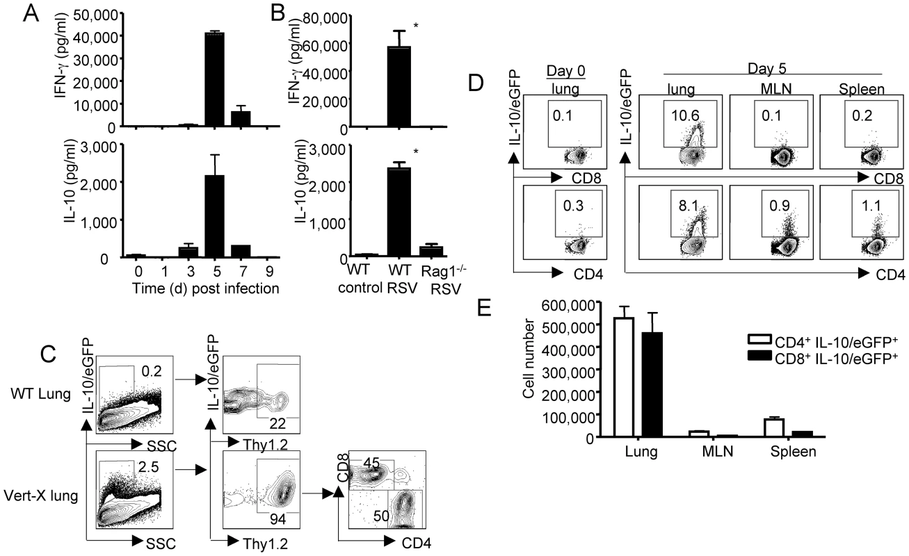
(A) BALB/c mice were infected with RSV. At the indicated days p.i., mice were sacrificed, and IL-10 and IFN-γ levels in the airway were measured by ELISA. (B) WT and Rag1-/- mice were infected with RSV. At d5 p.i., mice were sacrificed, and IL-10 and IFN-γ levels in the airway were measured by ELISA. P value was determined by unpaired two-tailed Student t test. * indicates P< = 0.05. (C–E) Vert-X mice were infected with RSV, at d5 p.i., the expression of IL-10/eGFP by lung cells was measured by flow cytometry. (C) The phenotype of IL-10/eGFP+ cells in the lungs was analyzed by flow cytometry. Numbers are the percentages of cells in gated population. (D) IL-10/eGFP expression by CD8+ and CD4+ T cells from lungs, MLN or spleen of uninfected (Day 0) mice or d5 infected mice are measured by flow cytometry. Numbers are the percentages of cells in gated populations. (E) The absolute numbers of IL-10/eGFP+ CD8+ and IL-10/eGFP+ CD4+ T cells in the lungs, MLN and spleens are depicted. Data are representative of two to three independent experiments. The kinetics of IL-10 and IFN-γ release into the BALF and its tight association with the influx of RSV-specific effector T cells into lungs raised the possibility that these 2 cytokines may be products of adaptive immune cells, or at least that their expression is linked to the recruitment of virus-specific adaptive immune cells into the infected lungs. To initially explore the contribution of adaptive immune (T and/or B) cells to the IL-10 response in the infected respiratory tract, we infected WT or Rag1-/- mice (which lack T and B cells) with RSV and measured IL-10 and IFN-γ levels in the BALF of these mice. We found that high levels of both cytokines were released into the airways of WT but not Rag1-/- mice after RSV infection (Figure 1B). Uninfected WT (Figure 1B) or Rag1-/- (data not shown) mice had negligible levels of this regulatory cytokine. These data suggest that the release of both regulatory cytokine IL-10 and effector cytokine IFN-γ following infection is dependent on the adaptive immune T and/or B cells.
T cells are the main source of IL-10 produced in the respiratory tract during RSV infection
To further define the cellular source(s) of IL-10 in vivo during RSV infection, we infected the IL-10/eGFP reporter mice (Vert-X) with RSV [16]. At day 5 p.i. (i.e at the peak of IL-10 detection in the BALF) we harvested lung cells and measured IL-10/eGFP expression by the liberated lung cell populations using flow cytometry. We found that, compared to the background fluorescence observed in lung cells from RSV infected WT mice, a significant percentage of lung cells from infected Vert-X mice express IL-10/eGFP (Figure 1C). More importantly we found that IL-10/eGFP expression in the Vert-X lungs was restricted almost exclusively (>90%) to Thy-1+ cells infiltrating the infected lungs and that the frequency of IL-10/eGFP+ CD4+ T cells among the Thy-1+ lymphocytes roughly equaled that of IL-10/eGFP+ CD8+ T cells (Figure 1C). These data further reinforced the view that CD4+ and CD8+ T lymphocytes are the major cellular sources of IL-10 in the lung during RSV infection. Next, we examined the IL-10/eGFP expression by CD8+ and CD4+ T cells from the uninfected (day 0) Vert-X lungs and from the lungs, the draining mediastinal lymph nodes (MLN) and the spleens of RSV infected Vert-X mice. We found that IL-10/eGFP-expressing cells are highly enriched in the RSV infected lungs, but not in the uninfected lungs nor in the MLN or the spleen of infected mice (Figure 1D, E). We also investigated the kinetics of accumulation of IL-10-expressing cells in the lungs following RSV infection and found that the IL-10-expressing cells were restricted to the acute phase of infection (Figure S1). Notably, even though the absolute number of IL-10/eGFP-expressing (IL-10 mRNA +) CD8+ and CD4+ continued to increase in the lungs from d5 to d7 post infection, the in vivo release of IL-10 protein in the airway peaks at d5 post infection, which coincides with the fall in lung virus titers and so the viral antigen load in the lung. This observation is consistent with our previously reported findings [10], [17] and likely reflects dependence of IL-10 protein synthesis and release on TCR engagement and viral antigen recognition.
IL-10 is produced in the lungs during RSV infection primarily by effector T-cells
The coordinated release of IL-10 and IFN-γ in the respiratory tract suggests that the IL-10 producing T cells in the RSV infected lungs may be anti-viral effector T cells capable of producing IFN-γ as well. To address this possibility, we first examined the expression of several cell surface molecules associated with T-cell activation and/or effector differentiation by the lung T cells from uninfected Vert-X mice (control), along with lung IL-10/eGFP+ and IL-10/eGFP− T cells from RSV infected Vert-X mice. We found that both IL-10/eGFP+ as well as IL-10/eGFP− CD8+ T cells from infected Vert-X lungs express high levels of T cell activation/effector markers such as CD44, CD43, ICOS, and low levels of naïve T cell marker CD62L suggesting that, like the eGFP− CD8+ T cells, the IL-10/eGFP+ CD8+ T cells likely represent activated effector CD8+ T cells (Figure 2A). Likewise, both IL-10/eGFP+ and IL-10/eGFP− CD4+ T cells express higher levels of those activation/effector markers compared to control CD4+ T cells (Figure 2A). We also FACS-sorted IL-10/eGFP+ and IL-10/eGFP− T cells from infected lungs and measured the gene expression of signature molecules of effector cells. We found that both CD8+ IL-10/eGFP+ cells and CD4+ IL-10/eGFP+ cells express high levels of effector molecules such as IFN-γ, Granzyme B and the type 1 effector cell lineage specific transcription factor T-bet [18], suggesting that the IL-10 expressing CD8+ and CD4+ cells are indeed type 1 effector cells (Figure 2B, C). Consistent with this idea, we observed that all IL-10-producing CD8+ and most IL-10-producing CD4+ T cells simultaneously produce IFN-γ in response to mitogenic or antigenic stimulation in vitro in the intracellular staining assay (ICS) (Figure 2D, E and data not shown). Furthermore, we analyzed by ICS assay the expression of T-bet and the regulatory T cell specific transcription factor Foxp-3 [19], in IL-10− IFN-γ+ (IFN-γ single positive, IFN-γSP) CD8+ T cells, IL-10− IFN-γ+ (IFN-γSP) CD4+ T cells , IL-10+ CD8+ T cells and IL-10+ CD4+ T cells from the infected lungs directly ex vivo. Consistent with the mRNA levels (Figure 2B, C), we found that IL-10 positive CD8+ or CD4+ T cells express T-bet at levels as high as the (IL-10−) IFN-γ single-positive CD8+ or CD4+ type 1 like effector cells (Figure 2D, E). Furthermore, most of IL-10+ CD8+ cells and the majority of the IL-10+ CD4+ T cells are Foxp-3 negative (Figure 2F). Collectively, these data suggest that the vast majority of the IL-10-expressing CD8+ and CD4+ T cells are primarily type 1 effector cells. We did however observe that a minor proportion, up to 30%, of the IL-10+ CD4+ T-cells, expressed Foxp-3. These Foxp-3+ CD4+ T cells were uniformly negative for IFN-γ production (Figure 2F and data not shown).
Fig. 2. IL-10 expressing T cells are mainly type 1 effector T cells. 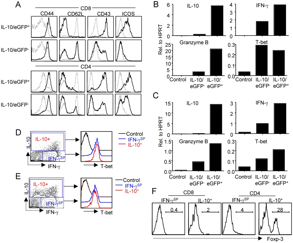
(A), Vert-X mice were infected with RSV, At d5 p.i., the expression of CD44, CD62L, CD43 and ICOS by IL-10/eGFP+ or IL-10/eGFP− T cells were measured by flow cytometry. Grey line, surface expression of the molecules displayed by T cells isolated from naïve lungs (Control). Black line, surface expression of the molecules displayed by IL-10/eGFP+ or IL-10/eGFP− T cells isolated from d5 infected lungs. (B, C) Vert-X mice were infected with RSV, At d6 p.i., CD44hi IL-10/eGFP+ cells and CD44hi IL-10/eGFP− T cells were sorted from infected lungs. (B) The expression of IL-10, IFN-γ, Granzyme B and T-bet genes in control (naïve) CD8+ T-cells, CD8+ CD44hi IL-10/eGFP− T cells and CD8+ CD44hi IL-10/eGFP+ T cells was determined by quantitative RT-PCR. (C) The expression of IL-10, IFN-γ, Granzyme B and T-bet genes in control (naïve) CD4+ T cells, CD4+ CD44hi IL-10/eGFP− T cells and CD4+ CD44hi IL-10/eGFP+ T cells was determined by quantitative RT-PCR. (D. E. F.) BALB/c mice were infected with RSV. At d5 p.i., lung cells were stimulated with PMA/Ionomycin. (D, E) The expression of IL-10, IFN-γ and T-bet by CD8+ T cells (D) or CD4+ T cells (E) was measured through ICS. (F) The expression of Foxp-3 by indicated cell population was measured by ICS. Data are representative of at least two independent experiments. IL-10 derived from effector T cells controls host morbidity without affecting viral clearance
To determine if the effector T cell-derived IL-10 had any impact on the outcome of RSV infection, we examined the effect of blockade of the IL-10 receptor (IL-10R) by in vivo administration of a blocking anti-IL-10Rα (α-IL-10R) mAb on virus clearance, pulmonary function and lung inflammation. We found that the administration of α-IL-10R mAb in vivo significantly enhanced the weight loss of RSV infected mice, particularly at day 5 and thereafter when RSV-specific adaptive immune T cells began infiltrating into the respiratory tract (Figure 3A). We also determined whether three parameters of lung function, as described in Methods, were similarly affected after blocking the action of IL-10 in vivo. As Figure 3B demonstrates, the blockade of IL-10 function in vivo leads to a significant decrease in lung compliance (LC) and increase in pulmonary artery pressure (PAP) in the treated mice at d7 post infection. In addition, although it did not reach statistical significance, airway resistance (AR) was also increased in the mice treated with α-IL-10R mAb compared to the mice administered control mAb (Figure 3B).
Fig. 3. The blockade of effector T cell-derived-IL-10 enhances host morbidity without affecting viral clearance. 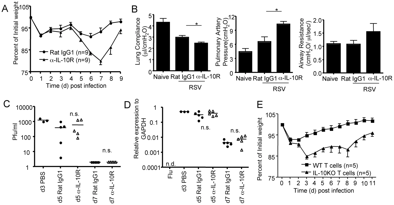
(A-D). BALB/c mice were infected with RSV and then were either injected with Rat IgG1 control mAb or α-IL-10R blocking mAb. (A) Mouse weight loss was monitored daily following infection. (B) Pulmonary function of naïve mice, Rat IgG1 mAb treated or α-IL-10R mAb treated d7 infected mice was monitored. P value was determined by unpaired two-tailed Student t test. * indicates P< = 0.05. (C) Airway virus titers from the indicated mice were determined by plaque assay. PBS, mice were infected with RSV and received PBS. P value was determined by unpaired two-tailed Student t test. n.s.(non-significant). (D) Lung RSV gene copies from the indicated mice were determined through quantitative RT-PCR. Flu, mice were infected with influenza A/PR8 virus. P value was determined by unpaired two-tailed Student t test. n.s.(non-significant). (E) Either WT or IL-10-/- naïve T cells were transferred into Rag1-/- mice which were then infected with RSV. Mouse weight loss was monitored daily after infection. A, B. Pooled data from three experiments are represented. C, D, E. Pooled data from two experiments are represented. We next compared viral clearance in RSV infected mice which were treated with either Rat IgG1 control mAb or α-IL-10R mAb by plaque assay and by quantitative RT-PCR for RSV genome copies. As Figure 3C, and D demonstrate, mice treated with α-IL-10R mAb had cleared virus by day 7 post infection as efficiently as the mice receiving Rat IgG1 control mAb, suggesting that the blockade of IL-10R signaling in vivo does not alter the kinetics of viral clearance in the lung during RSV infection. These results collectively demonstrated that the blockade of IL-10 function in vivo leads to enhanced host diseases without affecting viral clearance. Although we cannot exclude formally the contribution of IL-10 derived from the small fraction of Thy-1−/CD3− IL-10+ cells in the infected lungs (Figure 1C) to the control of inflammation, our results strongly suggest that effector T-cells are the major source and the most important source of this regulatory cytokine.
To determine the role of T cell-derived IL-10 in restraining host morbidity, we transferred naive WT or IL-10-/- Thy1+ cells (including both CD4+ and CD8+ T cells) into Rag1-/- mice and infected the recipient animals with RSV. We found that Rag1-/- mice reconstituted with IL-10-/- T cells had increased weight loss compared to Rag1-/- mice reconstituted with WT T cells following RSV infection (Figure 3E). These data suggested that IL-10 derived from T cells themselves is able to control host morbidity in response to RSV infection.
Blocking the action of IL-10 in vivo results in increased pulmonary inflammation
We next examined whether the blockade of IL-10 action in vivo leads to enhanced pulmonary inflammation following RSV infection. We first determined the impact of IL-10R blockade on the recruitment of innate inflammatory cells to the lung. We found that IL-10R blockade resulted in a substantial increase in the number of proinflammatory monocytic cells and notably neutrophils infiltrating the infected lungs (Figure 4A, B). This increase in inflammatory monocyte and neutrophil infiltration is accompanied by enhanced release of pro-inflammatory cytokines including IL-12/23 p40, IL-6, TNF-α and IFN-γ into the airways of α-IL-10R mAb-treated mice compared to control mAb-treated mice (Figure 4C–F). Taken together, these data suggest that a critical function of T-cell-derived IL-10 in vivo during RSV infection is to prevent the development of excessive pulmonary inflammation in the infected respiratory tract associated with virus infection and the host innate/adaptive immune response and to retain essential lung function in the infected host without inhibiting virus clearance.
Fig. 4. The blockade of effector T cell-derived-IL-10 leads to enhanced pulmonary innate and adaptive inflammation. 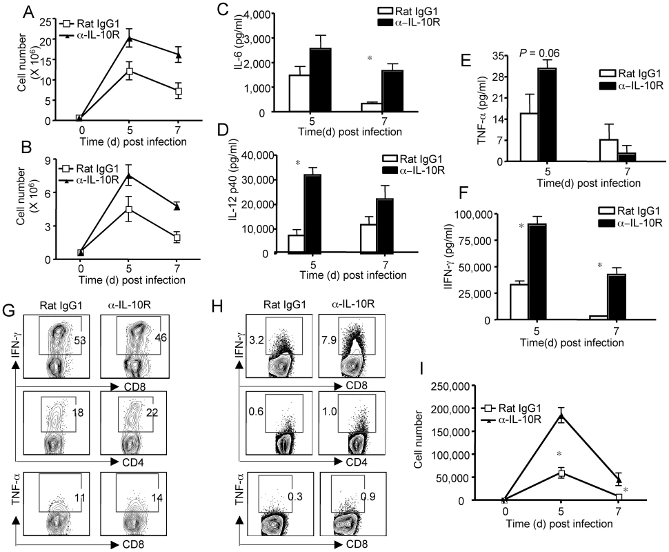
(A–I) BALB/c mice were infected with RSV and treated with either Rat IgG1 control mAb or α-IL-10R blocking mAb. P value was determined by unpaired two-tailed Student t test. * indicates P< = 0.05. (A, B) At the indicated days p.i., lung inflammatory monocytes (A) and neutrophils (B) were determined by flow cytometry. (C–F) At indicated days p.i., airway IL-6 (C), IL-12 p40 (D), TNF-α (E), IFN-γ (F) were determined by ELISA. (G) At d5 p.i., lung cells were collected and restimulated with PMA/Ionomycin. The production of IFN-γ by CD4+ and CD8+ T cells and TNF-α by CD8+ T cells was measured by ICS. Numbers are the percentages of cells in gated populations. (H. I) Mice were injected with monensin to block the in vivo release of cytokine. Lung cells were then collected and the in vivo production of IFN-γ, and TNF-α determined by ICS. (H) The percentages of IFN-γ+ cells in gated lung CD8+ or CD4+ T cells and TNF-α+ cells in lung CD8+ T cells at d5 p.i. are shown. Numbers are the percentages of cells in gated populations. (I) The numbers of IFN-γ+ lung CD8+ T cells at indicated days p.i. are shown. Data are representative of at least three independent experiments. IL-10R blockade in vivo results in enhanced proinflammatory activity of effector T cells
Along with their role in virus clearance [20], effector T cells have been shown to significantly contribute to lung inflammation and host morbidity in the murine model of primary RSV infection [3], [20], [21]. The enhanced morbidity, excess pulmonary inflammation/injury and altered pulmonary function observed with IL-10R blockade during RSV infection may reflect a normal function of IL-10 in regulating the induction, expansion and/or effector activity of effector T cells responding in the lungs to infection. We first investigated the impact of IL-10R blockade in vivo on the induction (in the draining MLN) and migration of effector T cells to the lung. Somewhat unexpectedly, the numbers of activated/effector CD8+ or CD4+ T cells in the infected lungs were comparable in mice treated either with control Rat IgG1 mAb or α-IL-10R mAb (Figure S2). Furthermore, effector T cells from either control or α-IL-10R-treated mice had comparable capability to produce IFN-γ and TNF-α in response to mitogenic or antigenic stimulation (Figure 4G and data not shown), suggesting that IL-10 does not affect the differentiation of T cells into RSV specific effector T-cells of the Th1 or Tc1 lineage.
In view of the elevated levels of lung proinflammatory cytokines, in particular IFN-γ, we next explored the impact of IL-10R blockade on the in vivo frequency and cytokine profile of virus-specific T cells responding in the infected respiratory tract. For this purpose we chose IFN-γ and TNF-α as representative effector cytokines and used the the in vivo ICS assay [22] to measure their release by CD8+ and CD4+ T cells in the respiratory tract. We found that blockade of IL-10 function in vivo triggered an increase in the percentage/number of CD4+ and particularly CD8+ T cells producing IFN-γ in vivo (Figure 4H, I). In addition, the in vivo production of TNF-α by CD8+ T cells was also modestly increased following IL-10R blockade during RSV infection (Figure 4H). The blockade of IL-10 function in vivo also leads to increased production of IFN-γ on a per cell basis (Figure S3). These data demonstrate that this effector T cell-derived IL-10 can act in vivo to suppress the production of proinflammatory mediators by effector T cells responding in the respiratory tract of virus infection.
Autocrine regulation of IL-10 to restrain host inflammation and morbidity
Cells of the myeloid, monocyte/macrophage/dendritic cell lineage are believed to be the major targets of IL-10 [7]. Therefore IL-10 would most likely be expected to diminish the effector activity of effector T cells in vivo by inhibiting the APC function of these inflammatory mononuclear cells infiltrating the infected lungs. Alternatively, the T-cell-derived IL-10 could also act in an autocrine fashion to suppress the activation/stimulation of CD8+ and CD4+ lung effector T cells through the engagement of the IL-10R on these T-cells. To explore this latter possibility, we first isolated CD8+ effector T cells from RSV infected lungs and examined whether they can respond to IL-10. As Figure 5A demonstrates, we found that CD8+ effector T cells isolated from infected lungs are able to respond to IL-10 treatment by phosphorylating STAT-3. Furthermore, IL-10 is able to inhibit the release of IFN-γ by CD8+ effector T cells in response to CD3 stimulation in the absence of antigen presenting cells (Figure 5B). Collectively, these data demonstrated that IL-10 is able to signal to effector T cells and suppress their proinflammatory activity. To directly explore in vivo the possible contribution of an autocrine mechanism of IL-10 action during RSV infection, we examined the response to primary RSV infection of mice with a conditional deletion of the IL-10Rα gene selectively in CD4+ and CD8+ T cells [23]. We confirmed that IL-10Rα was selectively deleted in T cells, particularly CD8+ T cells in the lung but not in lung monocytes/macrophages or NK cells etc (Figure 5C and data not shown). Notably, compared to CD8+ effector T cells, we failed to detect significant IL-10Rα expression in CD4+ effector T cells (Figure 5C). Importantly, following RSV infection, the deletion of IL-10Rα in T cells resulted in dramatically increased release of effector T cells derived cytokine IFN-γ into the airway (Figure 5D). Interestingly, the deletion of IL-10Rα in T cells also resulted in significantly increased infiltration of neutrophils and monocytes (but not T cells) into the infected lungs (Figure 5D, and Figure S4), suggesting a role for enhanced adaptive immune-mediated inflammation in promoting neutrophil and monocyte infiltration to the lung. Consistent with the finding of increased inflammation in the lung, we observed that conditional deletion of the IL-10Rα in T cells resulted in increased weight loss and delayed recovery following RSV infection of the knockout mice (Figure 5E).
Fig. 5. Autocrine regulation of pulmonary inflammation by effector T cell-derived IL-10 during RSV infection. 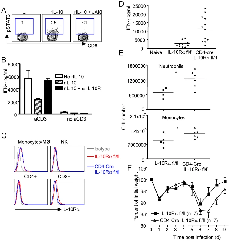
(A, B). BALB/c mice were infected with RSV. At d7 p.i., Lung CD8+ cells were purified and stimulated as indicated in vitro. (A) CD8+ T cells were stimulated with rIL-10 in the absence or presence of JAK inhibitor. STAT3 phophorylation was determined by ICS. (B) CD8+ T cells were left un-stimulated or stimulated with plate-bound a-CD3 overnight in the absence or presence of rIL-10 or rIL-10 plus α-IL-10R. IFN-γ release to the medium was determined by ELISA. (C – F) IL-10Rα fl/fl mice or CD4-Cre+ IL-10Rα fl/fl mice were infected with RSV. (C) The expression of IL-10Rα in lung monocytes, NK cells, CD4+ and CD8+ T cells at d6 post infection was determined by flow cytometry. (D) IFN-γ levels in the BALF were determined by ELISA. P value was determined by unpaired two-tailed Student t test. * indicates P< = 0.05. (E) Lung infiltrated neutrophils and inflammatory monocytes were determined by flow cytometry. P value was determined by unpaired two-tailed Student t test. * indicates P< = 0.05. (F) Mouse weight loss was monitored daily following infection. A, B, C. Data are representative of at least two independent experiments. D, E .Pooled data from four experiments are represented. F. Pooled data from three experiments are represented. Discussion
In this report, we examined the in vivo production, cellular sources and function of the regulatory cytokine IL-10 during acute pulmonary RSV infection. Importantly, the primary source of IL-10 is the anti-viral CD4+ and CD8+ effector T cells recruited to the RSV infected lungs. We demonstrate that this effector T cell-derived IL-10 plays a critical role in preventing excess inflammation during the innate and particularly the adaptive immune response to RSV. In the absence of IL-10 signaling, an enhanced inflammatory response and a concomitant alteration in pulmonary function ensues. Thus, IL-10 may play a critical role in maintaining lung function during infection. Of note, our findings suggest that effector T cells are not only the major source of IL-10 in the infected lungs but may also serve as important cellular targets for the action of this regulatory cytokine, reflecting a novel autocrine pathway for the action of IL-10 during infection.
Several lines of evidence in the current study implicate effector T cells as the primary source of IL-10 observed in the RSV infected respiratory tract in this model. Similar to our recently reported findings in the murine influenza infection model [10], we noted minimal IL-10 secretion in RSV infected Rag1-/- mice and localized IL-10 production by effector T cells in the infected lungs using the IL-10 reporter Vert-X mice for infection. However, we cannot formally exclude a major contribution of the small percentage (2%–5%) of Thy-1 – (negative) cells detected to the amelioration of pathology. In this case, however, we demonstrated that IL-10 derived solely from T-cells are sufficient to control disease development during RSV infection (Figure 3E) which also implicated T-cell-derived IL-10 in the control of excess pulmonary inflammation. The IL-10-producing effector T cells are, as we observed in this report, IL-4 and GATA-3 negative (data not shown) and thus show few features characteristic of type 2 lineage of T cells; but rather are T-bet positive type 1 effectors. Interestingly, we observed a minor fraction of T-bet+ IL-10 producers which co-express Foxp-3, a cell population that has been recently described in other systems [24]. Of note, CD25+ T regulatory cells have been recently demonstrated to modulate RSV specific CD8+ T cell responses and pulmonary inflammation during experimental RSV infection [25], [26], [27]. How these Tregs interact with the IL-10 secreting effector T cells responding in the respiratory tract to RSV infection to control excess inflammation will require further investigation.
It is of interest that RSV infection induced a lower level of IL-10 release into the infected airways relative to the release of effector T cells derived pro-inflammatory effector cytokine IFN-γ than detected in influenza infected lungs. The explanation for this discrepancy is unclear, but this could reflect an as yet unappreciated mechanism by which RSV infection results in an imbalance in the expression of pro-inflammatory (e.g. IFN-γ and regulatory (e.g. IL-10) cytokines by effector T-cells resulting in exaggerated inflammation/injury profile in the infected lungs out of proportion to the degree of RSV replication in the infected lungs.
Multiple lines of evidence both from human studies and murine models implicate myeloid cells of the monocyte/macrophage/dendritic cell lineage as the major targets of IL-10 action in vitro and (in model systems) in vivo [7], [9], [23], [28], [29]. Thus results from the current study suggest that effector T cell-derived IL-10 can act directly on these inflammatory mononuclear cells infiltrating the RSV infected lungs to decrease production of pro-inflammatory cytokines/chemokines by these inflammatory mononuclear cells as well as to modulate the expression of costimulatory ligands (e.g. CD40, 80, 86 etc.) and T-cell stimulatory cytokines (e.g. IL-12 etc.) [28]. The latter effects of IL-10 would be expected to diminish the efficiency of effector T cell triggering in response to contact with these APC populations and thus down regulating the effector activity of T cells. While our results are consistent with such a mechanism, our findings on the impact of conditional deletion of the IL-10Rα selectively in T-cells suggests the novel possibility that the effector T cell-derived IL-10 may also act in an autocrine fashion to regulate effector T cell activity directly. Of note, using animals deficient in IL-10Rβ chain, several recent reports have provided evidence that IL-10 may act directly on and suppress the function of T cells including Treg cells [30] and CD4+ T cells directly in vivo during acute LCMV infection [31] as well as inhibit robust memory CD8+ T cell development [32]. It should be noted, however, that IL-10Rβ is the common subunit for receptors recognizing several other cytokines including IL-22, IL-26 and IL-28 etc [33]. Our results using the conditional deletion of the IL-10 receptor a chain, which is unique to IL-10, firmly establish that effector T cells are able to respond to IL-10 directly in vivo during infection. Furthermore, we described a novel autocirne function of IL-10; that effector T cell-derived IL-10 is able to signal back to effector T cells, especially CD8+ effector T cells, to restrict the proinflammatory cytokine production by these effector T-cells. Importantly, this autocrine regulatory function of IL-10 acting on effector T cells modulates pulmonary inflammation and thereby results in diminished host morbidity. A similar autocrine mechanism has been reported recently for regulation of IL-10 producing macrophages during endotoxin challenge [23], [29]. The mechanisms through which IL-10R engagement on activated effector T cells acts to suppress effector T cell functions at sites of infection are currently under investigation.
IL-10 has been detected both in respiratory tract secretions and in the serum of infants and young children during the acute phase of RSV infection [34]. Although controversial [14], [35], several lines of evidence suggest that the level of IL-10 secretion may correlate inversely with disease severity [12], [13]. This may be particularly evident among children hospitalized for RSV infection where those children with symptoms of severe RSV bronchiolitis requiring mechanical ventilation express lower levels of IL-10 than hospitalized children with less severe disease [13]. Similarly, a recent study reported lower IL-10 levels in stimulated cord blood of children who were hospitalized for RSV infection before 6 months of age than in cord blood of infected infants who were treated as outpatients [36]. Furthermore, homozygosity for certain IL-10 alleles correlates with a higher risk of severe RSV bronchiolitis [12]. Such studies point to the importance of IL-10 in controlling the severity of acute infection with RSV.
The sources of IL-10 during acute primary RSV infection have not been clearly defined. While various cell types have been implicated as the source of this IL-10 [34], [37], [38], [39], [40], our results point to effector T cells as a potential major source of IL-10 produced during the acute phase of RSV infection. In this connection, we did observe a small increase of IL-10 released into the airways (BAL fluid) prior to the dramatic increase in IL-10 production observed in the respiratory tract at the time of effector T cell infiltration at day 5 post RSV infection. Thus, while one or more additional cell types may contribute to the pool of IL-10 observed during acute RSV infection, our results suggest that effector T-cells (both CD4+ and CD8+) may be a major source of IL-10 during human infection. Therefore the contribution of IL-10 from effector T cells should be rigorously evaluated in future studies of human RSV infection. Our analysis also suggests that the RSV specific effector T cells in the infected lungs serve as an important target of IL-10 action. Thus, the development and extent of immune mediated pathology in RSV infection may not only be linked to the level of IL-10 production by effector T cells (and potentially other cell types in the infected lungs) but also dependent upon the effectiveness of IL-10 signaling through the IL-10R on effector T cells leading to modulation of effector T cell function.
In summary, we have discovered a critical role of effector T cell-derived IL-10 in controlling the pulmonary inflammation and function during RSV infection. Furthermore, we established a previously unrecognized autocrine function of IL-10 in controlling proinflammatory activity of anti-viral effector cells. Our findings thus provide a cellular and mechanistic link to earlier clinical studies which implicate IL-10 in the pathogenesis of RSV disease and may provide the groundwork for future studies examining IL-10 as a therapeutic option in the treatment of RSV induced bronchiolitis in young infants.
Materials and Methods
Mice and infection
BALB/c mice were purchased from Taconic Farms and Rag1-/ - mice were purchased from The Jackson Laboratories. IL-10-eGFP reporter mice (Vert-X) were obtained from C.L. Karp from Children Hospital of Cincinnati. IL-10-/- mice were obtained from T. A. Wynn from NIAID, NIH. The T cell conditional IL-10Rα knock-out mice were generated through crossing IL-10Rα fl/fl mice to CD4-cre transgenic mice [23]. All mice were housed in a specific pathogen-free environment and all animal experiments were performed in accordance with protocols approved by the University of Virginia Animal Care and Use Committee. The A2 strain of RSV (obtained from P. L. Collins from NIAID, NIH) was grown in HEp-2 cells (ATCC) and titered for infectivity. We infected 10–12 week old BALB/c mice with a dose of ∼1–1.2×10⁷ pfu RSV in serum-free Iscove's medium (Invitrogen) intranasally after anesthesia with methyl ether (Matix Scientific). For the conditional IL-10Rα mice infection (B6 background), we infected 9–12 week transgenic mice with a dose of ∼2×10⁷ pfu RSV in serum-free Iscove's medium after anesthesia with ketamine and xylazine.
Ethics statement
This study was carried out in strict accordance with the Animal Welfare Act (Public Law 91-579) and the recommendations in the Guide for the Care and Use of Laboratory Animals of the National Institutes of Health (OLAW/NIH, 2002). The protocol was approved by the University of Virginia Animal Care and Use Committee (ACUC, Protocol Number: 2230).
T cell transfer and infection
We purified WT or IL-10-/ - Thy1 positive T cells (including both CD4+ and CD8+ cells) from spleen and lymph nodes by MACS separation. 15 million T cells were then transferred into Rag1-/ - mice i.v. 1 week after transfer, the recipient mice were infected with RSV.
Bronchoalveolar lavage fluid (BALF) for virus and cytokine determination
We obtained BALF by flushing the airway multiple times with a single use of 500 µl sterile PBS through a cannula attached to a syringe. Cells were recovered from the suspension by centrifugation at 4°C. Supernatants were collected and stored at –80°C until use for the cytokine and virus determinations. Cytokines were measured by ELISA (BD Biosciences) according to the manufacturer manuals. The viral titer in the BALF was determined through the plaque assay in serially diluted Hep-2 cell cultures.
Quantitative reverse-transcription PCR
To measure RSV-L gene expression, we isolated RNA from the infected lungs via Trizol (Invitrogen) and treated it with DNase I (Invitrogen). We used random primers (Invitrogen) and Superscript II (Invitrogen) to synthesize first-strand complementary DNAs from equivalent amounts of RNA from each sample. We performed real-time RT-PCR in a 7000 Real-Time PCR System (Applied Biosystems) with SYBR Green PCR Master Mix (Applied Biosystems). The sequence of RSV-L gene primers was previously reported [41]. Data were generated by the comparative threshold cycle (ΔCT) method by normalizing to GAPDH. For experiments to measure host genes in effector T cells from infected Vert-X mice. We isolated CD44hi IL-10/eGFP+ or CD44hi IL-10/eGFP- CD8+ T cells and CD44hi IL-10/eGFP+ or CD44hi IL-10/eGFP- CD4+ T cells by FACS-sorting. We then isolated RNA, synthesized cDNA and performed real-time RT-PCR as described above. The sequences of the primers are available upon request.
Intracellular cytokine staining (ICS)
Lung single cell suspensions were generated as described [42]. Lung cells were subsequently re-stimulated with either PMA (100 ng/ml) and ionomycin (1 µg/ml) in the presence of Golgi-Stop (1 µl/ml) for 5–6 h. Then cells were fixed and permeablized using the Cyto-Fix and Perm-Wash system (BD Biosciences). Cell surface CD4 and CD8 and intracellular IL-10 and IFN-γ were stained accordingly. Measurement of IL-10 and IFN-γ producing cells in vivo was based on a previously described protocol with modifications [22]. Briefly, at indicated days post RSV infection mice were injected i.v. with 500 µl of a PBS solution containing 500 µg Monensin (Sigma-Aldrich) 6 h before harvesting. Lung single cell suspensions were prepared in the presence of monensin. Cells were then fixed and permeablized and intracellular IL-10 and IFN-γ staining was as described [43].
Interleukin-10 receptor–specific monoclonal antibody administration in vivo
α-IL-10R blocking mAb (clone 1B1.3A) and isotype control Rat IgG1 mAb were obtained from Schering-plough Biopharma and Bio-express. We achieved IL-10 signaling blockade in vivo by injecting α-IL-10R blocking mAb on day 1 (0.75 mg intraperitoneally in 500 µl), day 3 (0.15 mg intranasally in 40 µl) and day 4 (0.75 mg intraperitoneally in 500 µl).
Measurement of pulmonary function
The functional properties of the lung i.e. the airway and vascular mechanics are characterized by different lung function parameters, including the pulmonary compliance, the airway resistance and pulmonary artery pressure. The airway resistance is an index for the resistive forces against the airflow in the airways and depends on the diameter and length of the airways. The airway resistance can be calculated from the relation between transpulmonary pressure and airflow velocity. The airway resistance increases as a consequence of narrowing of the airways due to bronchoconstriction or obstructive processes, e.g. bronchial edema or enhanced mucus deposition. The pulmonary compliance is a marker for the functional stiffness of the lung and can be calculated from the relation between tidal volume and transpulmonary pressure. The pulmonary compliance decreases during restrictive pathological processes e.g. atelectasis, fibrosis, pulmonary edema or disturbed surfactant secretion. The increase in pulmonary artery pressure is an index of vasoconstriction of lung and represents an underlying pathophysiology resulting in pulmonary edema or changes in pulmonary vascular resistance. We measured the lung function using a buffer-perfused mouse lung system (Hugo Sachs Elektronik) as previously described [44]. Briefly, at day 7 post RSV infection, mice were anesthetized with ketamine and xylazine and ventilated with room air at 100 strokes/min with a tidal volume of 7 ml/g body weight with a positive end expiratory pressure of 2 cm H2O using the MINIVENT mouse ventilator (Hugo Sachs Elektronik). The pulmonary artery was cannulated via the right ventricle, and the left ventricle was immediately tube-vented through a small incision at the apex of the heart. The lungs were then perfused at a constant flow of 60 µl·g body wt–1·min–1 with Krebs-Henseleit buffer containing 2% albumin, 0.1% glucose, and 0.3% HEPES. The perfusate buffer and isolated lungs were maintained at 37°C throughout the experiment. Isolated lungs were allowed to equilibrate on the apparatus during a 5-min stabilization period. After equilibration, data were recorded for an additional 10 minutes. Hemodynamic and pulmonary parameters were continuously recorded during this period by the PULMODYN data acquisition system (Hugo Sachs Elektronik).
STAT3 staining and inhibition of cytokine release of effector T cells
Effector T cells were purified from RSV infected lung at d7 p.i. and resuspended in complete media. Then T cells were stimulated with 20 ng/ml rIL-10 (eBioscience) in the presence or absence of JAK inhibitor (JAK inhibitor I, EMD Biosciences) and 15 min later the phosphorylation of STAT3 was determined through ICS following previousely described protocols [10]. To measure the inhibition of IFN-γ release by IL-10, we stimulated purified T cells (5×10⁵ cells/ml) with plate-bound α-CD3 (100 µl of 0.1 µg/ml α-CD3 for 4 h at 37°C) overnight. Then the supernatant of the culture were collected and IFN-γ concentration were determined through ELISA.
FACS analysis
All FACS antibodies are purchased from BD Biosciences or eBioscience. The dilution of surface staining antibodies was 1 in 200 and the dilution of intracellular staining antibodies was 1 in 100. After antibody staining, we examined cells using a six-color FACS-Canto system (BD Biosciences) and the data were analyzed by FlowJo software (Treestar). We characterized the various cell types according to their phenotypes as follows: neutrophils (Ly6G+CD11b+Ly6c–), monocytic cell lineage (Ly6G–CD11b+Ly6c+), natural killer cells (NK1.1+CD3−), CD8+ T lymphocytes (Thy1+CD8+) and CD4+ T lymphocytes (Thy1+CD4+).
Statistical analyses
Data are means ± SEM. We used two-tailed Student's t test for statistical analyses. We considered all P values >0.05 not to be significant.
Supporting Information
Zdroje
1. GlezenWPTaberLHFrankALKaselJA 1986 Risk of primary infection and reinfection with respiratory syncytial virus. Am J Dis Child 140 543 546
2. ShayDKHolmanRCNewmanRDLiuLLStoutJW 1999 Bronchiolitis-associated hospitalizations among US children, 1980-1996. JAMA 282 1440 1446
3. PeeblesRSJrGrahamBS 2005 Pathogenesis of respiratory syncytial virus infection in the murine model. Proc Am Thorac Soc 2 110 115
4. CollinsPLGrahamBS 2008 Viral and host factors in human respiratory syncytial virus pathogenesis. J Virol 82 2040 2055
5. OshanskyCMZhangWMooreETrippRA 2009 The host response and molecular pathogenesis associated with respiratory syncytial virus infection. Future Microbiol 4 279 297
6. MalleyRDeVincenzoJRamiloODennehyPHMeissnerHC 1998 Reduction of respiratory syncytial virus (RSV) in tracheal aspirates in intubated infants by use of humanized monoclonal antibody to RSV F protein. J Infect Dis 178 1555 1561
7. MooreKWde Waal MalefytRCoffmanRLO'GarraA 2001 Interleukin-10 and the interleukin-10 receptor. Annu Rev Immunol 19 683 765
8. CouperKNBlountDGRileyEM 2008 IL-10: the master regulator of immunity to infection. J Immunol 180 5771 5777
9. LiMOFlavellRA 2008 Contextual regulation of inflammation: a duet by transforming growth factor-beta and interleukin-10. Immunity 28 468 476
10. SunJMadanRKarpCLBracialeTJ 2009 Effector T cells control lung inflammation during acute influenza virus infection by producing IL-10. Nat Med. 15 277 284
11. GentileDADoyleWJZeeviAHowe-AdamsJKapadiaS 2003 Cytokine gene polymorphisms moderate illness severity in infants with respiratory syncytial virus infection. Hum Immunol 64 338 344
12. HoebeeBBontLRietveldEvan OostenMHodemaekersHM 2004 Influence of promoter variants of interleukin-10, interleukin-9, and tumor necrosis factor-alpha genes on respiratory syncytial virus bronchiolitis. J Infect Dis 189 239 247
13. WilsonJRowlandsKRockettKMooreCLockhartE 2005 Genetic variation at the IL10 gene locus is associated with severity of respiratory syncytial virus bronchiolitis. J Infect Dis 191 1705 1709
14. MiyairiIDeVincenzoJP 2008 Human genetic factors and respiratory syncytial virus disease severity. Clin Microbiol Rev 21 686 703
15. ChangJBracialeTJ 2002 Respiratory syncytial virus infection suppresses lung CD8+ T-cell effector activity and peripheral CD8+ T-cell memory in the respiratory tract. Nat Med 8 54 60
16. MadanRDemircikFSurianarayananSAllenJLDivanovicS 2009 Nonredundant roles for B cell-derived IL-10 in immune counter-regulation. J Immunol 183 2312 2320
17. HuffordMMKimTSSunJBracialeTJ 2011 Antiviral CD8+ T cell effector activities in situ are regulated by target cell type. J Exp Med 208 167 180
18. SzaboSJKimSTCostaGLZhangXFathmanCG 2000 A novel transcription factor, T-bet, directs Th1 lineage commitment. Cell 100 655 669
19. FontenotJDRudenskyAY 2005 A well adapted regulatory contrivance: regulatory T cell development and the forkhead family transcription factor Foxp3. Nat Immunol 6 331 337
20. CannonMJOpenshawPJAskonasBA 1988 Cytotoxic T cells clear virus but augment lung pathology in mice infected with respiratory syncytial virus. J Exp Med 168 1163 1168
21. GrahamBSBuntonLAWrightPFKarzonDT 1991 Role of T lymphocyte subsets in the pathogenesis of primary infection and rechallenge with respiratory syncytial virus in mice. J Clin Invest 88 1026 1033
22. LiuFWhittonJL 2005 Cutting edge: re-evaluating the in vivo cytokine responses of CD8+ T cells during primary and secondary viral infections. J Immunol 174 5936 5940
23. PilsMCPisanoFFasnachtNHeinrichJMGroebeL 2010 Monocytes/macrophages and/or neutrophils are the target of IL-10 in the LPS endotoxemia model. Eur J Immunol 40 443 448
24. KochMATucker-HeardGPerdueNRKillebrewJRUrdahlKB 2009 The transcription factor T-bet controls regulatory T cell homeostasis and function during type 1 inflammation. Nat Immunol 10 595 602
25. RuckwardtTJBonaparteKLNasonMCGrahamBS 2009 Regulatory T cells promote early influx of CD8+ T cells in the lungs of respiratory syncytial virus-infected mice and diminish immunodominance disparities. J Virol 83 3019 3028
26. FultonRBMeyerholzDKVargaSM 2010 Foxp3+ CD4 Regulatory T Cells Limit Pulmonary Immunopathology by Modulating the CD8 T Cell Response during Respiratory Syncytial Virus Infection. J Immunol 185 2382 2392
27. LeeDCHarkerJATregoningJSAtabaniSFJohanssonC 2010 CD25+ natural regulatory T cells are critical in limiting innate and adaptive immunity and resolving disease following respiratory syncytial virus infection. J Virol 84 8790 8798
28. DonnellyRPDickensheetsHFinbloomDS 1999 The interleukin-10 signal transduction pathway and regulation of gene expression in mononuclear phagocytes. J Interferon Cytokine Res 19 563 573
29. SieweLBollati-FogolinMWickenhauserCKriegTMullerW 2006 Interleukin-10 derived from macrophages and/or neutrophils regulates the inflammatory response to LPS but not the response to CpG DNA. Eur J Immunol 36 3248 3255
30. MuraiMTurovskayaOKimGMadanRKarpCL 2009 Interleukin 10 acts on regulatory T cells to maintain expression of the transcription factor Foxp3 and suppressive function in mice with colitis. Nat Immunol 10 1178 1184
31. BrooksDGWalshKBElsaesserHOldstoneMB 2010 IL-10 directly suppresses CD4 but not CD8 T cell effector and memory responses following acute viral infection. Proc Natl Acad Sci U S A 107 3018 3023
32. BiswasPSPedicordVPlossAMenetELeinerI 2007 Pathogen-specific CD8 T cell responses are directly inhibited by IL-10. J Immunol 179 4520 4528
33. ComminsSSteinkeJWBorishL 2008 The extended IL-10 superfamily: IL-10, IL-19, IL-20, IL-22, IL-24, IL-26, IL-28, and IL-29. J Allergy Clin Immunol 121 1108 1111
34. LeggJPHussainIRWarnerJAJohnstonSLWarnerJO 2003 Type 1 and type 2 cytokine imbalance in acute respiratory syncytial virus bronchiolitis. Am J Respir Crit Care Med 168 633 639
35. BennettBLGarofaloRPCronSGHosakoteYMAtmarRL 2007 Immunopathogenesis of respiratory syncytial virus bronchiolitis. J Infect Dis 195 1532 1540
36. JunttiHOsterlundPKokkonenJDunderTRenkoM 2009 Cytokine responses in cord blood predict the severity of later respiratory syncytial virus infection. J Allergy Clin Immunol 124 52 58 e51-52
37. HussellTSpenderLCGeorgiouAO'GarraAOpenshawPJ 1996 Th1 and Th2 cytokine induction in pulmonary T cells during infection with respiratory syncytial virus. J Gen Virol 77 Pt 10 2447 2455
38. BartzHBuning-PfaueFTurkelOSchauerU 2002 Respiratory syncytial virus induces prostaglandin E2, IL-10 and IL-11 generation in antigen presenting cells. Clin Exp Immunol 129 438 445
39. JarttiTPaul-AnttilaMLehtinenPParikkaVVuorinenT 2009 Systemic T-helper and T-regulatory cell type cytokine responses in rhinovirus vs. respiratory syncytial virus induced early wheezing: an observational study. Respir Res 10 85
40. ShireyKAPletnevaLMPucheACKeeganADPrinceGA 2010 Control of RSV-induced lung injury by alternatively activated macrophages is IL-4R alpha-, TLR4-, and IFN-beta-dependent. Mucosal Immunol 3 291 300
41. TregoningJSYamaguchiYHarkerJWangBOpenshawPJ 2008 The role of T cells in the enhancement of respiratory syncytial virus infection severity during adult reinfection of neonatally sensitized mice. J Virol 82 4115 4124
42. LawrenceCWBracialeTJ 2004 Activation, differentiation, and migration of naive virus-specific CD8+ T cells during pulmonary influenza virus infection. J Immunol 173 1209 1218
43. SunJPearceEJ 2007 Suppression of early IL-4 production underlies the failure of CD4 T cells activated by TLR-stimulated dendritic cells to differentiate into Th2 cells. J Immunol 178 1635 1644
44. YangZSharmaAKLindenJKronILLaubachVE 2009 CD4+ T lymphocytes mediate acute pulmonary ischemia-reperfusion injury. J Thorac Cardiovasc Surg 137 695 702 discussion 702
Štítky
Hygiena a epidemiologie Infekční lékařství Laboratoř
Článek Crystal Structure of Reovirus Attachment Protein σ1 in Complex with Sialylated OligosaccharidesČlánek A Protein Thermometer Controls Temperature-Dependent Transcription of Flagellar Motility Genes inČlánek Modulation of NKp30- and NKp46-Mediated Natural Killer Cell Responses by Poxviral Hemagglutinin
Článek vyšel v časopisePLOS Pathogens
Nejčtenější tento týden
2011 Číslo 8- Jak souvisí postcovidový syndrom s poškozením mozku?
- Měli bychom postcovidový syndrom léčit antidepresivy?
- Farmakovigilanční studie perorálních antivirotik indikovaných v léčbě COVID-19
- 10 bodů k očkování proti COVID-19: stanovisko České společnosti alergologie a klinické imunologie ČLS JEP
-
Všechny články tohoto čísla
- Phenotypic Screens, Chemical Genomics, and Antimalarial Lead Discovery
- Characterisation of Regulatory T Cells in Nasal Associated Lymphoid Tissue in Children: Relationships with Pneumococcal Colonization
- Crystal Structure of Reovirus Attachment Protein σ1 in Complex with Sialylated Oligosaccharides
- Absence of Cross-Presenting Cells in the Salivary Gland and Viral Immune Evasion Confine Cytomegalovirus Immune Control to Effector CD4 T Cells
- Transcriptomic Analysis of Host Immune and Cell Death Responses Associated with the Influenza A Virus PB1-F2 Protein
- A Quorum Sensing Regulated Small Volatile Molecule Reduces Acute Virulence and Promotes Chronic Infection Phenotypes
- Autocrine Regulation of Pulmonary Inflammation by Effector T-Cell Derived IL-10 during Infection with Respiratory Syncytial Virus
- A Protein Thermometer Controls Temperature-Dependent Transcription of Flagellar Motility Genes in
- Association of Human TLR1 and TLR6 Deficiency with Altered Immune Responses to BCG Vaccination in South African Infants
- Histo-Blood Group Antigens Act as Attachment Factors of Rabbit Hemorrhagic Disease Virus Infection in a Virus Strain-Dependent Manner
- MrkH, a Novel c-di-GMP-Dependent Transcriptional Activator, Controls Biofilm Formation by Regulating Type 3 Fimbriae Expression
- Beta-HPV 5 and 8 E6 Promote p300 Degradation by Blocking AKT/p300 Association
- Modulation of NKp30- and NKp46-Mediated Natural Killer Cell Responses by Poxviral Hemagglutinin
- Transportin 3 Promotes a Nuclear Maturation Step Required for Efficient HIV-1 Integration
- Coordination of KSHV Latent and Lytic Gene Control by CTCF-Cohesin Mediated Chromosome Conformation
- A Novel Persistence Associated EBV miRNA Expression Profile Is Disrupted in Neoplasia
- The Plant Pathogen pv. Is Genetically Monomorphic and under Strong Selection to Evade Tomato Immunity
- IL-10 Blocks the Development of Resistance to Re-Infection with
- Anti-Apoptotic Machinery Protects the Necrotrophic Fungus from Host-Induced Apoptotic-Like Cell Death during Plant Infection
- Crystal Structure of PrgI-SipD: Insight into a Secretion Competent State of the Type Three Secretion System Needle Tip and its Interaction with Host Ligands
- Evades Immune Recognition of Flagellin in Both Mammals and Plants
- Tumor Cell Marker PVRL4 (Nectin 4) Is an Epithelial Cell Receptor for Measles Virus
- Provides Insights into the Evolution of the Salmonellae
- B Cell Repertoire Analysis Identifies New Antigenic Domains on Glycoprotein B of Human Cytomegalovirus which Are Target of Neutralizing Antibodies
- Thy1 Nk Cells from Vaccinia Virus-Primed Mice Confer Protection against Vaccinia Virus Challenge in the Absence of Adaptive Lymphocytes
- The Cytokine Network of Acute HIV Infection: A Promising Target for Vaccines and Therapy to Reduce Viral Set-Point?
- Dendritic Cell Status Modulates the Outcome of HIV-Related B Cell Disease Progression
- Differential Contribution of PB1-F2 to the Virulence of Highly Pathogenic H5N1 Influenza A Virus in Mammalian and Avian Species
- A Communal Bacterial Adhesin Anchors Biofilm and Bystander Cells to Surfaces
- Two Group A Streptococcal Peptide Pheromones Act through Opposing Rgg Regulators to Control Biofilm Development
- Activation of HIV Transcription by the Viral Tat Protein Requires a Demethylation Step Mediated by Lysine-specific Demethylase 1 (LSD1/KDM1)
- Unique Evolution of the UPR Pathway with a Novel bZIP Transcription Factor, Hxl1, for Controlling Pathogenicity of
- Disruption of PML Nuclear Bodies Is Mediated by ORF61 SUMO-Interacting Motifs and Required for Varicella-Zoster Virus Pathogenesis in Skin
- Flagellar Motility Is Not Directly Required to Maintain Attachment to Surfaces
- Viral Infection Induces Expression of Novel Phased MicroRNAs from Conserved Cellular MicroRNA Precursors
- Functional Cure of SIVagm Infection in Rhesus Macaques Results in Complete Recovery of CD4 T Cells and Is Reverted by CD8 Cell Depletion
- Recruitment of the Major Vault Protein by InlK: A Strategy to Avoid Autophagy
- The Steroid Catabolic Pathway of the Intracellular Pathogen Is Important for Pathogenesis and a Target for Vaccine Development
- PLOS Pathogens
- Archiv čísel
- Aktuální číslo
- Informace o časopisu
Nejčtenější v tomto čísle- Tumor Cell Marker PVRL4 (Nectin 4) Is an Epithelial Cell Receptor for Measles Virus
- Two Group A Streptococcal Peptide Pheromones Act through Opposing Rgg Regulators to Control Biofilm Development
- Differential Contribution of PB1-F2 to the Virulence of Highly Pathogenic H5N1 Influenza A Virus in Mammalian and Avian Species
- Recruitment of the Major Vault Protein by InlK: A Strategy to Avoid Autophagy
Kurzy
Zvyšte si kvalifikaci online z pohodlí domova
Současné možnosti léčby obezity
nový kurzAutoři: MUDr. Martin Hrubý
Všechny kurzyPřihlášení#ADS_BOTTOM_SCRIPTS#Zapomenuté hesloZadejte e-mailovou adresu, se kterou jste vytvářel(a) účet, budou Vám na ni zaslány informace k nastavení nového hesla.
- Vzdělávání


