-
Články
Top novinky
Reklama- Vzdělávání
- Časopisy
Top články
Nové číslo
- Témata
Top novinky
Reklama- Videa
- Podcasty
Nové podcasty
Reklama- Kariéra
Doporučené pozice
Reklama- Praxe
Top novinky
ReklamaA LysM and SH3-Domain Containing Region of the p60 Protein Stimulates Accessory Cells to Promote Activation of Host NK Cells
Listeria monocytogenes (Lm) infection induces rapid and robust activation of host natural killer (NK) cells. Here we define a region of the abundantly secreted Lm endopeptidase, p60, that potently but indirectly stimulates NK cell activation in vitro and in vivo. Lm expression of p60 resulted in increased IFNγ production by naïve NK cells co-cultured with treated dendritic cells (DCs). Moreover, recombinant p60 protein stimulated activation of naive NK cells when co-cultured with TLR or cytokine primed DCs in the absence of Lm. Intact p60 protein weakly digested bacterial peptidoglycan (PGN), but neither muropeptide recognition by RIP2 nor the catalytic activity of p60 was required for NK cell activation. Rather, the immune stimulating activity mapped to an N-terminal region of p60, termed L1S. Treatment of DCs with a recombinant L1S polypeptide stimulated them to activate naïve NK cells in a cell culture model. Further, L1S treatment activated NK cells in vivo and increased host resistance to infection with Francisella tularensis live vaccine strain (LVS). These studies demonstrate an immune stimulating function for a bacterial LysM domain-containing polypeptide and suggest that recombinant versions of L1S or other p60 derivatives can be used to promote NK cell activation in therapeutic contexts.
Published in the journal: . PLoS Pathog 7(11): e32767. doi:10.1371/journal.ppat.1002368
Category: Research Article
doi: https://doi.org/10.1371/journal.ppat.1002368Summary
Listeria monocytogenes (Lm) infection induces rapid and robust activation of host natural killer (NK) cells. Here we define a region of the abundantly secreted Lm endopeptidase, p60, that potently but indirectly stimulates NK cell activation in vitro and in vivo. Lm expression of p60 resulted in increased IFNγ production by naïve NK cells co-cultured with treated dendritic cells (DCs). Moreover, recombinant p60 protein stimulated activation of naive NK cells when co-cultured with TLR or cytokine primed DCs in the absence of Lm. Intact p60 protein weakly digested bacterial peptidoglycan (PGN), but neither muropeptide recognition by RIP2 nor the catalytic activity of p60 was required for NK cell activation. Rather, the immune stimulating activity mapped to an N-terminal region of p60, termed L1S. Treatment of DCs with a recombinant L1S polypeptide stimulated them to activate naïve NK cells in a cell culture model. Further, L1S treatment activated NK cells in vivo and increased host resistance to infection with Francisella tularensis live vaccine strain (LVS). These studies demonstrate an immune stimulating function for a bacterial LysM domain-containing polypeptide and suggest that recombinant versions of L1S or other p60 derivatives can be used to promote NK cell activation in therapeutic contexts.
Introduction
NK cells are lymphocytes that help control infections and tumors and regulate autoimmune responses through both cytotoxic and cytokine-secreting effector functions. Through the use of Fas, TRAIL, and secretion of perforin and granzyme B, NK cells induce apoptosis or lysis of distressed somatic cells. NK cells can also activate or suppress other aspects of an immune response through lysis of antigen-presenting cells or production of immune regulatory cytokines such as IFNγ and IL-10 [1], [2], [3].
NK cells are activated by secreted cytokines and by proteins on the surface of target cells or other immune cells. NK cells recognize “self” class I major histocompatibility complex (MHCI) cell surface molecules by inhibitory receptors that prevent NK cell activation [4]. Reduced expression of MHCI by stressed or infected target cells can thus relieve this inhibition and lead to NK cell activation. Alternatively, NK cells can be directly activated by recognition of stress-induced ligands through a variety of activating NK cell receptors [5]. In addition to direct recognition of target cells, the activity of NK cells is regulated by cytokines released from macrophages and dendritic cells, such as TNFα, IFNγ, IL-15, IL-12, and IL-18 [6], [7]. IL-18 is a potent stimulus for IFNγ production by (NK) cells [8], [9] Full NK cell activation often requires direct contact of the NK cell with accessory cells, such as dendritic cells (DCs). In some cases, contact of NK cells and DCs permits signaling of co-stimulatory molecules [6], [10], [11], [12]. Contact may also promote efficient transmission of IL-18 and/or IL-12 [13], [14], and trans-presentation of IL-15 to “prime” the NK cell [15], [16]. The balance of both inhibitory and activating signals ultimately determines the extent of NK cell activation and possibly the nature of NK cell effector functions.
Systemic infection by numerous bacterial pathogens elicits potent NK cell activation and IFNγ production, but the mechanisms of NK cell activation during bacterial infections are incompletely understood. Infection by Listeria monocytogenes (Lm) rapidly activates a large population of NK cells to produce IFNγ [17], [18]. Lm is a facultative intracellular pathogen of humans and animals [19]. A number of secreted Lm virulence factors that contribute to pathogenicity. One of the two most abundantly secreted Lm proteins is a bacterial hemolysin (Hly) called listeriolysin O. Hly is essential for bacterial access to the cytosol of host cells and thus for intracellular bacterial growth and virulence during systemic infection of mice [19], [20]. The second most heavily secreted Lm protein is called p60. Expression of p60 also contributes to Lm virulence during systemic infections [17], [21], [22]. However, the virulence-promoting function of p60 has been enigmatic. The p60 sequence contains a C-terminal NLPC/p60 domain, two N-terminal LysM domains, and a single N-terminal SH3-like domain. Some NLPC/p60 domains have been associated with endopeptidase activity [23], [24], while LysM and bacterial SH3 domains generally bind glycans or proteins [25], [26], [27], [28]. Consistent with autolytic endopeptidase activity, semi-purified p60 protein digested Micrococcus luteus cells [29], [30], and crude Lm PGN [17]. We previously hypothesized that Lm expression of p60 might thus contribute to Lm pathogenicity by altering the production of immune modulating muropeptides [21]. Subsequently, an immune modulatory function was associated with Lm expression of p60. Namely, systemic infections by wt Lm promoted significantly increased NK cell activation when compared to infections by p60-deficient (Δp60) Lm [17].
Here, we confirm that p60 deficiency correlates with impaired NK cell activation in a recently developed cell culture assay system. Furthermore, using recombinant p60 protein and p60-derived polypeptides, we show that p60 protein can indirectly enhance NK cell activation in the absence of additional Lm factors. Purified p60 protein binds to DCs and induces IL-18 secretion, which is required for NK cell activation by p60 in co-culture. The ability of p60 to stimulate DCs for NK cell activation mapped to the first LysM and SH3 domains (L1S) of the p60 protein. The L1S region was also sufficient to promote activation of NK cells in vivo when given to naïve mice. In vivo treatment with p60 increased serum IFNγ and reduced susceptibility of recipient mice to infection by the heterologous NK cell-sensitive bacterial pathogen, Francisella tularensis. These data demonstrate that p60 protein boosts NK cell activation during Lm infection through appropriate stimulation of accessory cells and suggest that L1S may be useful to therapeutically manipulate immune responses.
Results
Lm expression of p60 enhances IFNγ production in cell cultures containing NK cells and infected DCs
Systemic infections with Δp60 Lm strains elicit weak IFNγ production by NK cells [17]. Likewise, bone marrow dendritic cells (BMDCs) infected with Δp60 Lm elicited significantly less IFNγ from co-cultured naïve splenic lymphocytes (Figure 1A). Intracellular staining revealed that NK1.1+ cells were responsible for nearly all IFNγ production in these cultures (Figure 1B-E). Multiple independently generated p60 deletion mutants showed a similarly poor ability to induce IFNγ production in these co-cultures (Figure 1F). This weak IFNγ production was restored to wt levels when expression of p60 was restored in the Lm Δp60 mutant using an integrated vector coding for His tagged p60 protein (Figure 1G). The complemented Δp60+p60 strain secreted p60 at levels similar to wild-type Lm based on immunoblotting of culture supernatants (not shown). Reduced NK cell activation in response to Δp60 Lm infection might conceivably reflect reduced bacterial burdens within the infected BMDCs. However, microscopy and cfu plating revealed that the growth rate was identical for wt and Δp60 Lm, as was the percent of infected BMDCs over the course of infection (not shown). Finally, the ratio of cytosolic (actin-associated) versus phagosome localized Lm was also similar for the two strains (not shown). Thus, expression of p60 was not required for the invasion or cytosolic replication of Lm in BMDCs, but nonetheless increased the activation of neighboring NK cells.
Fig. 1. A secreted bacterial protein enhances activation of naïve mouse NK cells by L. monocytogenes-infected DCs. 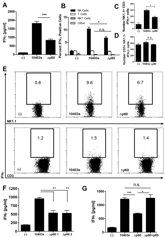
(A) BMDCs were infected in triplicate with wildtype Lm (10403s) or a mutant Δp60 Lm strain. NK-enriched NWNA splenocytes were added 2 h post-infection, and supernatants were harvested 21 h post-infection. The average ± SEM concentrations of IFNγ produced are plotted. (B-E) BMDC were infected with 10403s or Δp60 Lm, and NWNA cells were added 2 h post-infection. At 10 h post-infection, the NWNA cells were stained for CD3, NK1.1, and intracellular IFNγ. The average percentage (B) and Number (C,D) of IFNγ positive cells in gated NK1.1+CD3- (NK), NK1.1-CD3+ (T), CD3+NK1.1+ (NKT), or CD3-NK1.1- (other) cells are graphed ± SEM. (E) Representative dot plots are shown. (F) BMDC were infected in triplicate with wt Lm (10403s) or one of two independent Δp60 deletion mutants, and co-culture IFNγ was measured as in (A). (G) BMDCs were infected with wt or Δp60 Lm, or a Δp60 Lm strain complemented with His-tagged p60. Average ± SEM concentrations of IFNγ produced are shown. Data are representative of at least three (A-E,G) or two (F) experiments. Purified p60 protein stimulates IFNγ production by NK cells in culture with primed DCs
We expressed and purified recombinant His-tagged p60 protein from E. coli using nickel affinity and cation exchange columns. When added to co-cultures of BMDCs and nylon wool non-adherent cells (NWNA) prepared from naïve mouse spleens, the purified protein induced IFNγ production (Figure 2A). The recombinant p60 protein was associated with ∼1 ng of E. coli LPS per 1 µg of protein. However, this amount of LPS was insufficient to stimulate IFNγ production when added to the co-cultures without p60 protein (Figure 2A). Moreover, production of IFNγ was not seen in response to treatments with BSA or a His-tagged phage autolysin (HPL511) that was purified from E. coli using a similar procedure and also contained ∼1 ng LPS per µg protein (Figure 2A). To further exclude possible artifacts due to LPS, polymyxin B columns were used to remove LPS from the purified p60 protein. The detoxified p60 was initially insufficient to activate IFNγ production (Figure 2B), suggesting that activation by p60 required priming or maturation of the BMDCs. To test this, BMDCs were pretreated with TLR agonists for three hours before addition of p60. Pre-stimulation of co-cultures with LPS, the non-toxic LPS analog monophosphoryl LipidA (MPA), or poly I∶C (PIC) each sufficed to elicit IFNγ production following p60 stimulation (Figure 2B). None of the priming agents tested stimulated IFNγ production on their own.
Fig. 2. Primed DCs activate naïve NK cells when treated with purified p60 protein. 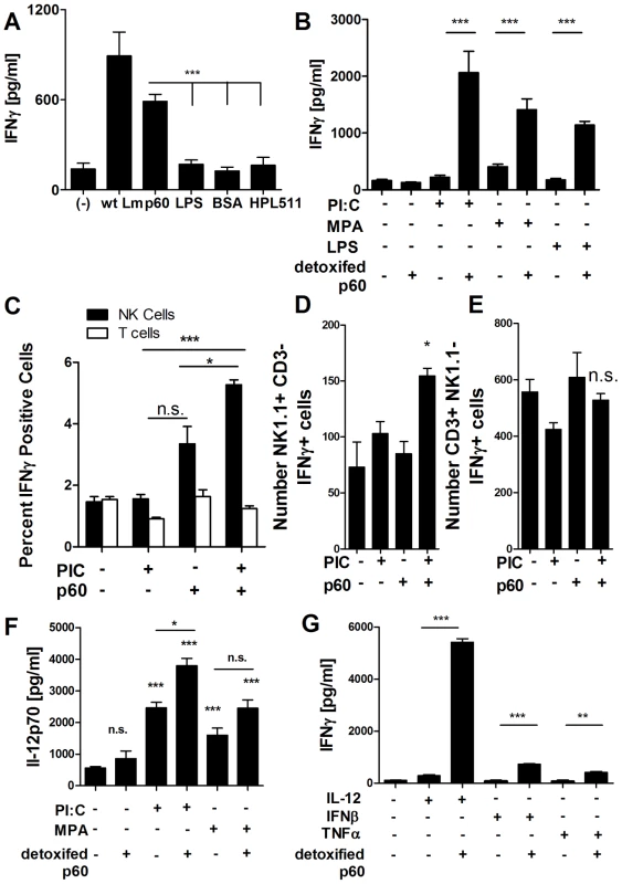
(A) NWNA splenocytes were co-cultured in triplicate with BMDC infected with wt Lm or treated with 10 µg of recombinant His-tagged p60 purified from E.coli, 10 ng LPS, 10 µg BSA, or 10 µg of a His-tagged control protein, the phage autolysin HPL511. Average ± SEM concentrations of IFNγ produced are shown. (B) Purified p60 was detoxified of LPS using a polymyxin B column. BMDC in triplicate were primed for 3 h with 20 µg/ml poly I∶C, 10 ng/ml LPS, or 10 ng/ml MPA, and then treated with 10 µg of detoxified protein. NWNA splenocytes were co-cultured 2 h post-infection, and IFNγ was measured by ELISA 21 h post-infection/treatment. Average ± SEM concentrations of IFNγ produced are shown. (C-E) BMDC were pretreated 3 h with 20 µg/ml PIC, then stimulated with 10 µg detoxified p60. Two hours after p60 treatment, NWNA cells were added to the BMDC. Eight hours later, the NWNA cells were collected and stained for CD3, NK1.1, and IFNγ. NK cells are NK1.1 positive, CD3 negative, while T cells are CD positive, NK1.1 negative. The average percent (C) and number (D,E) of positive IFNγ cells are shown ± SEM. (F) Supernatants from BMDC and NWNA co-culture as in (B) were analyzed for IL-12 secretion by ELISA. Average ± SEM concentrations of IL-12 produced are shown. (G) BMDC were treated with 2 ng IL-12, 100 units IFNβ, or 2 ng TNFα with or without 10 µg detoxified p60. NWNA cells were added 2 h post-treatment, and IFNγ levels were measured by ELISA 21 h post-treatment. Average ± SEM concentrations of IFNγ produced are shown. (A-G) Data are representative of at least three experiments. Based on flow cytometry using intracellular IFNγ staining, NK cells were the major source of IFNγ produced in the co-cultures with primed and p60-stimulated BMDCs (Figure 2C-E). To test whether these NK cells responded directly to the stimulated BMDC, NWNA splenocytes were stained and flow sorted to obtain 97–98% pure populations of NK1.1+CD3 - NK cells, CD3+NK1.1 - T cells, and “other” cells (negative for both NK1.1 and CD3). Each sorted population was added to BMDCs (>90% CD11c+) that had previously been treated with LPS and a p60-derived peptide (peptide described further below). As previously shown for Lm-infected co-cultures [8], the purified NK cells produce IFNγ when cultured alone with stimulated BMDCs (Figure S1). The amount of IFNγ was not significantly affected by adding back either or both other cell populations present in NWNA splenocyte preparations (Figure S1). Although T cells did not impact IFNγ production by the NK cells, we observed small amounts of IFNγ production when purified splenic T cells were cultured alone with the stimulated BMDCs (Figure S1). This likely reflects the ability of memory CD8+ T cells to respond to IL-12 and IL-18 in the cultures [31] (see below for further discussion of cytokines present in the cultures). We conclude that the LPS and p60-stimulated BMDCs were sufficient to activate NK cells in these in co-cultures, and that the other cells present in the NWNA population did not significantly modulate this activation.
Stimulation of BMDC with LPS and other TLR stimuli elicits production of cytokines that stimulate DC and NK cells. Detoxified p60, failed to stimulate IFNγ production by the co-cultures in the absence of priming agents and also failed to induce significant levels of IL-12p70 secretion by BMDC. However, the priming agents PIC and MPA both elicited strong IL-12 production in the co-cultures containing NK cells and BMDC (Figure 2F). In some cases, but not universally, this IL-12p70 secretion was further enhanced by p60 stimulation. Recombinant IL-12p70, IFNβ, and TNFα each sufficed to prime the production IFNγ by detoxified p60 protein in the absence of TLR agonists (Figure 2G). IL-12 was by far the most potent priming agent, most likely due both to BMDC priming and the enhancement of IFNγ transcription in NK cells [32]. These findings suggested that cytokines produced in response to TLR agonists mediate priming or maturation of the BMDCs, which can then respond to recombinant p60 protein or mediate activation of naïve NK cells in NWNA splenocytes in response to this protein.
Stimulation of IFNγ production from NWNA splenocytes by purified p60 protein requires co-culture with BMDCs and correlates with binding of the p60 protein to BMDCs
We next asked how p60 might mediate NK cell activation in co-culture by examining the role of accessory DCs. Addition of p60 protein did not stimulate IFNγ production in the absence of NK cells or when added to NWNA cells in the absence of BMDCs ([17] and Figure 3A). This result suggested two possibilities. Either p60 protein might act on DCs to induce the ability of DCs to activate NK cells, or the protein might be presented to NK cells by DCs for NK cell activation. To investigate whether p60 protein bound to BMDCs, the cells were treated or not with p60 protein, fluorescent beads, or p60 plus beads. After washing, the treated and untreated BMDCs were stained using anti-p60 rabbit polyclonal antisera and a secondary Cy3-labeled anti-rabbit Fab (Figure 3B-E). A punctuate staining pattern was seen on the stained p60-treated BMDCs (Figure 3C and E). Identical results were obtained using two independent anti-p60 polyclonal antibodies (data not shown). This punctuate staining was not observed on stained untreated cells or cells treated with beads alone (Figure 3B and D), nor on sorted NK cells, T cells, or other NWNA splenocytes (not shown). The punctuate staining for p60 did not require detergent permeation of the BMDC membrane (not shown), nor did p60 puncta co-localize with phagocytosed FITC-labeled latex beads (Figure 3E). These data suggest that p60 protein binds to an unknown receptor/s present at or near the surface of BMDCs.
Fig. 3. Purified p60 binds to BMDCs to active NK cells. 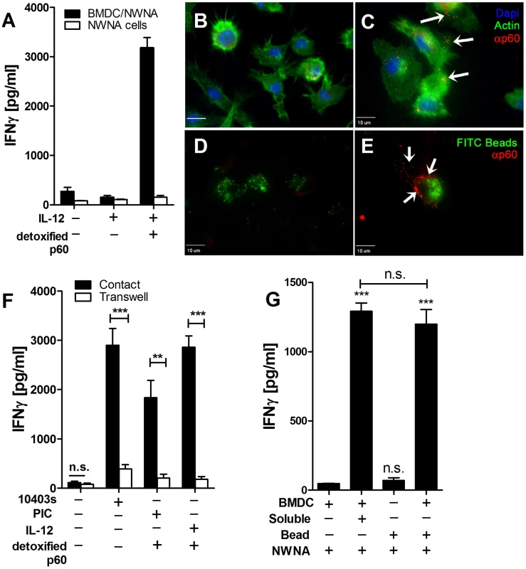
(A) NWNA splenocytes were treated in triplicate with 2 ng IL-12 and 10 µg detoxified p60, in the presence or absence of BMDC. IFNγ in the supernatant was measured by ELISA 21 hours post-treatment. Average ± SEM concentrations of IFNγ produced are shown. (B-E) Scale bars represent 10 µm. BMDCs were untreated (B) or stimulated with 30 µg/ml p60 (C) for 4 h at 37°C and stained with polyclonal p60 antibody (red). Actin stained with Alexa-488 is shown in green, and nuclei (DAPI) are shown in blue. (D,E) BMDCs were treated with FITC-latex beads (green) alone (D) or with 30 µg/ml p60 protein (E) for 4 h at 37°C and stained with polyclonal p60 antibody (red). Arrows indicate p60 puncta. (F) BMDC were infected with WT Lm or primed with 20 µg/ml poly I∶C or 2 ng/ml IL-12 and treated with 10 µg detoxified p60. NWNA cells were added to the co-culture either in contact with the BMDC or separated by a 0.4 µm transwell support. Average ± SEM concentrations of IFNγ produced are shown. (G) NWNA splenocytes were cultured with BMDC alone or treated with 10 ng soluble LPS + 10 µg soluble p60, or with Ni beads and an equal amount of p60 and LPS (Beads). IFNγ in the supernatant was measured by ELISA 21 hours post-treatment. Data shown are representative of two (G) or at least three experiments (A-F). We previously reported that contact between DCs and NK cells was required for NK cell activation during Lm-infection [8]. Similarly, contact between the DC and NWNA splenoctyes was required for p60-induced NK cell activation (Figure 3F). It was conceivable that binding of p60 to the DC surface might permit presentation of this protein to NK cells. However, nickel beads coated with a His-tagged p60 were not able to stimulate NWNA cells in the absence of BMDC (Figure 3G). Together, these data suggested that p60 primarily stimulates NK cell activation indirectly, due to its effect on DCs.
NK cells might respond to altered MHC I expression and/or upregulation of stress ligands by BMDCs treated with p60 protein [5], [33]. Thus, we stained BMDCs that had been primed with LPS plus or minus an active p60-derived peptide (described further below) and assessed their expression of activation markers (MHCII) and several known ligands for NK cell surface receptors (Figure S2). MHCII expression increased after protein treatment, consistent activation of the BMDC. No down regulation of MHC I was observed and the expression of NKG2D ligands RAE1γ, RAE1δ, and MULT1 were unchanged. There was no change in staining levels for the SLAM family members 1, 2, 3, and 6. SLAMF 5 staining was slightly reduced after protein treatment, which is likely due to DC activation. These data suggested that NK cell activation by p60 was due to effects of p60 on DCs that were independent of altering expression of these known ligands for NK cell activating and inhibitory receptors.
Treatment with p60 causes BMDCs to secrete IL-18, which is required for IFNγ production by co-cultures containing NK cells
Both cell contact and inflammatory cytokines such as IL-12 and IL-18 modulate NK cell activation and IFNγ production [34]. IL-12 production by BMDC infected with wildtype versus Δp60 Lm was not significantly different (data not shown). Since IL-18 production is essential for NK cell activation by Lm infected BMDCs [8], we asked whether bacterial expression of p60 effected IL-18 production in infected BMDCs. We found that secretion of IL-18 was significantly reduced in the supernatants of C57BL/6 BMDCs infected with Δp60 Lm (Figure 4A). Consistent with this observation, detoxified p60 protein in combination with PIC strongly simulated IL-18 secretion from BMDCs (Figure 4B). We next evaluated the effects of IL-18 production on IFNγ production in cultures of infected BMDC and NWNA splenocytes. In response to Lm infection, IL-18-/- BMDCs stimulated very little IFNγ production (Figure 4C). Moreover, the amount of residual IFNγ produced in these co-cultures was no longer affected by bacterial expression of p60. Further, IL-18 expression in BMDCs was additionally required to elicit IFNγ production in co-cultures primed with PIC or MPA and stimulated with detoxified p60 protein (Figure 4D). Together, these data suggest that binding of p60 to BMDC elicits IL-18 secretion, which is required for activation of NWNA splenocytes.
Fig. 4. IL-18 produced by BMDCs is required for p60-elicited IFNγ from NK cells in co-culture. 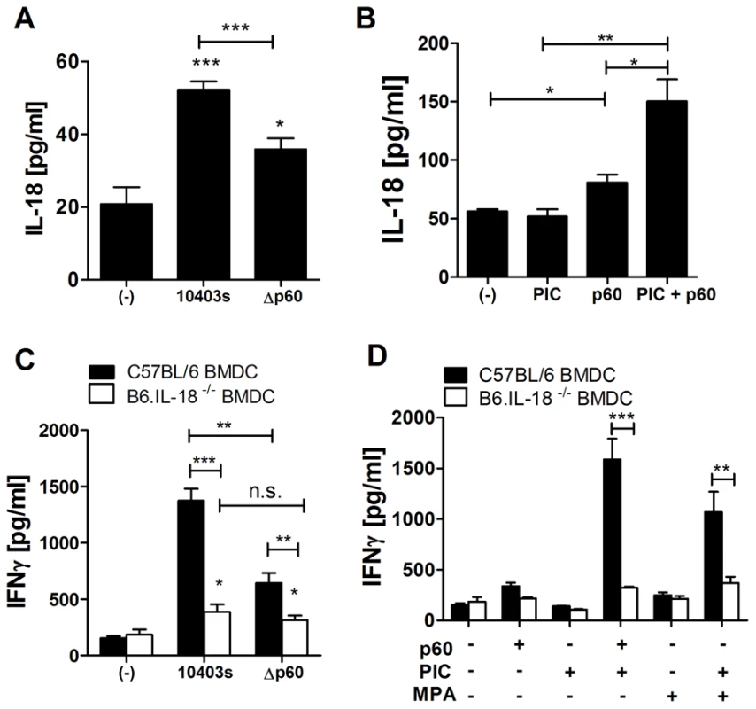
All treatments were performed in triplicate; average cytokine concentrations ± SEM are shown. (A) BMDCs were infected with 10403s or Δp60 Lm, and supernatant levels of IL-18 were assessed by ELISA 21 hours post-infection. (B) BMDC were treated with 20 µg/ml PIC and 10 µg detoxified p60 alone or in combination, and supernatant levels of IL-18 were assessed by ELISA 21 hours post-infection. (C,D) BMDC from C57B6 or IL-18-/- mice were infected with 10403s or Δp60 Lm (C) or treated with 10 ng/ml MPA or 20 µg/ml PIC alone or in combination with 10 µg detoxified p60 (D). Two hours post treatment, C57B6 NK-enriched NWNA splenocytes were co-cultured with the BMDCs. IFNγ levels were measured by ELISA 21 hours post-infection. Data shown are representative of at least three experiments. The enzymatic activity of p60 is not required for its ability to stimulate IFNγ production in co-cultures of BMDCs and NWNA cells
The p60 protein has been shown to weakly digest peptidoglycan (PGN) [21], [29], hence, we previously hypothesized that PGN cleavage by p60 might release muramyl di-peptide (MDP) or other bioactive muropeptides [21], [29]. MDP is detected by NOD2, which signals through the RIP2 kinase [35], [36], [37], [38]. To test whether MDP generation by p60 might stimulate NK cell activation, we compared the ability of Lm infected B6 and B6.RIP2-/- BMDC to activate NK cells from B6 mice. Bacterial expression of p60 enhanced IFNγ production in NWNA splenocytes co-cultured with RIP2-/- BMDCs to the same extent as C57B6 BMDCs (Figure S3A). Additionally, purified recombinant p60 stimulated BMDC and NK cell enriched splenocytes co-cultures in the absence of added Listeria PGN. Therefore, generation and detection of the MDP PGN fragment was not required for NK cell activation nor for the ability of p60 to enhance such activation.
Like the Bacillius subtilis LytF protein, p60 contains a C-terminal NLPC/p60 domain with a putative catalytic triad of two histidines and a single cysteine residue (Figure 5A). In LytF, the cysteine is essential for endopeptidase activity and permits cleavage of the cross-linking peptide chains in peptidoglycan (PGN) [24]. However, NLPC/p60 domains have also been associated with other catalytic functions. To formally test whether the enzymatic activity of p60 was required for stimulation of NK cell activation, we engineered and purified a p60 derivative in which the catalytic cysteine residue was mutated to alanine. The resulting p60C389A mutant protein was purified as for wt p60 and tested for digestion of heat-killed Lm and crude Lm PGN substrates using zymography (Figure S3B and not shown). As previously published [21], [29], the wt p60 protein cleaved PGN, although this activity was much weaker than that seen with a control phage lysin (HPL511). The p60C389A was completely inactive in this assay (Figure S3B), confirming that the cysteine residue was required for PGN digestion by p60. Nonetheless, the purified p60C389A was as efficient as the catalytically active wt protein for stimulating IFNγ production in co-cultures of NWNA splenocytes and BMDCs (Figure S3C, Figure 5B). These data demonstrate that the enzymatic activity of p60 is not required for its ability to promote NK cell activation.
Fig. 5. The p60 L1S fragment is sufficient to activate DC/NK cell co-cultures. 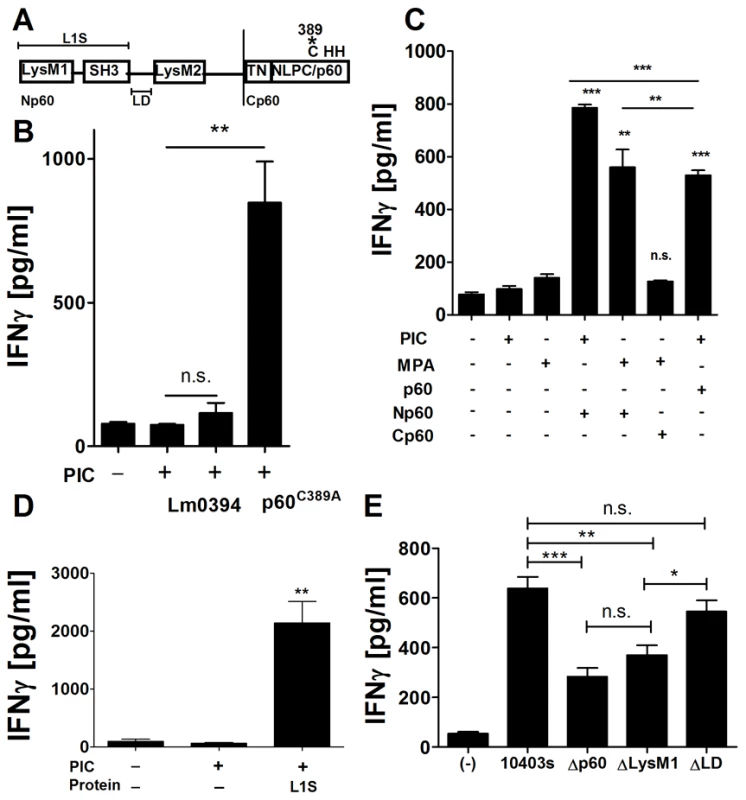
(A) p60 domain map. The p60 protein consists of two LysM domains on either side of an SH3 domain in the N terminal portion. The C-terminus consists of an NLPC/p60 domain preceded by a TN repeat region. C389, H439 and H465 are the predicted catalytic triad. Recombinant versions of (B) p60C389A, the Lm p60 homolog 0394, and (C) the p60 fragments Np60, Cp60, and (D) LysM1-SH3 (L1S) were purified and detoxified of LPS using a polymyxin B column. BMDC were primed for 3 h with 20 µg/ml poly I∶C or (C,D) 10 ng/ml MPA (C) then treated with 10 µg of detoxified protein. NWNA splenocytes were co-cultured 2 h post-treatment, and IFNγ was measured by ELISA 21 h post-treatment. (E) BMDC were infected with wt 10403s, Δp60, ΔLysM1, or ΔLD Lm. NWNA splenocytes were added at 2 hr post-treatment. IFNγ levels were assessed 21 hr post-treatment by ELISA. Data are representative of at least three (B-D) or two (E) experiments. All treatments were performed in triplicate. The N-terminal LysM-SH3 region of p60 is sufficient to stimulate IFNγ production by NWNA cells
Given that enzymatic activity was dispensable for NWNA splenocyte activation by p60, we asked whether this activation was associated with NLPC/p60 or other domains. The Lm genome contains a homolog of p60 (Lm0394) with both an SH3 domain and a C-terminal NLPC/p60 domain but lacking the N-terminal LysM domains found in p60. A His-tagged recombinant Lm0394 protein was unable to activate NWNA splenocytes in co-culture (Figure 5B). Thus, the presence of SH3 and NLPC/p60 domains was not sufficient to confer the ability to activate co-cultures. Additional p60 derivatives were engineered and purified, including an N-terminal fragment (Np60) truncated immediately before the TN repeat region and a C-terminal fragment (Cp60) that comprised the TN repeats and NLPC/p60 domain (Figure 5A). These truncated proteins were purified, detoxified, and tested as for full length p60. Np60 induced IFNγ production in co-cultures pre-stimulated with either PIC or MPA, while Cp60 failed to induce IFNγ (Figure 5C). Further truncation of the N-terminal region mapped the stimulating activity to a fragment containing the LysM1 and SH3 domains, termed L1S (Figure 5D). The results of our experiments with SH3-domain-containing Lm0394 indicate that the LysM1 domain may be responsible for the activity of L1S. However, efforts to purify the LysM1 or SH3 domains alone have thus far been unsuccessful, suggesting that both domains may be required for conformation and stability. Given that the L1S polypeptide was the minimal active component of p60 identified in our studies, we tested whether the LysM1 domain was necessary for p60-induced co-culture activation during Lm infection. We compared Δp60 mutants complemented with p60 constructs that lacked the LysM1 domain or the linker domain (LD) between the SH3 and LysM2 domains (Figure 5A). Both complemented strains expressed and secreted the p60 mutant proteins at levels comparable to wildtype Lm based on immunoblotting of precipitated culture supernatants (not shown). The ΔLysM1 complementation mutant induced low IFNγ levels in co-culture similar to Δp60 Lm infection, while the ΔLD complementation mutant induced IFNγ similar to wild type Lm infection (Figure 5E). Thus, the LysM1 domain appears to be largely responsible for p60-mediated activation of BMDC/NWNA splenocyte co-cultures.
L1S activates NK cells in vivo
The regulation of NK cell activation and responses in vivo may differ from their regulation in our cell culture system. We thus asked whether purified, LPS-associated L1S was sufficient to activate NK cells in vivo when administered to mice by intraperitoneal (i.p.) injection. LPS was administered to a second group of mice as a negative control. At 24 h after injecting the L1S or LPS, IFNγ production by both splenic and peritoneal infiltrating NK cells was assessed using intracellular cytokine staining. The data showed that LPS treatment failed to stimulate NK cell activation in the absence of L1S polypeptide. However, there were significant increases in the percentage of NK1.1+CD3- cells staining positive for IFNγ in both peritoneum (Figure 6A, 6C) and spleen (Figure 6B). The activation of splenic NK cells was more modest than seen in the peritoneum, suggesting the NK cell activation largely occurred locally at the site of L1S injection (Figure 6B). The NK cell activation by LPS-associated L1S was dose-dependent (Figure S4). We additionally observed that the percent granzyme B positive NK1.1+CD3 - NK cells was increased in the peritoneal cells in response to L1S treatment (Figure 6D). Hence, we measured cytotoxicity from NWNA splenocytes after co-culture with BMDCs stimulated with LPS with or without L1S. Consistent with the increased granzyme B staining in vivo, L1S significantly enhanced the cytolytic activity of NWNA splenocytes against NK cell-sensitive B16F10 melanoma target cells in vitro (Figure 6E). These data confirmed that the p60-derived polypeptide was bioactive in the treated animals and suggested that L1S might be useful for therapeutic stimulation of both cytokine and cytoxicity-based immune responses.
Fig. 6. L1S activates NK cells in vivo to secrete IFNγ and increase cytotoxicity. 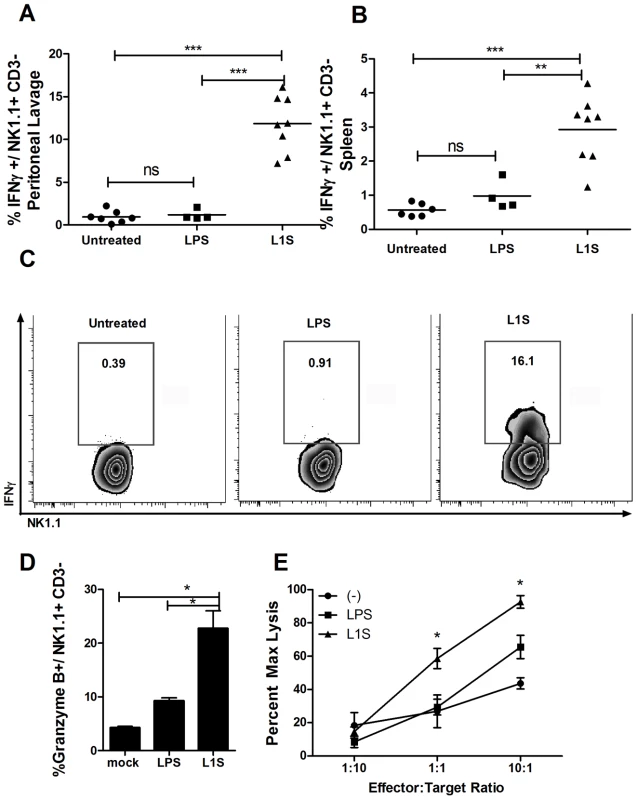
Mice were injected i.p. with 500 µg L1S or 500 ng LPS in 250 µl PBS. After 24 hours (A) peritoneal cells harvested by lavage and (B) splenocytes were stained for CD3, NK1.1, and intracellular IFNγ. Shown are graphical representations of the NK1.1+, CD3- cells that stained positive for IFNγ. Symbols represent individual mice. (C) FACS plots showing the IFNγ positive gate used for (A and B). Gated NK cells from peritoneal lavage are depicted. Data are pooled from two independent experiments; n = two to four treated mice per experimental group. (D) The peritoneal cells from (A) were stained for granzyme B. The average percent granzyme B-positive NK1.1+/CD3- cells are shown. (E) NK-enriched splenocytes were co-cultured with BMDC that were treated with LPS with or without L1S for 21 hours. The NWNA splenocytes were added to B16F10 melanoma target cells at the effector∶target ratios indicated, based on estimated 5% NK cells in the splenocytes. Cytotoxicity was assessed after 4 hours incubation. Conditions were assessed in triplicate, and results are representative of two experiments. In vivo administration of L1S confers protection against Francisella infection
Secretion of IFNγ by NK cells is thought to promote clearance of the bacterial pathogen Francisella tularensis [39], [40], [41]. However, this cytosolic intracellular bacterial pathogen normally suppresses innate immune responses [41], [42], [43]. We thus hypothesized that boosting of NK cell activation during F. tularensis infection might reduce host susceptibility to this pathogen. To test this hypothesis, we administered purified, LPS-associated L1S or PBS alone by a single i.p. injection 24 hours prior to an i.p. infection with the attenuated live vaccine strain of Francisella tularensis holarctica LVS (Ft). Bacterial burdens in the infected spleens (Figure 7A) and livers (Figure 7B) were assessed 96 hours post Ft infection. Colony-forming units (CFU) recovered from spleens and livers of the L1S treated mice were significantly reduced when compared to the control mice. Consistent with the increase in IFNγ+ NK1.1+CD3 - cells seen after in vivo L1S stimulation (Figure 6A, 6B), we observed a significant increase in serum IFNγ levels in the mice treated with L1S prior to Ft infection (Figure 7C). To control for the potential effects of LPS associated with purified L1S, we pre-treated mice with LPS or LPS-associated L1S 24 hours prior to Ft LVS infection as above. The CFUs recovered 4 days post-infection were significantly lower in mice pre-treated with LPS-associated L1S compared to LPS alone (Figure 7D). Serum levels of IFNγ were also significantly higher in the L1S versus LPS pre-treated mice (not shown), which correlates with the observed minimal effect of LPS on IFNγ levels in NK cells in vivo (Figure 6A, 6B). These findings suggest that p60 and its derivatives enhance NK cell activation in a biologically relevant manner and may be useful for further development as a therapeutic for immune stimulation.
Fig. 7. In vivo administration of L1S is protective for Francisella infection. 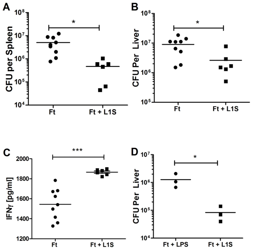
(A-C) Mice were pretreated with 300 µl PBS control or 500 µg purified L1S injected i.p. After 24 hours, the mice were infected i.p with 104 live F. tularensis LVS. CFUs were counted from the spleens (A) and livers (B) of the infected mice 4 days post-infection. (C) IFNγ from serum was measured by ELISA. Data are pooled from two independent experiments; n = three to six mice per experimental group. (D) Mice were pretreated with 500 ng LPS with or without 500 µg L1S in 300 µl PBS injected i.p. 24 hours before infection with 104 live F. tularensis LVS, administered i.p. CFUs from liver were assessed 4 days post-infection. Discussion
Bacterial pathogens have developed numerous strategies to interfere with or subvert host immune responses [44], [45]. Our findings here demonstrated an indirect role for the abundantly secreted L. monocytogenes (Lm) p60 protein in modulation of NK cell activity. We showed that Lm secretion of the p60 protein during infection of cultured BMDCs stimulated enhanced activation of naïve NK cells in cell co-culture assays. Moreover, endotoxin-free purified p60 protein was sufficient to stimulate IFNγ production from NK cells in co-cultures containing BMDCs primed with TLR agonists or inflammatory cytokines such as IL-12. Purified p60 protein bound to the BMDCs and in the presence of priming stimuli this binding correlated with BMDC secretion of the NK cell activating cytokine IL-18. These findings support the model that p60 indirectly activates NK cells by stimulating a DC surface receptor in a manner that induces secretion of IL-18. Consistent with this model, IL-18 production by the BMDCs was essential for eliciting IFNγ production by NK cells and cultures of NWNA splenocytes. The known endopeptidase enzymatic activity of p60 was not required for this biological response and stimulation of NK cell activation by p60 or its derivatives was independent of bacterial PGN and muropeptide detection systems dependent on the RIP2 kinase. Rather, the ability to stimulate DC-dependent NK cell responses mapped to an N-terminal fragment of p60 that contains a LysM domain and a bacterial SH3 domain. A polypeptide containing just these domains (L1S) was sufficient to stimulate DC-dependent NK cell activation both in cell culture assays and when administered to mice in the absence of Lm infection. These results thus revealed a novel role for a bacterial LysM domain-containing protein in the modulation of mammalian innate immune responses.
Our studies here demonstrated that extracellular delivery of p60 protein or the L1S polypeptide in cell culture acted in concert with DCs to stimulate NK cell activation. However, the p60 protein was not sufficient to activate NK cells in the absence of primed BMDCs. In addition, soluble L1S polypeptide triggered NK cell activation when injected into mice without any known mechanism for uptake into the cytosol of host cells. These data suggest that p60 and/or L1S act extracellularly to increase the ability of DCs to promote NK cell activation. Consistent with this interpretation, we observed that p60 protein bound to the surface of BMDCs but not NK cells. Staining of p60 on BMDCs that were fed latex beads suggested that aggregates of p60 are not simply phagocytosed. Furthermore, delivery of p60 into the cytosol of cultured BMDCs using transfection protocols did not improve NK cell activation (not shown). These data suggest that p60 protein acts extracellularly to promote NK cell activation. The fact that infected individuals develop antibodies against p60 further suggests this protein may be abundant extracellularly during Lm infection [31]. Potential sources of extracellular p60 include production by extracellular bacteria, which are known to be present at early and later times of infection [46], or release of protein upon lysis of infected cells. However, we cannot exclude the possibility that p60 present in the cytosol after phagosomal escape of Lm also contributes to NK cell activation. Indeed, p60 protein is abundant in the cytosol of Lm infected macrophages and stimulates protective cytotoxic T cell (CTL) responses [47], [48]. Since cytokines and TLR agonists are also present during Lm infections, soluble extracellular p60 protein that interacts with DCs or other infected cells during in vivo Lm infection is likely an important stimulus for NK cell activation during in vivo Lm infection. However, our data here (Figure 1) and in a prior publication [17] clearly indicate that there are also p60-independent mechanisms for NK cell activation.
The activation of naïve NK cells by DCs infected with live Lm bacteria was previously shown by us and others to require both direct contact between DCs and NK cells and the production of IL-12 and IL-18 [8], [18]. Lm bacteria obviously contain TLR agonists that can induce IL-12 production to prime NK cell activation during in vivo infection. However, it has not been clear whether specific bacterial factors stimulate IL-18 production and/or cell contact between naïve NK cells and DCs. Our data here implicate the L1S region of p60 as a bacterial factor that promotes IL-18 production by DCs. Specifically, we showed that priming of BMDCs with TLR agonists stimulated IL-12p70 production by these cells and that IL-12p70 could substitute for TLR agonists. In some experiments, we also observed a modest p60-induced enhancement of IL-12 secretion from BMDCs that were already primed with TLR agonists, which is consistent with the ability of IL-18 to positively regulate IL-12 production. However, neither TLR agonists nor IL-12 were sufficient to stimulate NK activation in the absence of p60 protein and the IL-18 production elicited by p60. Moreover, despite the presence of IL-12 and IL-18, stimulation of BMDCs with TLR agonists and p60 was insufficient to stimulate NK cell activation when there was not direct cell-cell contact between the BMDCs and the NK cells. The p60 treatment appeared to induce a more activated phenotype in BMDCs but it did not alter the expression by BMDCs of several known ligands for NK cell activating and inhibitory receptors. Thus, there exist at least three possible explanations for the contact requirement: (1) The p60 stimulation triggers both IL-18 secretion and expression of an activating ligand by the BMDCs. This ligand is not one we have tested and may be novel. (2) Contact merely serves to increase the local concentration of IL-18 (and perhaps IL-12) above some threshold that normally prevents activation of the naïve NK cells. This may be facilitated by immunological synapses formed between the DC and NK cells, as previously suggested [13], [14]. (3) BMDCs constitutively express (or are induced to express e.g. by p60 or IL-12) a surface associated “co-stimulatory” factor that is required to “prime” the NK cells for responsiveness to IL-18. Ongoing and future studies focused on identification of putative ligands or co-stimulatory factors may resolve which, if any, of these possible explanations is correct.
NK cells are the major source of IFNγ production early after viral and bacterial infections. IFNγ normally plays a protective role in immunity to Lm and other pathogens, including F. tularensis (Ft). IFNγ induces CD4 Th1 differentiation, stimulates cytotoxic CD8 cells, and activates macrophages to become more bactericidal [49], [50]. During Ft infection, IFNγ-positive NK cells are quickly recruited to sites of infection, where they promote granuloma formation and limit bacterial spread [39], [41]. We found that injection of L1S polypeptide into mice was sufficient to activate NK cells to produce IFNγ, particularly at the site of injection. We also found increased serum levels of IFNγ persisting through infection in mice pre-treated with L1S polypeptide. Presumably, the IFNγ produced by these NK cells created a non-permissive environment for Ft expansion. Thus, when Ft was inoculated at the same site as the L1S polypeptide, its growth was significantly reduced compared to inoculations in the absence of L1S. It will be important to determine whether L1S polypeptide injection might also protect against other routes of Ft infection and against other pathogens.
In contrast to Ft infection, the results of in vivo depletion studies suggest that NK cells are associated with increased susceptibility of mice to Lm [17], [51], [52], [53], [54] and the expression of p60 by Lm increased host susceptibility to systemic Lm infection [17], [21]. Thus, production by Lm of a protein that promotes NK cell activation correlates with the fact that NK cell activation increases susceptibility to Lm. It was also previously reported that IFNγ production by NK cells fails to protect mice against systemic Lm infections [55]. This may be due to suppression of macrophage responsiveness to IFNγ during early stages of Lm infection [56]. Thus, Lm produces a protein that enhances NK cell activation and also has been shown to be more pathogenic in the presence of NK cells. It will thus be of interest in future studies to understand the mechanisms by which activated NK cells promote Lm pathogenicity.
In contrast to Lm, Ft normally suppresses host inflammatory responses during the initial stages of infection [42], [43]. The Ft genome contains several LysM-containing proteins, but using BLAST searches we failed to identify any Ft proteins whose LysM-domains showed more than 20% identify to the LysM1 region of p60. Thus, it is possible that the LysM proteins present in Ft have evolved to lack residues critical for binding to DCs or activation of IL-18 secretion by DCs. Consistent with this model, we found that no IFNγ was produced by NWNA splenocytes cultured with Ft-infected BMDCs (data not shown). However, this issue will need to be further investigated, since it is also possible that Ft LysM proteins are not secreted and thus accessible to bind DCs in the same manner as the Lm p60 L1S region.
NK cells are attractive targets for therapy in cancer and infectious diseases as they can directly kill target cells. NK cells also regulate immune and autoimmune B cell and T cell responses through production of IFNγ or inhibitory cytokines such as TGFβ and IL-10 [57], [58]. NK cells have additionally been shown to impact Type I diabetes, multiple sclerosis, and other diseases associated with inflammation [59], [60]. Our findings demonstrated use of the p60 protein to stimulate activation of cultured NK cells. L1S also demonstrated effective NK cell activation when administered in vivo. With refinement, p60 or L1S may be adapted to therapeutic use to harness anti-cancer or immune regulatory effector mechanisms of NK cells. Further experimentation on the clinical and biological effects of p60 protein may thus provide novel approaches to manipulate host immune responses. Additionally, it will be of interest to determine whether and how LysM-containing proteins from other pathogens modulate innate immune responses. Such studies should improve our understanding of bacterial pathogenesis and the role of NK cells in immune responses.
Methods
Ethics statement
This study was carried out in strict accordance with the recommendations of the Public Health Service Policy on the Humane Care and Use of Laboratory Animals, the Guide for the Care and Use of Laboratory Animals, and the Association for Assessment and Accreditation of Laboratory Animal Care. The protocols used were approved by the Institutional Animal Care and Use Committee at National Jewish Health (Protocol Permit AS2682-9-13). All efforts were made to minimize suffering.
Mice
C57BL/6 and B6.IL-18-/- mice were obtained from Jackson labs. Breeders of B6.Rip2-/- mice were generously provided by K. Kobayashi (Dana-Farber/Harvard, Boston, MA). Mice were bred and housed in the Biological Research Center of National Jewish Health. Studies were performed with the approval of the National Jewish Health Institutional Animal Care and Use Committee.
Bacterial strains
Wild type Listeria monocytogenes 10403s was used in these studies. In-frame deletion of p60 in 10403s was done by allelic exchange, as described [21]. The full p60 complementation mutant expresses a secreted His-tagged p60 protein expressed from the pPL2-derived vector pIMK2, a generous gift from C.G.M. Gahan described in [61]. The ΔLysM1-p60 complementation mutant lacks the first LysM1 domain, residues 26-69, and is also expressed from the pIMK2 vector. SOE PCR primer sequences are provided in Table S1. 10403s Δp60 was transformed with the His-p60 construct or ΔLysM1and p60 protein secretion was assayed by immunoblot of TCA precipitated of supernatants from overnight Lm cultures. Plasmid DNA encoding the ΔLD-p60, lacking residues 138-179, was provided by E. Pamer (Sloan-Kettering, NY) and described in [62]. The mutated gene was amplified with primers described in Table S1 and subcloned into the pPL-2 vector for transformation into 10403s Δp60 Lm. The Francisella tularensis live vaccine strain (LVS) holarctica type b was obtained from ATCC BEI Resources (Manassas, VA). Escherichia coli TOP10 cells were obtained from Invitrogen (Carlsbad, CA) and were used to clone and express all His-tagged purified proteins in this study.
BMDC culture and infection
Femoral bone marrow was flushed and cultured in RPMI 1640 (high glucose) (Gibco, Invitrogen) with 10% FBS, .1% betamercapto-ethanol, 1%L-glutamine, 1% sodium pyruvate, 1% penicillin/streptomycin, and 2 ng/ml GM-CSF. BMDC were washed on days 2 and 4, and harvested on day 7. 3×10⁵ cells were plated per well of a 24-well plate in triplicate for >12 hours in antibiotic-free media, then infected with log phase 10403s wt or Δp60 at MOI of 1 for 1 hour. Cells were then washed and treated with 10 µg/ml gentamycin. For protein stimulation, 3×10⁵ BMDC were treated with 10 µg purified protein plus or minus pre-treatment with 10 ng/ml ultra-pure LPS, 10 ng/ml mono-phosporo-Lipid A (MPA) (Sigma-Alderich, St. Louis, MO), or 20 µg/ml Polyinosine-polycytidylic acid (PIC) (Invivogen, San Diego, CA) for 3 hours.
Co-Cultures for NK cell activation
Splenocytes were prepared and enriched for lymphoctyes by nylon wool non-adherence (NWNA) as described [8]. Lymphocytes were 5-6% CD3- NK cells based on staining with NK1.1 (PK136) and CD3 (145-2C11) (BD Biosciences Franklin Lakes, NJ and eBioscience San Diego, CA). The splenocytes were added to the BMDC at a 0.1∶1 NK cell∶BMDC ratio at 2 hours post-infection. To obtain purified NK and T cells from NWNA splenocytes, cells were stained with NK1.1 and CD3 and sorted by flow cytometry on the Synergy (Icyt, Champaign, IL). Purified NK1.1+/CD3 - NK cells (3×10⁴), CD3+/NK1.1 - T cells (5×10⁴) and NK1.1-/CD3 - cells (3×10⁴) were added to 3×10⁵ BMDC per well. To test NWNA splenocytes activation in the absence of BMDC, a 50% bead slurry of Ni-NTA agarose beads (Invitrogen) was washed 5 times with PBS, associated with 50 µg L1S/well, washed 2 times with PBS, and then was added to NWNA splenocytes in the presence or absence of BMDC.
Protein purification
DNA coding for the mature p60, p60C389A , Lm 0394, Np60, Cp60, and L1S were cloned into the pTrcHis-TOPO TA cloning vector (Invitrogen, Carlsbad, CA) for IPTG-induced expression in TOP 10 E.coli. Primers are listed in Table S1. The phage autolysin HPL511 was purified from a construct supplied by M. Loessner (Zurich). E.coli were lysed with BugBuster (Novagen, Gibbstown, NJ) in 20 mM Na phosphate, 0.5 M NaCl, and 20 mM imidazole, pH 7.4, containing protease inhibitor and 2 mg/ml lysozyme. Proteins were purified using HisTrap FF 5 ml affinity columns (GE, Piscataway, NJ) on an Akta FPLC (GE). Further purification was achieved with Hi-Trap FF or HP (GE) cationic exchange in 50 mM HEPES buffer. LPS was removed from the proteins using polymyxin B columns as indicated by the manufacturer (Thermo Scientific, Waltham, MA).
ELISA
Supernatant levels of murine IFNγ, IL-12p70, and IL-18 were measured at 21 hours post-infection using commercial ELISA kits (BD Biosciences, MBL International, Woburn, MA).
Microscopy
BMDCs (3×10⁵ per coverslip) were treated with 30 µg/ml purified p60 with or without 1×10⁸ FITC-labeled 0.5 um latex beads (Polysciences, Inc, Warrington, PA). p60 was probed with PFII rabbit anti-p60 (supplied by E. Pamer, New York) and Fab (ab′)2 goat-anti-rabbit Cy3 (Invitrogen). Actin was visualized with Alexa-488 or Alexa-680 phalloidin and nuclei were stained with DAPI (Invtrogen). Slides were viewed with the Leica DMRXA (Leica Microsystems Inc., Bannockburn, IL). Data were collected at 100x and 40x magnification in oil at room temperature. Lenses were 100x oil, numerical aperture 1.4 - 0.7, and 40x oil numerical aperture 1.25-0.75. Images were taken using the Coolsnap XQ camera (Photometrics, Tucson, AZ) and processed with Slidebook 5 (Intelligent Imaging Innovations, Inc., Denver, CO). Minimal contrast adjustment was applied equally to experimental and control merged images. Images were sized and annotated using Photoshop (Adobe Systems, Inc., San Jose, CA).
BMDC phenotype staining
BMDCs were plated in triplicate and primed for 3 hours with 30 ng/ml LPS and then treated with 30 µg/ml purified L1S p60 protein-derived peptide for 4 hours. The cells were then lifted and surface stained for Kb (AF6-88.5.5.3), Db (28-14-8), MHC-II (M5/114.15.2), RAE1γ (CX1), RAE1δ (RD-41), MULT-1 (5D10), CD229/Ly9/SLAMF3 (Ly9ab3), Ly-108/SLAMF6 (eBio13G3-19D), CD150/SLAMF1 (9D1), CD84/SLAMF5 (mCD84.7), and CD48/SLAMF2 (HM48-1). All antibodies were from eBioscience (San Diego, CA). Cells were run on a LSRII (BD Biosciences) and 50,000 events were collected. FlowJo software (Tree Star Inc, Ashland, OR) was used to analyze samples.
Zymography
10 µg each of p60, p60C389A, and 0.25 µg of phage autolysin HPL511 were loaded into native 7.5% PAGE gels with .02% heat-killed Lm as PGN substrate. The gels were re-natured in 25 mM Tris ph 7 with 1 mM DTT and 10 mM CaCl2, shaking overnight at 37°C. Zymography activity was visualized by staining with 0.01% methylene blue in 0.1%KOH.
In vivo L1S treatment
Female mice between ages 8–10 weeks were treated intraperitonally with 500 µg purified L1S or 10 ng/ml LPS in 300 µl 0.2 M sodium phosphate buffer. For NK cell IFNγ intracellular staining, peritoneal infiltrates were harvested by injecting the peritoneum with 10 ml ice cold PBS with 5 mM EDTA. After light shaking, the fluid was recovered, and cells were stained as described below. Spleens were harvested at 24 into RPMI 1640 (Gibco, Invitrogen). Spleens were treated with 1 mg/ml collagenase in Hank's Buffered Salt Solution (HBSS) plus cations (Invitrogen, Carlsbad, CA) for 30 minutes, mashed through a cell strainer into a single cell suspension and treated with RBC Lysis Buffer (0.15 M NH4Cl, 10 mM KHCO3, 0.1 mM Na2EDTA, pH 7.4) and stained as described below.
Splenocyte and peritoneal lavage staining
Splenocytes and Peritoneal infiltrates were counted and 2×10⁶ cells were incubated in RP-10 media (RPMI 1640, 10% FBS, 1% L-glutamine, 1% Sodium Pyruvate, 1% Penicillin, 1% Streptomycin and 0.1% β-mercaptoethanol) plus GolgiPlug (BD Biosciences, Franklin Lakes, NJ) for 3 hours. Cells were then incubated in anti-CD16/32 (2.4G2 hybridoma supernatant) to block Fc receptors. Surface staining was performed first and included anti-CD3 (clone 145 2C11) and anti-NK1.1 (clone PK136). Cells were then fixed and permeabilzed in a 4% paraformaldehyde and saponin solution and stained with anti-IFNγ (clone XMG1.2) and anti-granzyme B (16G6) (eBioscience, San Diego, CA). Cells were run on a LSRII (BD Biosciences) and 100,000 events were collected. FlowJo software (Tree Star Inc, Ashland, OR) was used to analyze samples. Splenocytes from co-culture experiments were collected 10 hours post-infection, cultured with GolgiPlug (BD Biosciences) for 3 hours, and stained as above.
Cytotoxicity assays
BMDCs (3×10⁴ per well) were treated or not with 10 ng LPS with or without 10 µg L1S per well for 2 hours. NK-enriched NWNA splenocytes were added to the BMDCs at 2 hours as described in NK-activation and Co-culture. After 21 hours of co-culture, the NWNA splenocytes were collected from co-culture, counted, and added to 5×10⁴ B16F10 mouse melanoma cells (ATCC, Manassas, VA) at Effector∶Target ratios of 1∶10, 1∶10, and 10∶1 based on the estimated number of NK cells in the NWNA splenocytes (5%). The effector and target cells were incubated for 4 hours and cytotoxicity based on LDH release was measured using the Cytotox96 cytotoxicity kit as per manufacturer instructions (Promega, Madison, WI).
Francisella infection
6–8 week old female mice were pre-treated with 300 µl PBS alone or with 500 ng LPS with or without 500 µg purified L1S, injected i.p. After 24 hours, the mice were infected i.p. with ∼104 LVS strain of F. tularensis ssp. holarctica LVS (Ft). Livers and spleens were harvested at 96 hours post Ft infection into 0.02% Nonidet P-40. Livers and spleens were homogenized in a protected fume hood for 1 minute and 2 serial dilutions of homogenate were plated on BHI (Brain and Heart Infusion)(BD Biosciences) agar plates. Plates were incubated at 37°C, 7.5% CO2 with humidity for 72 hours and colonies were counted to determine colony forming units per organ. Serum levels of IFNγ were measured by ELISA.
Statistics
Statistical analysis was performed using Graph Pad Prism 5 (La Jolla, CA). P values were assessed using unpaired, two-tailed Student's t tests (α = 0.05). In the figures, * denotes P values between 0.05 and 0.01, ** denotes P values between 0.01 and 0.001, and *** denotes P values < or = 0.001.
Accession numbers
p60 (NCBI accession ZP_05235088.1), Lm 0394 (NCBI accession ZP_05235264.1).
Supporting Information
Zdroje
1. Perona-WrightGMohrsKSzabaFMKummerLWMadanR 2009 Systemic but not local infections elicit immunosuppressive IL-10 production by natural killer cells. Cell Host Microbe 6 503 512
2. SmythMJCretneyETakedaKWiltroutRHSedgerLM 2001 Tumor necrosis factor-related apoptosis-inducing ligand (TRAIL) contributes to interferon gamma-dependent natural killer cell protection from tumor metastasis. J Exp Med 193 661 670
3. TrapaniJASmythMJ 2002 Functional significance of the perforin/granzyme cell death pathway. Nat Rev Immunol 2 735 747
4. LjunggrenHGMalmbergKJ 2007 Prospects for the use of NK cells in immunotherapy of human cancer. Nat Rev Immunol 7 329 339
5. VivierETomaselloEBaratinMWalzerTUgoliniS 2008 Functions of natural killer cells. Nat Immunol 9 503 510
6. Degli-EspostiMASmythMJ 2005 Close encounters of different kinds: dendritic cells and NK cells take centre stage. Nat Rev Immunol 5 112 124
7. NewmanKCRileyEM 2007 Whatever turns you on: accessory-cell-dependent activation of NK cells by pathogens. Nat Rev Immunol 7 279 291
8. HumannJLenzLL 2010 Activation of naive NK cells in response to Listeria monocytogenes requires IL-18 and contact with infected dendritic cells. J Immunol 184 5172 5178
9. TakedaKTsutsuiHYoshimotoTAdachiOYoshidaN 1998 Defective NK cell activity and Th1 response in IL-18-deficient mice. Immunity 8 383 390
10. FerlazzoGMunzC 2009 Dendritic cell interactions with NK cells from different tissues. J Clin Immunol 29 265 273
11. GerosaFBaldani-GuerraBNisiiCMarchesiniVCarraG 2002 Reciprocal activating interaction between natural killer cells and dendritic cells. J Exp Med 195 327 333
12. PiccioliDSbranaSMelandriEValianteNM 2002 Contact-dependent stimulation and inhibition of dendritic cells by natural killer cells. J Exp Med 195 335 341
13. BorgCJalilALaderachDMaruyamaKWakasugiH 2004 NK cell activation by dendritic cells (DCs) requires the formation of a synapse leading to IL-12 polarization in DCs. Blood 104 3267 3275
14. SeminoCAngeliniGPoggiARubartelliA 2005 NK/iDC interaction results in IL-18 secretion by DCs at the synaptic cleft followed by NK cell activation and release of the DC maturation factor HMGB1. Blood 106 609 616
15. BurkettPRKokaRChienMChaiSBooneDL 2004 Coordinate expression and trans presentation of interleukin (IL)-15Ralpha and IL-15 supports natural killer cell and memory CD8+ T cell homeostasis. J Exp Med 200 825 834
16. LucasMSchachterleWOberleKAichelePDiefenbachA 2007 Dendritic cells prime natural killer cells by trans-presenting interleukin 15. Immunity 26 503 517
17. HumannJBjordahlRAndreasenKLenzLL 2007 Expression of the p60 autolysin enhances NK cell activation and is required for listeria monocytogenes expansion in IFN-gamma-responsive mice. J Immunol 178 2407 2414
18. KangSJLiangHEReizisBLocksleyRM 2008 Regulation of hierarchical clustering and activation of innate immune cells by dendritic cells. Immunity 29 819 833
19. HamonMBierneHCossartP 2006 Listeria monocytogenes: a multifaceted model. Nat Rev Microbiol 4 423 434
20. VicenteMFMengaudJChenevertJPerez-DiazJCGeoffroyC 1989 Reacquisition of virulence of haemolysin-negative Listeria monocytogenes mutants by complementation with a plasmid carrying the hlyA gene. Acta Microbiol Hung 36 199 203
21. LenzLLMohammadiSGeisslerAPortnoyDA 2003 SecA2-dependent secretion of autolytic enzymes promotes Listeria monocytogenes pathogenesis. Proc Natl Acad Sci U S A 100 12432 12437
22. LenzLLPortnoyDA 2002 Identification of a second Listeria secA gene associated with protein secretion and the rough phenotype. Mol Microbiol 45 1043 1056
23. AnantharamanVAravindL 2003 Evolutionary history, structural features and biochemical diversity of the NlpC/P60 superfamily of enzymes. Genome Biol 4 R11
24. OhnishiRIshikawaSSekiguchiJ 1999 Peptidoglycan hydrolase LytF plays a role in cell separation with CwlF during vegetative growth of Bacillus subtilis. J Bacteriol 181 3178 3184
25. BatemanABycroftM 2000 The structure of a LysM domain from E. coli membrane-bound lytic murein transglycosylase D (MltD). J Mol Biol 299 1113 1119
26. BuistGSteenAKokJKuipersOP 2008 LysM, a widely distributed protein motif for binding to (peptido)glycans. Mol Microbiol 68 838 847
27. LeonENavarro-AvilesGSantiveriCMFlores-FloresCRicoM 2010 A bacterial antirepressor with SH3 domain topology mimics operator DNA in sequestering the repressor DNA recognition helix. Nucleic Acids Res 38 5226 5241
28. MortonCJCampbellID 1994 SH3 domains. Molecular ‘Velcro’. Curr Biol 4 615 617
29. WuenscherMDKohlerSBubertAGerikeUGoebelW 1993 The iap gene of Listeria monocytogenes is essential for cell viability, and its gene product, p60, has bacteriolytic activity. J Bacteriol 175 3491 3501
30. VillanuevaMSFischerPFeenKPamerEG 1994 Efficiency of MHC class I antigen processing: a quantitative analysis. Immunity 1 479 489
31. BergRECordesCJFormanJ 2002 Contribution of CD8+ T cells to innate immunity: IFN-gamma secretion induced by IL-12 and IL-18. Eur J Immunol 32 2807 2816
32. WatfordWTMoriguchiMMorinobuAO'SheaJJ 2003 The biology of IL-12: coordinating innate and adaptive immune responses. Cytokine Growth Factor Rev 14 361 368
33. VeilletteA 2006 NK cell regulation by SLAM family receptors and SAP-related adapters. Immunol Rev 214 22 34
34. VivierERauletDHMorettaACaligiuriMAZitvogelL 2010 Innate or adaptive immunity? The example of natural killer cells. Science 331 44 49
35. GirardinSEBonecaIGVialaJChamaillardMLabigneA 2003 Nod2 is a general sensor of peptidoglycan through muramyl dipeptide (MDP) detection. J Biol Chem 278 8869 8872
36. OguraYInoharaNBenitoAChenFFYamaokaS 2001 Nod2, a Nod1/Apaf-1 family member that is restricted to monocytes and activates NF-kappaB. J Biol Chem 276 4812 4818
37. Tigno-AranjuezJTAsaraJMAbbott 2010 DW Inhibition of RIP2's tyrosine kinase activity limits NOD2-driven cytokine responses. Genes Dev 24 2666 2677
38. WindheimMLangCPeggieMPlaterLACohenP 2007 Molecular mechanisms involved in the regulation of cytokine production by muramyl dipeptide. Biochem J 404 179 190
39. BokhariSMKimKJPinsonDMSlusserJYehHW 2008 NK cells and gamma interferon coordinate the formation and function of hepatic granulomas in mice infected with the Francisella tularensis live vaccine strain. Infect Immun 76 1379 1389
40. ElkinsKLColombiniSMKriegAMDe PascalisR 2009 NK cells activated in vivo by bacterial DNA control the intracellular growth of Francisella tularensis LVS. Microbes Infect 11 49 56
41. KirimanjeswaraGSOlmosSBakshiCSMetzgerDW 2008 Humoral and cell-mediated immunity to the intracellular pathogen Francisella tularensis. Immunol Rev 225 244 255
42. BosioCMBielefeldt-OhmannHBelisleJT 2007 Active suppression of the pulmonary immune response by Francisella tularensis Schu4. J Immunol 178 4538 4547
43. WoolardMDWilsonJEHensleyLLJaniaLAKawulaTH 2007 Francisella tularensis-infected macrophages release prostaglandin E2 that blocks T cell proliferation and promotes a Th2-like response. J Immunol 178 2065 2074
44. BhavsarAPGuttmanJAFinlayBB 2007 Manipulation of host-cell pathways by bacterial pathogens. Nature 449 827 834
45. DiacovichLGorvelJP 2010 Bacterial manipulation of innate immunity to promote infection. Nat Rev Microbiol 8 117 128
46. GlomskiIJDecaturALPortnoyDA 2003 Listeria monocytogenes mutants that fail to compartmentalize listerolysin O activity are cytotoxic, avirulent, and unable to evade host extracellular defenses. Infect Immun 71 6754 6765
47. BhuniaAK 1997 Antibodies to Listeria monocytogenes. Crit Rev Microbiol 23 77 107
48. GentschevISokolovicZKohlerSKrohneGFHofH 1992 Identification of p60 antibodies in human sera and presentation of this listerial antigen on the surface of attenuated salmonellae by the HlyB-HlyD secretion system. Infect Immun 60 5091 5098
49. DaiWJBartensWKohlerGHufnagelMKopfM 1997 Impaired macrophage listericidal and cytokine activities are responsible for the rapid death of Listeria monocytogenes-infected IFN-gamma receptor-deficient mice. J Immunol 158 5297 5304
50. SchoenbornJRWilsonCB 2007 Regulation of interferon-gamma during innate and adaptive immune responses. Adv Immunol 96 41 101
51. GoldmannOChhatwalGSMedinaE 2005 Contribution of natural killer cells to the pathogenesis of septic shock induced by Streptococcus pyogenes in mice. J Infect Dis 191 1280 1286
52. KamodaYUematsuHYoshiharaAMiyazakiHSenpukuH 2008 Role of activated natural killer cells in oral diseases. Jpn J Infect Dis 61 469 474
53. SchultheisRJKearnsRJ 1990 In vivo administration of anti-asialo-GM1 antibody enhances splenic clearance of Listeria monocytogenes. Nat Immun Cell Growth Regul 9 376 386
54. TeixeiraHCKaufmannSH 1994 Role of NK1.1+ cells in experimental listeriosis. NK1+ cells are early IFN-gamma producers but impair resistance to Listeria monocytogenes infection. J Immunol 152 1873 1882
55. BergRECrossleyEMurraySFormanJ 2005 Relative contributions of NK and CD8 T cells to IFN-gamma mediated innate immune protection against Listeria monocytogenes. J Immunol 175 1751 1757
56. RayamajhiMHumannJPenheiterKAndreasenKLenzLL 2010 Induction of IFN-alphabeta enables Listeria monocytogenes to suppress macrophage activation by IFN-gamma. J Exp Med 207 327 337
57. LuLIkizawaKHuDWerneckMBWucherpfennigKW 2007 Regulation of activated CD4+ T cells by NK cells via the Qa-1-NKG2A inhibitory pathway. Immunity 26 593 604
58. TakedaKDennertG 1993 The development of autoimmunity in C57BL/6 lpr mice correlates with the disappearance of natural killer type 1-positive cells: evidence for their suppressive action on bone marrow stem cell proliferation, B cell immunoglobulin secretion, and autoimmune symptoms. J Exp Med 177 155 164
59. DottaFFondelliCFalorniA 2008 Can NK cells be a therapeutic target in human type 1 diabetes? Eur J Immunol 38 2961 2963
60. ZhangBYamamuraTKondoTFujiwaraMTabiraT 1997 Regulation of experimental autoimmune encephalomyelitis by natural killer (NK) cells. J Exp Med 186 1677 1687
61. MonkIRGahanCGHillC 2008 Tools for functional postgenomic analysis of listeria monocytogenes. Appl Environ Microbiol 74 3921 3934
62. SijtsAJPilipIPamerEG 1997 The Listeria monocytogenes-secreted p60 protein is an N-end rule substrate in the cytosol of infected cells. Implications for major histocompatibility complex class I antigen processing of bacterial proteins. J Biol Chem 272 19261 19268
Štítky
Hygiena a epidemiologie Infekční lékařství Laboratoř
Článek Microbial Spy Games and Host Response: Roles of a Small Molecule in Communication with Other SpeciesČlánek Sequence-Based Analysis Uncovers an Abundance of Non-Coding RNA in the Total Transcriptome ofČlánek The Splicing Factor Proline-Glutamine Rich (SFPQ/PSF) Is Involved in Influenza Virus Transcription
Článek vyšel v časopisePLOS Pathogens
Nejčtenější tento týden
2011 Číslo 11- Jak souvisí postcovidový syndrom s poškozením mozku?
- Měli bychom postcovidový syndrom léčit antidepresivy?
- Farmakovigilanční studie perorálních antivirotik indikovaných v léčbě COVID-19
- 10 bodů k očkování proti COVID-19: stanovisko České společnosti alergologie a klinické imunologie ČLS JEP
-
Všechny články tohoto čísla
- Microbial Spy Games and Host Response: Roles of a Small Molecule in Communication with Other Species
- Simple Rapid Near-Patient Diagnostics for Tuberculosis Remain Elusive—Is a “Treat-to-Test” Strategy More Realistic?
- Ultra-Efficient PrP Amplification Highlights Potentialities and Pitfalls of PMCA Technology
- Assessing Predicted HIV-1 Replicative Capacity in a Clinical Setting
- Inhibition of IL-10 Production by Maternal Antibodies against Group B Streptococcus GAPDH Confers Immunity to Offspring by Favoring Neutrophil Recruitment
- Anti-filarial Activity of Antibiotic Therapy Is Due to Extensive Apoptosis after Depletion from Filarial Nematodes
- West Nile Virus Experimental Evolution and the Trade-off Hypothesis
- Shedding Light on the Elusive Role of Endothelial Cells in Cytomegalovirus Dissemination
- Galactosaminogalactan, a New Immunosuppressive Polysaccharide of
- Fatal Prion Disease in a Mouse Model of Genetic E200K Creutzfeldt-Jakob Disease
- BST2/Tetherin Enhances Entry of Human Cytomegalovirus
- Metagenomic Analysis of Fever, Thrombocytopenia and Leukopenia Syndrome (FTLS) in Henan Province, China: Discovery of a New Bunyavirus
- Neurons are MHC Class I-Dependent Targets for CD8 T Cells upon Neurotropic Viral Infection
- Sap Transporter Mediated Import and Subsequent Degradation of Antimicrobial Peptides in
- A Molecular Mechanism for Bacterial Susceptibility to Zinc
- Genomic Transition to Pathogenicity in Chytrid Fungi
- Evolution of Multidrug Resistance during Infection Involves Mutation of the Essential Two Component Regulator WalKR
- ChemR23 Dampens Lung Inflammation and Enhances Anti-viral Immunity in a Mouse Model of Acute Viral Pneumonia
- SH3 Domain-Mediated Recruitment of Host Cell Amphiphysins by Alphavirus nsP3 Promotes Viral RNA Replication
- A Gammaherpesvirus Cooperates with Interferon-alpha/beta-Induced IRF2 to Halt Viral Replication, Control Reactivation, and Minimize Host Lethality
- Early Secreted Antigen ESAT-6 of Promotes Protective T Helper 17 Cell Responses in a Toll-Like Receptor-2-dependent Manner
- CD4 T Cell Immunity Is Critical for the Control of Simian Varicella Virus Infection in a Nonhuman Primate Model of VZV Infection
- The Role of the P2X Receptor in Infectious Diseases
- Down-Regulation of Shadoo in Prion Infections Traces a Pre-Clinical Event Inversely Related to PrP Accumulation
- Cross-Reactive T Cells Are Involved in Rapid Clearance of 2009 Pandemic H1N1 Influenza Virus in Nonhuman Primates
- Single Molecule Analysis of Replicated DNA Reveals the Usage of Multiple KSHV Genome Regions for Latent Replication
- The Critical Role of Notch Ligand Delta-like 1 in the Pathogenesis of Influenza A Virus (H1N1) Infection
- Sequence-Based Analysis Uncovers an Abundance of Non-Coding RNA in the Total Transcriptome of
- Murine Gamma Herpesvirus 68 Hijacks MAVS and IKKβ to Abrogate NFκB Activation and Antiviral Cytokine Production
- EBV Tegument Protein BNRF1 Disrupts DAXX-ATRX to Activate Viral Early Gene Transcription
- SAG101 Forms a Ternary Complex with EDS1 and PAD4 and Is Required for Resistance Signaling against Turnip Crinkle Virus
- Multiple Candidate Effectors from the Oomycete Pathogen Suppress Host Plant Immunity
- Rab7A Is Required for Efficient Production of Infectious HIV-1
- A TNF-Regulated Recombinatorial Macrophage Immune Receptor Implicated in Granuloma Formation in Tuberculosis
- Novel Anti-bacterial Activities of β-defensin 1 in Human Platelets: Suppression of Pathogen Growth and Signaling of Neutrophil Extracellular Trap Formation
- CD11b, Ly6G Cells Produce Type I Interferon and Exhibit Tissue Protective Properties Following Peripheral Virus Infection
- The Splicing Factor Proline-Glutamine Rich (SFPQ/PSF) Is Involved in Influenza Virus Transcription
- A Kinase Chaperones Hepatitis B Virus Capsid Assembly and Captures Capsid Dynamics
- Towards a Structural Comprehension of Bacterial Type VI Secretion Systems: Characterization of the TssJ-TssM Complex of an Pathovar
- Indirect DNA Readout by an H-NS Related Protein: Structure of the DNA Complex of the C-Terminal Domain of Ler
- The Pore-Forming Toxin Listeriolysin O Mediates a Novel Entry Pathway of into Human Hepatocytes
- The Human Herpesvirus-7 (HHV-7) U21 Immunoevasin Subverts NK-Mediated Cytoxicity through Modulation of MICA and MICB
- Avirulence Effector Avr3b is a Secreted NADH and ADP-ribose Pyrophosphorylase that Modulates Plant Immunity
- Murid Herpesvirus-4 Exploits Dendritic Cells to Infect B Cells
- Unique Type I Interferon Responses Determine the Functional Fate of Migratory Lung Dendritic Cells during Influenza Virus Infection
- Evolution of a Species-Specific Determinant within Human CRM1 that Regulates the Post-transcriptional Phases of HIV-1 Replication
- Transcriptome Analysis of Transgenic Mosquitoes with Altered Immunity
- Antibody Evasion by a Gammaherpesvirus O-Glycan Shield
- UDP-glucose 4, 6-dehydratase Activity Plays an Important Role in Maintaining Cell Wall Integrity and Virulence of
- Protease-Resistant Prions Selectively Decrease Shadoo Protein
- A LysM and SH3-Domain Containing Region of the p60 Protein Stimulates Accessory Cells to Promote Activation of Host NK Cells
- Deletion of AIF Ortholog Promotes Chromosome Aneuploidy and Fluconazole-Resistance in a Metacaspase-Independent Manner
- PLOS Pathogens
- Archiv čísel
- Aktuální číslo
- Informace o časopisu
Nejčtenější v tomto čísle- Multiple Candidate Effectors from the Oomycete Pathogen Suppress Host Plant Immunity
- The Splicing Factor Proline-Glutamine Rich (SFPQ/PSF) Is Involved in Influenza Virus Transcription
- A TNF-Regulated Recombinatorial Macrophage Immune Receptor Implicated in Granuloma Formation in Tuberculosis
- SH3 Domain-Mediated Recruitment of Host Cell Amphiphysins by Alphavirus nsP3 Promotes Viral RNA Replication
Kurzy
Zvyšte si kvalifikaci online z pohodlí domova
Současné možnosti léčby obezity
nový kurzAutoři: MUDr. Martin Hrubý
Všechny kurzyPřihlášení#ADS_BOTTOM_SCRIPTS#Zapomenuté hesloZadejte e-mailovou adresu, se kterou jste vytvářel(a) účet, budou Vám na ni zaslány informace k nastavení nového hesla.
- Vzdělávání


