-
Články
Top novinky
Reklama- Vzdělávání
- Časopisy
Top články
Nové číslo
- Témata
Top novinky
Reklama- Videa
- Podcasty
Nové podcasty
Reklama- Kariéra
Doporučené pozice
Reklama- Praxe
Top novinky
ReklamaContribution of Intragenic DNA Methylation in Mouse Gametic DNA Methylomes to Establish Oocyte-Specific Heritable Marks
Genome-wide dynamic changes in DNA methylation are indispensable for germline development and genomic imprinting in mammals. Here, we report single-base resolution DNA methylome and transcriptome maps of mouse germ cells, generated using whole-genome shotgun bisulfite sequencing and cDNA sequencing (mRNA-seq). Oocyte genomes showed a significant positive correlation between mRNA transcript levels and methylation of the transcribed region. Sperm genomes had nearly complete coverage of methylation, except in the CpG-rich regions, and showed a significant negative correlation between gene expression and promoter methylation. Thus, these methylome maps revealed that oocytes and sperms are widely different in the extent and distribution of DNA methylation. Furthermore, a comparison of oocyte and sperm methylomes identified more than 1,600 CpG islands differentially methylated in oocytes and sperm (germline differentially methylated regions, gDMRs), in addition to the known imprinting control regions (ICRs). About half of these differentially methylated DNA sequences appear to be at least partially resistant to the global DNA demethylation that occurs during preimplantation development. In the absence of Dnmt3L, neither methylation of most oocyte-methylated gDMRs nor intragenic methylation was observed. There was also genome-wide hypomethylation, and partial methylation at particular retrotransposons, while maintaining global gene expression, in oocytes. Along with the identification of the many Dnmt3L-dependent gDMRs at intragenic regions, the present results suggest that oocyte methylation can be divided into 2 types: Dnmt3L-dependent methylation, which is required for maternal methylation imprinting, and Dnmt3L-independent methylation, which might be essential for endogenous retroviral DNA silencing. The present data provide entirely new perspectives on the evaluation of epigenetic markers in germline cells.
Published in the journal: . PLoS Genet 8(1): e32767. doi:10.1371/journal.pgen.1002440
Category: Research Article
doi: https://doi.org/10.1371/journal.pgen.1002440Summary
Genome-wide dynamic changes in DNA methylation are indispensable for germline development and genomic imprinting in mammals. Here, we report single-base resolution DNA methylome and transcriptome maps of mouse germ cells, generated using whole-genome shotgun bisulfite sequencing and cDNA sequencing (mRNA-seq). Oocyte genomes showed a significant positive correlation between mRNA transcript levels and methylation of the transcribed region. Sperm genomes had nearly complete coverage of methylation, except in the CpG-rich regions, and showed a significant negative correlation between gene expression and promoter methylation. Thus, these methylome maps revealed that oocytes and sperms are widely different in the extent and distribution of DNA methylation. Furthermore, a comparison of oocyte and sperm methylomes identified more than 1,600 CpG islands differentially methylated in oocytes and sperm (germline differentially methylated regions, gDMRs), in addition to the known imprinting control regions (ICRs). About half of these differentially methylated DNA sequences appear to be at least partially resistant to the global DNA demethylation that occurs during preimplantation development. In the absence of Dnmt3L, neither methylation of most oocyte-methylated gDMRs nor intragenic methylation was observed. There was also genome-wide hypomethylation, and partial methylation at particular retrotransposons, while maintaining global gene expression, in oocytes. Along with the identification of the many Dnmt3L-dependent gDMRs at intragenic regions, the present results suggest that oocyte methylation can be divided into 2 types: Dnmt3L-dependent methylation, which is required for maternal methylation imprinting, and Dnmt3L-independent methylation, which might be essential for endogenous retroviral DNA silencing. The present data provide entirely new perspectives on the evaluation of epigenetic markers in germline cells.
Introduction
Throughout mammalian gametogenesis, dynamic DNA methylation changes occur in a sex - and sequence-specific manner. These changes result in the establishment of oocyte - and sperm-specific genomic imprints and unique methylation patterns of repetitive elements via DNA methyltransferase activity [1]–[4]. This process is indispensable for functional gamete and embryo development. For example, sex-specific methylation imprints are maintained throughout cell division after fertilization, despite genome-wide demethylation and de novo methylation during embryogenesis. These imprints control parent-of-origin specific monoallelic expression of a subset of genes, which are known as imprinted genes [5]–[9]. In addition, DNA methylation during spermatogenesis plays a crucial role in meiotic progression and retrotransposon silencing [10]–[14]. However, little is known about the profile and functional role of DNA methylation during oogenesis, except for the establishment of genomic imprints.
Recently, the epigenetic modifications which are responsible for regulating cell differentiation and embryo development have been studied in detail by using high-throughput sequencing: bisulfite sequencing (“BS-seq”); “Methyl-seq” with a methyl-sensitive restriction enzyme; “MeDIP-seq” with methylated DNA immunoprecipitation; and “MBD-seq” with a methyl-DNA binding domain protein antibody [15]–[26]. However, a major limitation of epigenomic studies is the lack of a standard methodology for DNA methylome analysis. Ideally, the gold standard is high resolution and genome-wide methylome analysis of germ cells. However, genome-wide methylome analysis of female germ cells has almost never been performed due to the limited availability of samples. Shotgun bisulfite sequencing (SBS) may be able to overcome this limitation and enable the determination of the cytosine methylation status of individual CpG sites at a whole-genome level without a bias toward CpG-rich regions [22], [23], [26] and with only relatively small-scale DNA samples [24], [27]. As a result, in this study, an improved SBS method for small-scale DNA samples was used to analyze the DNA methylome of mouse germ cells. In addition, the mouse germ cell transcriptome was investigated using high-throughput cDNA sequencing (mRNA-seq) to reveal relationships between DNA methylation and gene transcription in both male and female germ cells.
Results
Genome sequencing
We performed SBS analysis by using MethylC-seq [22] and a new SBS method called “whole bisulfitome-amplified DNA sequencing” (WBA-seq). The MethylC-seq and WBA-seq libraries were generated as shown in Figure S1. The MethylC-seq method generated 1010 and 1085 million tags (reads) from germinal vesicle (GV) stage oocytes and epididymal sperm, respectively. Oocyte DNA libraries generated by MethylC-seq showed higher redundancies than sperm DNA libraries. For example, 33.0% and 81.7% of the 21 million cytosines of CpGs in the mouse genome were covered by at least 1 sequence read from GV oocytes and sperm, respectively; whereas the average read depth (i.e., the number of hits of reads that were mapped to a given position) was over 10× for both germ cells (Figure S2). The WBA-seq method generated 307 and 397 million tags from GV oocytes obtained from wild-type and Dnmt3L-deficient (Dnmt3L−/−) mice, respectively. WBA-seq libraries for GV oocytes showed higher genome coverage (60% of genomic CpGs were covered by at least 1 read) but with smaller average read depth (7.4×) than MethylC-seq library. Some reads from the oocyte libraries strongly matched mitochondrial DNA (mtDNA), satellite, low complexity, or simple repeat sequences (Figure S3), which might have been due to a distinct genomic copy number bias in the mitochondria of germ cells or an over-amplification bias. Thus, SBS results were simplified by removing the redundancy information (only mtDNA was separately examined for DNA methylation) and combining MethylC-seq and WBA-seq results for wild-type oocytes. Consequently, the average read depth was 18.8×, 4.4×, and 12.5× for wild-type and Dnmt3L−/− oocytes, and sperm, respectively, and 70.8%, 45.6%, and 79.9% of genomic CpGs were covered by at least 1 sequence read from each cell type (Table 1 and Figure S3). Furthermore, the average read depths of MethylC-seq of mouse blastocysts and embryonic stem cells (ESCs), which served as zygote and stem cell controls, were 12.8× and 6.1×, respectively (Table 1).
Tab. 1. Summary of shotgun bisulfite sequencing data. 
Methylome of mouse germ cells
The average methylation level of wild-type oocytes (40.0%) was less than half that of sperm (89.4%) (Figure S4). This difference in global DNA methylation between male and female germ cells was consistent with results from the previous studies [28], [29]. The Dnmt3L−/− oocyte genome was observed to be hypomethylated, exhibiting a methylation level of only 5.5%. Furthermore, blastocysts showed a lesser extent of methylation (21.3%) than did wild-type oocytes; ESCs, on the other hand, showed relatively high levels of methylation (70.6%). To elucidate the distribution of methylation levels on CpG sites, on regional and genome-wide scales, we created dot plots of CpG methylation for individual chromosomes and histograms of the methylation levels for all CpGs. These graphs revealed that hypermethylated CpGs in oocytes tended to cluster in transcribed regions of particular genes (e.g., Kcnq1 or Rlim genes, known to be expressed in oocytes [30], [31]); the sperm genome was almost entirely hypermethylated, except at most CpG-rich regions (Figure 1 and Figure S5). Specifically, 55.7% of the CpGs in the oocyte genome exhibited <10% methylation, whereas another 32.0% of CpGs exhibited ≥90% methylation (Figure 2A). The Dnmt3L−/− oocyte genome was also hypomethylated in almost all chromosomal regions (Figure S6). The methylation level of the mtDNA genome in Dnmt3L−/− oocytes (4.4%) was lower than that observed in wild-type oocytes (6.6%). Sperm methylation levels, by comparison, were relatively high (14.7%), whereas those of the blastocysts and ESCs were quite low (1.3% and 2.1%, respectively) (Figure S4).
Fig. 1. High-resolution DNA methylome map of mouse distal chromosome 7 imprinting cluster. 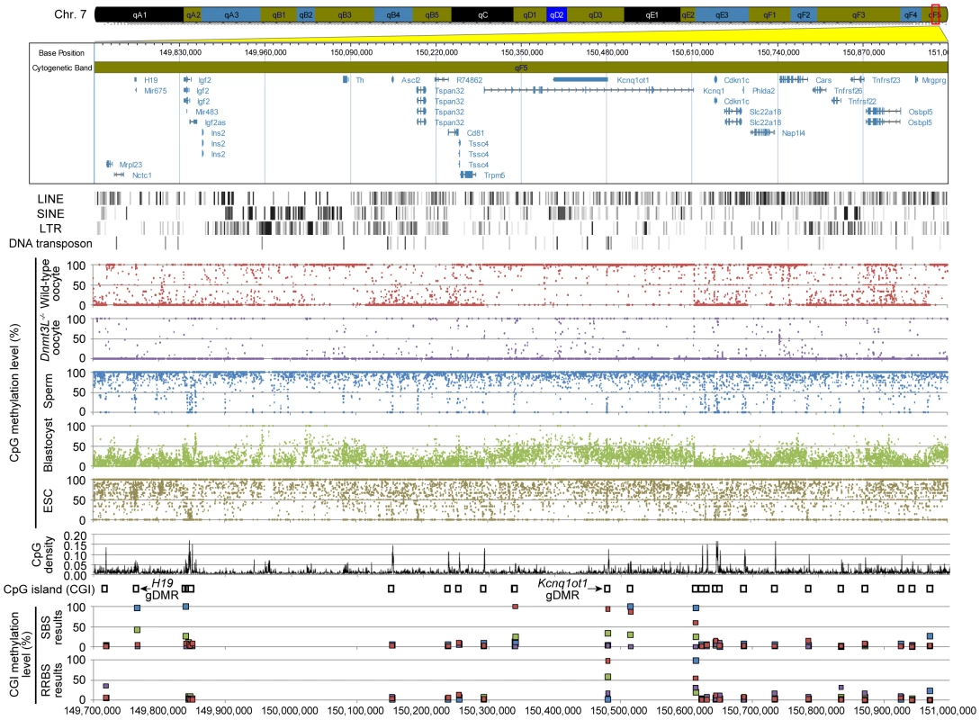
Illumina GenomeStudio viewer displays the locations of genes in distal chromosome 7 (149,700,000–151,000,000). Black vertical bars represent the location of 4 repetitive elements: LINE, SINE, LTR, and DNA transposons. Red, purple, blue, green, and khaki dots represent the methylation levels at individual CpGs in wild-type oocyte, Dnmt3L−/− oocyte, sperm, blastocyst, and ESC genomes, respectively. Black line plots depict the distribution of CpG densities (number of CpG per 200 nt) of individual CpGs. Open boxes represent the location of CpG islands (CGIs). Red, purple, blue, and green boxes represent the methylation levels at individual CGIs in wild-type oocyte, Dnmt3L−/− oocyte, sperm, and blastocyst genomes, respectively, determined by our results from shotgun bisulfite sequencing (SBS) method and Smallwood's results from reduced representation bisulfite sequencing (RRBS) method [38]. Fig. 2. Genome-wide methylation profiling of mouse germ cells. 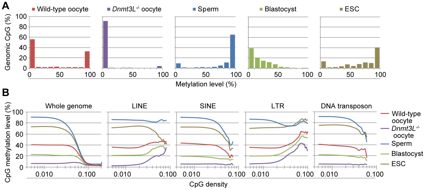
(A) Histograms of methylation levels of genomic CpGs in wild-type oocyte, Dnmt3L−/− oocyte, sperm, blastocyst, and embryonic stem cell (ESC) genomes. (B) CpG methylation levels are plotted as a function of CpG density for the whole genome and 4 families of transposable elements (long interspersed nuclear element (LINE), short interspersed nuclear element (SINE), long terminal repeat (LTR), and DNA transposon). Since previous studies revealed a significant correlation between CpG frequency and methylation within intra - and intergenic regions in somatic cells [32], [33], the CpG density and methylation levels were compared to identify genome-wide differential methylation patterns in germ cells. CpG density was defined as the number of CpG dinucleotides in 200 nucleotide (nt) windows (e.g., 1 CpG dinucleotide per 200 nt corresponds to a density of 0.005). At low CpG densities (range, 0.005–0.05), the oocyte genome was about 50% methylated, whereas the sperm genome was 80–90% methylated. At moderate to high CpG densities (range, 0.05–0.2), both male and female germ cells were hypomethylated (Figure 2B). Furthermore, 4 families of transposable elements (long interspersed nuclear elements (LINEs), short interspersed nuclear elements (SINEs), long terminal repeats (LTRs), and DNA transposons) were moderately methylated in oocyte genomes but were hypermethylated in sperm. In addition, a general trend towards higher methylation levels at higher CpG densities in the oocyte genome occurred in LTRs. Conversely, a trend toward lower CpG methylation levels at higher CpG densities in the wild-type oocyte and sperm genomes was observed in SINEs and DNA transposons. In contrast, all of these transposable elements were hypomethylated in Dnmt3L−/− oocytes. Interestingly, however, there was partial CpG methylation in LINEs and LTRs at relatively high CpG densities (range, 0.03–0.1). These complete or partial undermethylations were confirmed by bisulfite sequencing in L1 LINEs, B1/Alu SINEs, and intracisternal A particle (IAP) LTRs (Figure S7). These results suggested that each germ cell has a unique sequence - and CpG-density-dependent methylation pattern. In addition, oocyte CpG methylation, except in a subset of retrotransposons, appears to be Dnmt3L dependent.
We also characterized the methylation patterns of 15 germline-differentially methylated regions (gDMRs). The differential (between oocyte and sperm) methylation occurs at imprinted gene loci (also called imprinting control regions (ICRs)). The ICRs of maternally methylated imprinted genes (e.g., Nespas-Gnas) were shown to be hypermethylated in oocytes but hypomethylated in sperm, while the converse was true in ICRs of paternally-methylated imprinted genes (e.g., H19) (Figure 3 and Figure S8). Interestingly, only the Snrpn gDMR was partially methylated (35.7%), whereas all other maternal ICRs were hypomethylated in Dnmt3L−/− oocytes (Table 2). This residual methylation might result in the stochastic acquisition of the maternal imprint in the progeny of Dnmt3L−/− females [34]. These results strongly suggested that the methylation level of individual CpGs can be determined from DNA methylome maps with a high degree of accuracy.
Fig. 3. High-resolution genome-wide mRNA expression and CpG methylation profiling. 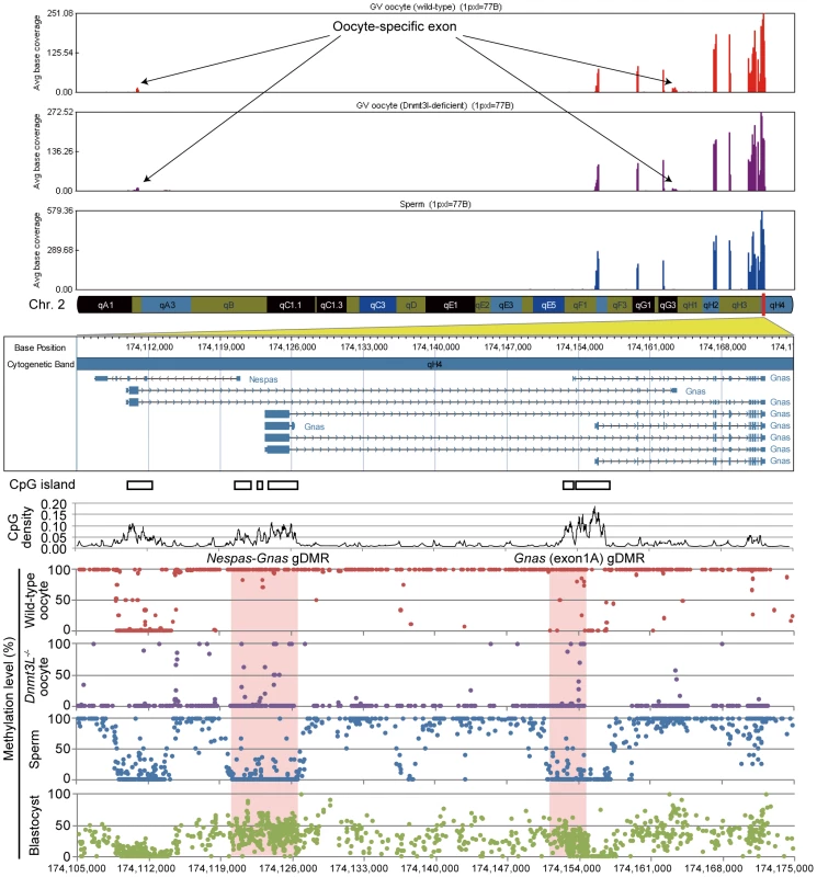
GenomeStudio view of mRNA-seq data and CpG methylation map of the genomic region spanning the Nespas-Gnas maternally imprinted locus. (Top) Genomic stacked alignment plots of wild-type oocytes, Dnmt3L−/− oocytes, and sperm. (Middle) Open boxes and black line plots represent the location of CGIs and the distribution of CpG densities of individual CpGs, respectively. (Bottom) Red, purple, blue, and green dots represent the methylation levels at individual CpGs in wild-type oocyte, Dnmt3L−/− oocyte, sperm, and blastocyst genomes, respectively. The red shaded areas show the extent of two maternal imprinting control regions (ICRs). Tab. 2. CpG methylation profiling of 12 maternal and 3 paternal imprinting control regions. 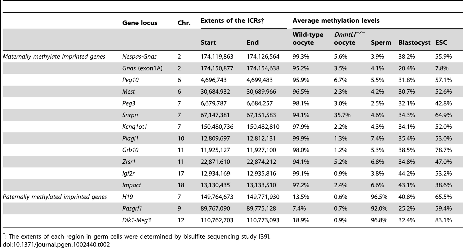
: The extents of each region in germ cells were determined by bisulfite sequencing study [39]. The study of mammalian DNA methylation patterns has previously suggested that methylation predominantly occurs at CpG sites; however, more recent studies, based on SBS methods, have indicated that methylation at non-CpG sites also occurs in human ESCs [22], [23]. Detection of non-CpG methylation is one of the applications of the bisulfite-based methylation analysis but is problematic due to the incomplete conversion of cytosine, and overestimates of such cytosine by PCR amplification, which cannot be discriminated from true methylation. In order to evaluate the methylation status of non-CpG sites and avoid these problems, additional SBS analysis of mouse GV oocytes, sperm, blastocysts, and ESCs was performed by a non-amplification technique, termed Post-Bisulfite Adapter Tagging (PBAT) [Miura F. & Ito T, personal communication]. All C (originally methylated cytosine) and T (originally unmethylated cytosine) that mapped to genomic CpG and CpH sites (H = A, T, or C) were counted. The PBAT results showed CpG methylation ratios (C ratios = 0.395, 0.748, 0.137, 0.615 in oocytes, sperm. blastocysts, and ESCs) which are similar to the average methylation levels of individual DNA methylome maps obtained by MethylC-seq and WBA-seq among all examined cells. Interestingly, a relatively high fold enrichment of non-CpG methylation was observed in GV oocytes (C ratio = 0.034–0.038), but not in the other cell types, including mouse ESCs (C ratio <0.01) (Figure S11).
Relationship between the DNA methylome and transcriptome of mouse germ cells
To elucidate the interaction between intragenic DNA methylation and gene transcription, the correlation between promoter and gene-body methylation and expression levels for 20,854 different genes was examined. The mRNA-seq profiles for germ cells and ESCs are shown in Table S1. The results showed that mRNA transcript levels in oocytes were strongly correlated to gene-body methylation levels (Spearman's ρ>0.5, p<1×10⁻⁹) but were not significantly correlated to promoter methylation levels (|ρ|<0.1) (Figure 4A). For example, the regions +2 to +5 kb from the transcription start site (TSS) and 0 to −5 kb from the transcription termination site (TTS) were hypermethylated (60–90% methylation) for the top 20% of expressed genes but were hypomethylated (10–30% methylation) for the bottom 20% of expressed genes. However, areas near the TSS (±500 base pairs (bp)) were hypomethylated (10–20% methylation) in all genes, regardless of their expression level. In contrast, in the Dnmt3L−/− oocyte genome, the correlation between gene expression and gene-body methylation was very weak (|ρ|<0.1) (Figure 4B). In the sperm genome, promoter methylation was negatively correlated (Spearman's ρ = −0.36, p<1×10⁻⁹) with gene expression, whereas gene-body methylation was positively correlated (Spearman's ρ = 0.14–0.16, p<1×10⁻⁹) to gene expression; the latter correlation was weaker than that observed in the oocyte genome (Figure 4C).
Fig. 4. Relationship between gene expression and methylation in promoter and gene-body regions in mouse germ cells. 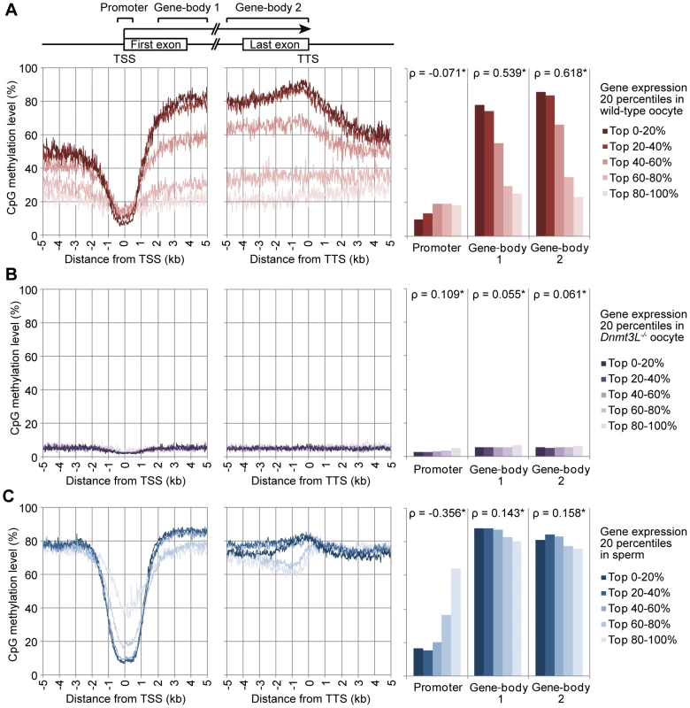
The expression level of genes in wild-type oocytes (A), sperm (B), and Dnmt3L−/− oocytes (C) were divided into 5 percentile groups. The distribution of methylation is shown ±5 kb from the transcription termination site (TTS; left) and transcription start site (TSS; middle). The graphs on the right show the average methylation levels in the promoter and gene-body regions. Spearman's rank correlation coefficient (ρ) was used to test the statistical significance of the correlation between gene expression and DNA methylation levels (*: p<1×10⁻⁹). Role of Dnmt3L in the DNA methylome/transcriptome relationship
Further investigation of gene expression patterns in oocyte genomes revealed that the mRNA transcript levels between wild-type and Dnmt3L−/− oocytes were very highly correlated (R2 = 0.9611) (Figure 5A). In fact, there were no significant differences in the expression levels of representative oocyte-specific genes (e.g., Gdf9, Bmp15, Bcl2l10, Zp1, Zp2, Zp3, Zar1, Npm2, Nlrp5, and Dppa3, which are responsible for ovarian follicle formation, reproduction, and early development [35]) and DNA methyltransferase genes (e.g., Dnmt1, a maintenance methyltransferase, and Dnmt3a and Dnmt3b de novo methyltransferases); the expected difference in the expression level of Dnmt3L between wild-type and Dnmt3L−/− oocytes was observed (Figure 5B, 5C). These results suggested that changes in gene expression did not occur during oogenesis, despite global intragenic hypomethylation in Dnmt3L−/− oocytes. Furthermore, the expression levels and exon patterns of maternally-methylated imprinted genes across each ICR were not altered in Dnmt3L−/− oocytes (Figure 3 and Figure 5D). This result suggested that the disruption of maternal methylation imprints in the Dnmt3L−/− oocyte genome was not due to the lack of their transcription [36]. On the other hand, maternal methylation imprints at ICRs (and many other hypermethylations at transcribed regions) in wild-type oocyte genomes might be the result of gene transcription via Dnmt3L-mediated intragenic methylation.
Fig. 5. Comparison of gene expression profiles between wild-type and Dnmt3L−/− oocytes. 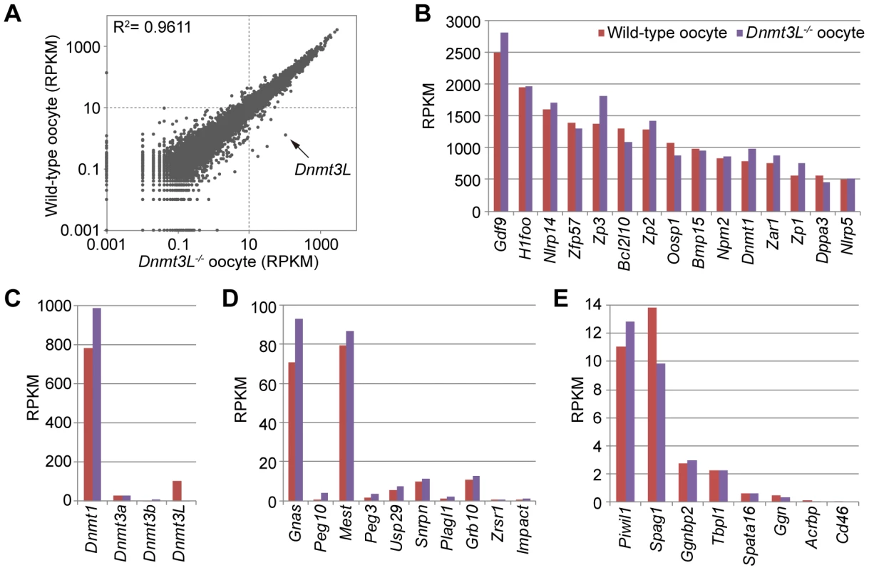
(A) Scatter plot and correlation coefficient (R2) of RPKM values of 20,854 genes in wild-type and Dnmt3L−/− oocytes. Expression levels of oocyte-specific genes (B), DNA methyltransferase genes (C), maternally-imprinted genes that are potentially necessary to establish methylation imprints (D), and male germline-specific genes that contain oocyte-specific methylated CpG islands (CGIs) (E). Surprisingly, gene expression in ESC genomes was negatively correlated with promoter methylation and was not positively correlated with gene-body methylation (Figure S12). Meanwhile, these ESCs showed the apparent expression of all DNA methyltransferase gene families including Dnmt3L (Figure S13). Previous studies indicated that the zygotic and somatic functioning of Dnmt3L is not essential for global methylation in ESCs in mice [6]. Thus, unlike oocytes, the functional role of Dnmt3L in gene-body methylation after fertilization is unclear. However, the expression of pluripotency-associated genes, Pou5f1, Klf4, Sox2, Myc, Nanog, and Lin28a, was clearly observed in ESCs. The expression of Pou5f1, Lin28a, and Glis1, recently identified as maternal reprogramming factors, were also observed in oocytes (Figure S14). While differential expression of the pluripotency genes among germ and stem cells was observed, the promoter regions of these genes demonstrated low-level methylation in almost all of the examined cells. In sperm cells, only the Nanog promoter was hypermethylated (this result was similar to a previous study [29]).
Identification and characterization of germline differentially methylated regions
To identify gDMRs, the average CpG methylation levels of individual CpG islands (CGIs), which are CpG-rich genomic regions often lacking DNA methylation, were calculated. Recently, Illingworth et al. determined the number of CGIs by deep sequencing of isolated, unmethylated DNA clusters [37]. Among the 23,021 mouse CGIs (22,974 CGIs were informative in both oocytes and sperm), 2014 were highly methylated (≥80% methylation) in oocytes, 818 were highly methylated in sperm, and 377 were highly methylated in both germ cells (Figure 6A). Furthermore, we also identified 1678 gDMRs (≥80% methylation in 1 gamete and ≤20% in the other), 1329 of which were oocyte-specific methylated CGIs, while the remaining 349 were sperm-specific methylated CGIs (Figure 6A, Figure S6, and Table S2). Among these gDMRs, 646 gDMRs were confirmed to show a differential methylation status between GV oocytes and sperm (by similar criteria: ≥75% methylation in 1 gamete and ≤25% in the other); the methylation status was previously examined by performing large-scale bisulfite sequencing of CpG-rich regions of the genome (reduced representation bisulfite sequencing: RRBS) (Table S3) [38]. Additionally, almost all known ICRs except Zdbf2 DMRs (which do not have any CGIs) were re-identified from our gDMR list (Table S2).
Fig. 6. Identification of germline differentially methylated CGIs from DNA methylome profiles. 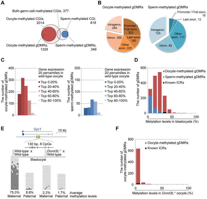
(A) Venn-like diagram of two groups of CGIs, namely, oocyte-methylated CGIs (light pink) and sperm-methylated CGIs (light blue) and two groups of gDMRs, namely, oocyte-methylated gDMRs (red) and sperm-methylated gDMRs (blue). (B) The genomic distribution of 1329 oocyte-methylated (left) and 349 sperm-methylated gDMRs (right). The gDMRs were classified into 5 genomic locations; promoter (within 500-bp upstream from the first exon) or first exon, last exon, other exon, intron, and intergenic region. (C) The locations of the intragenic 1045 oocyte-methylated (left) and 229 sperm-methylated gDMRs (right). The gDMRs were classified into 5 gene group locations; the genes were divided into 5 percentile groups according to their expression levels in wild-type oocytes and sperm, as shown in Figure 3. (D) Histograms of the methylation levels of the gDMRs in blastocysts. The number of newly identified oocyte-specific, sperm-specific methylated gDMRs, and known ICRs are shown in black, red, and blue, respectively. (E) Bisulfite sequencing at the Gpr1 gDMR in mouse blastocysts. (Top) Schematic representation of paternally-expressed Gpr1. The gene and gDMRs are shown in blue and green, respectively, and CpG sites are represented by vertical bars. (Bottom) Methylated and unmethylated CpGs are indicated by open and closed circles, respectively. The maternal and paternal alleles were distinguished by three polymorphisms between C57BL/6N and JF1 mice (G/A at 63,247,064; T/A at 63,247,072; and TA/AG at 63,247,089–63,247,090 on chromosome 1). (F) Histograms of the methylation levels of the demethylation-resistant oocyte-methylated gDMRs in Dnmt3L−/− oocytes. The number of newly identified oocyte-specific, sperm-specific methylated gDMRs, and known ICRs are shown in black, red, and blue, respectively. A total of 78% oocyte-methylated gDMRs (n = 1045) were located within the intragenic regions. Approximately 25% of the oocyte-methylated gDMRs (n = 322) overlap with either the first exon or the proximal promoter regions of the genes, as has been observed with most of the described maternal ICRs [39]; only 5% of the sperm-methylated gDMR (n = 18) showed such overlap. Alternatively, 34% of sperm-methylated gDMRs (n = 120) overlap with intergenic regions, as in all known paternal ICRs (Figure 6B). Interestingly, oocyte-methylated gDMRs in transcribed regions tended to be more abundant within highly expressed genes, but such a trend was not observed in the sperm genome (Figure 6C). Oocyte-methylated gDMRs were also identified in non-imprinted genes, such as the DNA methyltransferase genes (e.g., Dnmt1 and Dnmt3b) and some male germline-specific genes (e.g., Piwil1, Spag1, Ggnbp2, Tbpl1, Spata16, Ggn, Acrbp, and Cd46). The oocyte-methylated gDMR in Dnmt1 was located in spermatocyte - and somatic-specific exons, while oocyte-specific exons were hypomethylated in oocytes (Figure S9). Dnmt3L−/− oocytes also showed hypomethylation in most of these gDMRs. Significant changes in the expression levels of genes with alternative splicing patterns were not observed in the Dnmt3L−/− oocyte genome (Figure 3, Figure 5E, and Figure S9). These results indicate that these oocyte-specific methylated gDMRs do not regulate gene expression or alternative splicing during the oocyte stage.
To determine whether or not these germ cell-specific methylations are maintained after fertilization, when the genomes undergo global demethylation, the individual CGI methylation levels in blastocyst genomes were calculated. In blastocysts, all ICRs demonstrated low to moderate methylation (25.1–64.3%), whereas many gDMRs were demethylated (0–20%) (Figure 6D). Furthermore, 817 oocyte-methylated gDMRs (including Piwil1, despite being a non-imprinted gene locus) and 34 sperm-specific gDMRs were resistant to demethylation during early embryogenesis (≥20% methylation in blastocysts) (Figure 6D and Table S2). Among the demethylation-resistant gDMRs, a novel gDMR in the intron of Gpr1 (Figure S10) was found to be a tissue-specific, paternally-expressed imprinted gene [40]. Bisulfite sequencing analysis showed that this gDMR was hypomethylated in Dnmt3L−/− oocytes and maternal allele-specific methylation was detected in this region in blastocysts (Figure 6E). Methylation profiles in ESCs showed that 26% (n = 213) of the demethylation-resistant gDMRs became less methylated (0–20%) whereas the other gDMRs maintained or increased DNA methylation (Figure S15). Among ICRs, only Gnas exon1A ICR was demethylated (7.8%), whereas the other ICRs developed partial or high methylation levels (range, 38.6–83.1%) in ESCs (Table 2). Among other demethylation-sensitive gDMRs, which were demethylated (<20% methylation) in blastocysts, many (76%, n = 264) sperm-methylated gDMRs were re-methylated (≥20% methylation); most (81%, n = 416) of the oocyte-methylated gDMRs maintained low methylation (0–20%) in ESCs (Figure S15). Finally, out of 704 demethylation-resistant (in blastocysts) oocyte-methylated gDMRs which were informative in Dnmt3L−/− oocytes, only 4 remained hypermethylated (80–100% methylation) in the Dnmt3L−/− oocyte genome. However, almost all other oocyte-specific methylation marks at gDMRs were Dnmt3L-dependent (Figure 6F). These results suggest that Dnmt3L-mediated methylation during oogenesis regulates the establishment of most heritable oocyte-specific marks, including genomic imprints.
Discussion
To the best of our knowledge, this is the first study to generate single-base resolution maps of DNA methylomes spanning the entire genome of mouse germ cells. The oocyte maps are particularly valuable and informative because, in the past, such an analysis was prohibitive due to the need for large quantities of DNA. Recently, Smallwood et al. [38] reported large-scale DNA methylation patterns in mouse germ cells by using the RRBS method, which targets only CpG-rich regions. However, our more comprehensive results provide strong evidence that gene expression was positively correlated to Dnmt3L-dependent intragenic methylation in oocytes, and that methylation patterns in oocytes differed from those in sperm and non-germline cells.
The functional role of gene-body methylation has been an enigma despite its conservation in plants and animals [41]–[43]. Maunakea et al. [44] suggested that gene-body methylation is involved in the regulation of alternative splicing events. Although methylated gDMRs were detected in the alternative exons of Dnmt1 and Gnas in mouse oocytes, loss of oocyte-specific methylation marks in the Dnmt3L−/− oocytes did not affect the expression patterns of alternatively spliced transcripts. Therefore, our results indicate that gene-body methylation is not involved in alternative splicing in oocytes.
Previously, Chotalia et al. [36] showed that transcription during the oocyte stage is required for the establishment of maternal methylation marks on an imprinted gene. The present results show that Dnmt3L−/− oocytes lost almost all of their maternal methylation imprints while maintaining a constant amount of mRNA through each ICR despite the global loss of intragenic methylation. Thus, these results strongly suggest that the establishment of genomic imprints via transcription is mediated by Dnmt3L-dependent intragenic methylation.
A possible mechanism for gene-body methylation involves the exposure of intragenic regions to DNA methyltransferases, considering that RNA polymerase disrupts the chromatin structure during transcription. However, not all transcripts across gDMRs corresponded to highly expressed genes in oocytes (Figure 6C). Therefore, other epigenetic marks with an open chromatin structure might also be important for DNA methylation in oocytes. For instance, a recent knockout study showed that Kdm1b, which encodes histone H3K4 demethylase, is required for the establishment of some maternal methylation imprints [45]. Thus, several factors, including transcriptional and epigenetic modifications, might be involved in Dnmt3L-mediated intragenic methylation.
The results of this study show that gene-body methylation was correlated to gene expression in sperm. However, the extent of that correlation is much less than in oocytes due to genome-wide hypermethylation, including in low-CpG-density regions. In male germline cells, global methylation acquisition begins during late embryonic development and before birth [3]. To more clearly show this correlation, analysis of early-stage germ cells in fetal or neonatal animals might be required. Surprisingly, a positive correlation between mRNA expression and gene-body methylation was not observed in mouse ESCs. In addition, the accumulation of non-CpG methylation was not observed in mouse ESCs. These results contradict the results of another study, which showed that active transcription was associated with intragenic DNA methylation with non-CpG methylation in human ESCs [22], [23]. This discrepancy might reflect the differences between human and mouse ESCs, the precise cell derivations or culture conditions [46], [47]. However, further comparative studies on germ cell epigenomes from other species are required to further elucidate the functional role of epigenetic marking systems.
In this study, a large number of heritable oocyte-specific methylation marks were identified within a set of novel CpG islands [37]. The difference in the number of oocyte - and sperm-specific gDMRs reflects the fact that only 3 or 4 paternally-methylated imprinted loci were observed, as compared to approximately 20 maternally-methylated imprinted loci. The reason for the relative abundance of oocyte-specific methylated CGIs might be related to the intragenic methylation of CpG-rich regions, which are hypomethylated in sperm. The results show that most of the oocyte-specific marks are Dnmt3L-dependent, similar to results recently obtained by RRBS-based analysis [38]. However, whether all of these CpG-rich regions serve as imprinting methylation marks is unclear. For instance, although many genes with oocyte-specific methylation marks were identified (Figure 6B), the evidence that these genes were imprinted was lacking (e.g., Piwil1 and Dnmt1). These methylation marks might not be involved in the formation of a fertile oocyte but might play crucial roles in gene expression after fertilization. Furthermore, ESC methylomes showed that many gDMRs, especially sperm-specific gDMRs, acquired new methylation patterns after implantation. Methylation of these CGIs might control tissue-specific gene expression [48], [49]. Partial alternation of imprinted methylation patterns in ESCs were observed in the present study, potentially caused by significant differences in the extent of the ICRs during embryo development [39]. A fuller understanding of epigenetic stability will require further methylome profiling during early embryogenesis and stem cell differentiation. The present study also identified a gDMR as a novel ICR candidate in the intron of the imprinted Gpr1 gene. Thus, traditional promoter arrays may not identify all ICRs. However, further analyses are needed to determine which gDMRs, identified in the CpG methylome maps, are true ICRs at the imprinted Gpr1-Zdbf2 locus [40], [50].
mRNA-seq results showed that the expression levels of most genes in the wild-type and Dnmt3L−/− oocytes were similar. For instance, the expression level of almost all oocyte-specific genes, which regulate ovarian follicle formation, reproduction, and early development, were not significantly altered (Figure 5B and Table S1). These results are consistent with the findings of previous studies, which showed that Dnmt3L−/− female mice were capable of producing fertile oocytes (however, their offspring were not viable due to the lack of imprinting) [5], [6]. Thus, regulation of oocyte-specific genes must be beyond the control of Dnmt3L-dependent cytosine methylation.
Although Dnmt3L−/− oocytes showed global hypomethylation at low to high CpG densities, some families of retrotransposons, such as LINEs and LTRs, were partially methylated at moderate to high CpG densities. Therefore, Dnmt3L-independent methylation might be involved in the silencing of retrotransposons and completion of oocyte meiosis. Previously, De La Fuente et al. [51] showed that Hells (also known as Lsh), which encodes a member of the sucrose non-fermenter 2 (SNF2) family of chromatin remodeling proteins, is required for DNA methylation of IAP and pericentromeric satellite repeats as well as repression of IAP retrotransposition in pachytene oocytes. Unfortunately, measurement of the methylation levels of satellite DNA, which is abundant in the pericentromeric regions, was not possible because these sequences were excluded from our analysis. However, a previous sequencing study showed that methylation levels of satellite DNA did not differ between the wild-type and Dnmt3L−/− oocytes [52]. Combined, these results suggest the presence of 2 types of oocyte methylation patterns: (i) Dnmt3L-mediated intragenic methylation that is essential for early embryogenesis and (ii) Dnmt3L-independent retroviral and pericentromeric methylation, which may be mediated by Hells activity, is crucial for oocyte meiosis [51]. Further studies on Hells-mediated oocyte methylation are required to elucidate the details of this mechanism.
Previous studies on the cytosine methylation of mtDNA have been highly controversial. A recent study by Shock et al. [53] reported cytosine methylation and hydroxymethylation in mammalian mitochondria. Our results indicated that mtDNA is unmethylated in blastocysts and ESCs, but is partially methylated in germ cells. Whether or not 5-hydroxymethylcytosine (5-hmC) exists in mitochondrial or genomic chromosomes of germ cells remains unclear. Meanwhile, rapid hydroxylation of 5-methylcytosine (5-mC) in the paternal pronucleus during zygotic development was also recently reported [54], [55]. Currently, it is difficult to assess hydroxymethylation profiles in oocyte genomes due to the limited DNA recovery. Further investigation of cytosine modification during germ cell and zygote development will be required in the future to better understand this process.
The DNA methylome maps of mouse germ cells, in this study, were derived from SBS data and, therefore, accurately represent methylation levels of individual CpGs on a whole-genome level. The adaptation of the SBS method for small-scale DNA analysis, described in the present report, has the potential to enable further analyses of germline lineages. The current work examined SBS library construction using 3 methods, MethylC-seq, WBA-seq, and PBAT. MethylC-seq basically required only microograms of DNA [22], [23], [56], thus over amplification might cause redundancy in oocyte libraries. The latter methods allow comprehensive methylome analysis in samples with low amounts of starting DNA by avoiding DNA damage due to sodium bisulfite treatment (after adapter ligation, in the case of MethylC-Seq). Recent studies using BS sequencing have shown that methylated cytosine is abundant in the non-CpG regions of human pluripotent stem cells and mouse oocytes [22], [23], [39], [56]; however, the function of non-CpG methylation in mammalian genomes remains unclear. The PBAT results also showed an abundance of non-CpG methylation in oocytes, with results similar to a previous sequencing study on imprinted loci [39]. However, accurate assessment of non-CpG methylation is required using increased sequencing depths because methylation levels of the non-CpG sites were much lower than those of the CpG sites. SBS library construction was conducted by WBA-seq from 2000 fully matured (metaphase II stage) oocytes; sufficient quantities for sequencing were not obtained. During oogenesis, most of the oocyte specific imprinted methylation marks were established during the GV stage. This contrasted to a previous study where a continuous increase in methylation levels was observed [38]. Further improvement of SBS methods, requiring smaller amounts of DNA, is needed to provide complete germ cell methylome maps and to elucidate the exact function of non-CpG methylation in germ cells.
In conclusion, we constructed the first extensive, high-resolution maps of DNA methylomes of mouse oocytes and sperm. These maps described the epigenetic properties of these DNA methylomes. Our data could serve as a platform for future studies to elucidate the role of epigenetic modifications in the development and functioning of germ and stem cells. Such studies are anticipated to improve our understanding of epigenetic reprogramming.
Materials and Methods
Preparation of MethylC-seq libraries
Five thousand germinal vesicle (GV)-stage oocytes were collected from the ovarian follicles of adult (7 - to 9-week-old) female C57BL/6N mice (Clea Japan, Tokyo, Japan) 44–48 h after they were injected with equine chorionic gonadotropin. Three hundred blastocysts at embryonic day 3.5 were obtained from superovulated adult female C57BL/6N mice by flushing the uterus. Genomic DNA was extracted using the QIAamp DNA Mini Kit (Qiagen, Valencia, CA). Sperm were released from the cauda epididymises of adult male C57BL/6N mice. Sperm DNA was isolated by a standard phenol-chloroform extraction procedure with dithiothreitol (DTT). Genomic DNA from 2 lines of ESCs derived from C57BL/6J mice (Clea Japan) was extracted using the DNeasy Blood & Tissue Kit (Qiagen). DNA samples were sheared into 100-bp fragments in oocytes and 200-bp fragments in other samples using the Covaris S2 focused acoustic system (Covaris, Woburn, MA). Cytosine-methylated adapters (Illumina, San Diego, CA) were ligated to DNA by using the Paired-End DNA Sample Prep Kit or ChIP-Seq DNA Sample Prep Kit (Illumina). DNA fragments were isolated by 2–3% agarose gel electrophoresis and purified using the QIAquick Gel Extraction Kit (Qiagen). Sodium bisulfite conversion was performed using the Epitect Bisulfite Kit (Qiagen).
All bisulfite-converted DNA molecules were polymerase chain reaction (PCR)-amplified as follows: 2.5 U of Hot Start Taq polymerase (TaKaRa, Tokyo, Japan), 5 µL 10× PCR buffer, 25 µM dNTPs, 1 µL of each PCR Primer PE 1.0 and 2.0 (Illumina) (50 µL final). Thermocycling parameters were: initial denaturation at 94°C for 1 min, 15–25 cycles of denaturation at 94°C for 30 s, annealing at 65°C for 30 s, and extension at 72°C for 30 s, followed by a final extension at 72°C for 5 min. PCR reaction products were purified using the QIAquick kit (Qiagen).
Preparation of whole WBA-seq libraries
Two thousand GV-stage oocytes were collected from 7 - to 9-week-old female C57BL/6N mice (Clea Japan) and, 2300 GV-stage oocytes were collected from 7–15-week-old Dnmt3L−/− female mice (129SvJae×C57BL/6N hybrid genetic background) [6], [57]. Genomic DNA was extracted using the QIAamp DNA Mini Kit (Qiagen), and then bisulfite-treated with Epitect Bisulfite Kit (Qiagen). Subsequently, the bisulfite-converted DNA was amplified using Epitect Whole Bisulfitome Kit (Qiagen). The collected DNA was sheared into 200-bp fragments using Covaris S2. Unmodified Paired-End adapters (Illumina) were ligated to the DNA by using the Paired-End DNA Sample Prep Kit (Illumina). DNA fragments were isolated by 2% agarose gel electrophoresis and purified using the QIAquick Kit (Qiagen). All DNA was PCR amplified and purified in the same manner as the MethylC-seq method, except the number of PCR cycles was reduced to 7.
Preparation of PBAT libraries
GV-stage oocytes (400) and blastocysts (100) were obtained from 7 - to 9-week-old female C57BL/6N mice (Clea Japan), and genomic DNA was extracted using the QIAamp DNA Mini Kit (Qiagen). The isolated oocyte and blastocyst genomic DNA and 100 ng of genomic DNA from sperm, blastocysts, and ESCs containing 1∶200 amount of unmethylated lambda DNA (Invitrogen, Carlsbad, CA) were bisulfite-treated using the MethylCode Bisulfite Conversion Kit (Invitrogen). Details of the PBAT method are unpublished [Miura F & Ito T, personal communication]. Briefly, bisulfite-treated DNA were double-stranded using Klenow Fragments (3′ - 5′ exo-) (New England Biolabs, Ipswich, MA) with random primers containing 5′ biotin tags and Illumina PE adaptors. The biotinylated molecules (first strand) were captured using Dynabeads M280 Streptavidin (Invitrogen) and double-stranded using Klenow Fragments (3′-5′ exo-) with random primers containing Illumina PE adaptors (second strand). Finally, template DNA strands were synthesized as complementary DNA with a second strand (unmethylated C is converted to T) using Phusion Hot Start High-Fidelity DNA Polymerase (New England Biolabs) with PCR Primer PE 1.0 (Illumina).
Preparation of mRNA sequencing libraries
Total RNA from 1000 wild-type GV oocytes, 500 Dnmt3L−/− GV oocytes, sperm, and ESCs was extracted using the RNeasy Mini Kit (Qiagen) and treated with DNase I (Promega, Madison, WI). RNA-Seq libraries were constructed using the mRNA-Seq Sample Preparation Kit (Illumina).
Sequencing
The MethylC-seq for blastocysts, WBA-seq, and PBAT libraries were sequenced on a HiSeq 2000 sequencing system (Illumina); the other MethylC-seq and mRNA-seq libraries were sequenced on a Genome Analyzer II (Illumina). Sample preparation, cluster generation, and sequencing were performed using the Paired-End Cluster Generation Kit-HS and the TruSeq SBS Kit-HS for the HiSeq 2000. Similarly, the Paired-End Cluster Generation Kits v2 and v4 and 18 - and 36-Cycle Sequencing Kits v3 and v4 were used for the Genome Analyzer II. All kits were from Illumina.
Gene mapping
All sequenced reads were processed using the standard Illumina base-calling pipeline (v1.4–1.7). Generated sequence tags were mapped onto the mouse genome (mm9, UCSC Genome Browser, July 2007, Build 37.1) by using the Illumina ELAND program.
MethylC-seq tags (36 or 76 nt) were mapped with a custom Perl program, as described previously [17], [22]. Briefly, all cytosines in the tags were replaced by thymines. Next, these tags were aligned to 2 mouse genome reference sequences (mm9), such that the antisense strand had cytosines replaced by thymines and the sense strand had guanines replaced by adenines. Finally, all tags (32–76 nt) that mapped uniquely without any mismatches to both strands were compiled and used for further analyses.
The 76 nt WBA-seq tags were mapped as follows. All tags were converted to 2 types of reads; in 1 read (“For” read), cytosines were replaced by thymines and in the other read (“Rev” read), guanines were replaced by adenines. Both “For” and “Rev” reads were aligned to sense and antisense mm9 strands. A total of 793, 397, 948, 480, and 238 million tags were aligned in wild-type oocytes, Dnmt3L−/− oocytes, sperm, blastocysts, and ESC genomes, respectively. To avoid bias, tags mapped with multiple hits or matched chromosome M (mitochondria), chromosome Y, or 3 types of repetitive sequences (simple repeat, low complexity repeat, and satellite DNA sequences) were omitted from further analyses.
The 47 nt PBAT tags (trimmed first 4 nt and last 1 nt) were mapped as follows. All guanidines in the tags were replaced by adenines, and these tags were aligned to sense and antisense strands mm9.
For gene-level analysis, the concentrations of the perfectly matching 35 nt (trimmed first nt) mRNA-seq tags from wild-type oocytes, Dnmt3L−/− oocytes, sperm, and ESCs were calculated for the genomic regions corresponding to those covered by the RefSeq transcript models. The expression level of 20,854 unique genes was ranked by expression levels (calculated as RPKM values) in each library (Table S1). A total of 33, 28, 23, and 25 tags were aligned in 4 mRNA-seq libraries, respectively. mRNA-seq data analysis was performed and visualized using GenomeStudio Data Analysis software (Illumina).
Methylation analysis
The percentage of individual cytosines methylated at all CpG sites covered by at least 1 read was calculated as 100×(number of aligned cytosines (methylated cytosines))/(total number of aligned cytosines and thymines (originally unmethylated cytosines)). All genomic CpG methylation data are available on our website (http://www.nodai-genome.org/mouse_en.html). The CpG and non-CpG (CpH) methylation levels determined by PBAT results were calculated as the ratio between the total read C and the total read T mapped to genomic cytosines. Bisulfite conversion failure rates were calculated by read C∶T ratios from lambda DNA mapping data. The failure rates were as follows: GV oocyte, 0.009; sperm, 0.008; blastocysts, 0.011; and ESCs, 0.006. Locations of transposable elements in the mouse genome (mm9) were obtained from the UCSC Genome Browser, and the average methylation levels of the whole genome and each transposable element were recalculated from the ratio of the aligned cytosines and thymines in each sequence. Lists of 23,021 CGIs were obtained from a previous report [37]. Around the TSS and TTS (±5 kb), genomic regions were divided into 20-bp bins. For each bin, the average methylation value was calculated for each gene. The expression level of 20,854 genes was divided into 5 percentile groups ranked by RPKM values, and the average methylation level for each group was mapped onto the gene structure model. These computational analyses were performed using a custom Perl program. Supercomputing resources were provided by the Human Genome Center, Institute of Medical Science, University of Tokyo.
Statistical analysis
Correlations between gene expression ranks and average methylation levels in the promoter (±500 bp from the TTS) or gene-body regions (gene-body 1: +2 to +5 kb from the TSS; gene-body 2 : 0 to −5 kb from the TTS) were calculated using Spearman's rank correlation coefficient (ρ). An R-squared value (R2) was calculated to evaluate the correlation of RPKM values between wild-type and Dnmt3L−/− oocytes. Statistical analysis was performed using the R statistical package.
Bisulfite sequencing
To analyze the methylation of the three transposable elements (L1 LINE, B1/Alu SINE, and IAP LTR), 20 wild-type GV oocytes were obtained from adult female C57BL/6N mice. Bisulfite sequencing conditions and primer sets for the three transposable elements were described, previously [52]. To analyze the methylation of the Gpr1 locus, 10 blastocysts were obtained from BJF1 (C57BL/6N×JF1) and Dnmt3Lmat−/− (Dnmt3L−/−×JF1) mice [6], [57]. Genomic DNA from blastocysts was isolated using the QIAamp DNA Mini Kit (Qiagen) and treated with sodium bisulfite with the Epitect Bisulfite Kit (Qiagen). The Gpr1 gDMR sequence was amplified with 2 rounds of nested PCR. The first-round PCR reaction contained 1 U of Hot Start Taq polymerase (TaKaRa), 1× PCR buffer, 200 µM dNTPs, 1 µM forward primer, and 1 µM reverse primer (20 µL final). Thermocycling parameters were as follows: initial denaturation at 94°C for 1 min, 35 cycles of denaturation at 94°C for 30 s, annealing at 50°C for 30 s, and extension at 72°C for 30 s, followed by a final extension at 72°C for 5 min. Subsequently, 2 µL of the product was used as the input for the second-round PCR, which was performed in the same manner. Primer sets for the nested PCR were as follows: Gpr1-BSF1 (5′-GATTAGATTAGGTTAGTTTGGAA-3′) and Gpr1-BSR1 (5′-ACTAAAACACTAATCACCAAATA-3′) for the first round; Gpr1-BSF2 (5′-AGATTAGGTTAGTTTGGAATT-3′) and Gpr1-BSR2 (5′-AACACTAATCACCAAATAATTC-3′) for the second round. The second-round PCR product was subcloned and sequenced, as described previously [50]. The percentage methylation was calculated as 100×(number of methylated CpG dinucleotides)/(total number of CpGs). At least 10 clones from each parental allele were sequenced. Sequence data were analyzed using the QUMA quantification tool for methylation analysis [58].
Accession number
The MethylC-seq, WBA-seq, PBAT, and mRNA-seq data in this study have been deposited in the DNA Data Bank of Japan (DDBJ) under accession number DRA000484.
Supporting Information
Zdroje
1. LiE 2002 Chromatin modification and epigenetic reprogramming in mammalian development. Nat Rev Genet 3 662 673
2. JaenischRBirdA 2003 Epigenetic regulation of gene expression: how the genome integrates intrinsic and environmental signals. Nat Genet 33 Suppl 245 254
3. Lees-MurdockDJWalshCP 2008 DNA methylation reprogramming in the germ line. Epigenetics 3 5 13
4. SasakiHMatsuiY 2008 Epigenetic events in mammalian germ-cell development: reprogramming and beyond. Nat Rev Genet 9 129 140
5. Bourc'hisDXuGLLinCSBollmanBBestorTH 2001 Dnmt3L and the establishment of maternal genomic imprints. Science 294 2536 2539
6. HataKOkanoMLeiHLiE 2002 Dnmt3L cooperates with the Dnmt3 family of de novo DNA methyltransferases to establish maternal imprints in mice. Development 129 1983 1993
7. KanedaMOkanoMHataKSadoTTsujimotoN 2004 Essential role for de novo DNA methyltransferase Dnmt3a in paternal and maternal imprinting. Nature 429 900 903
8. KonoTObataYWuQNiwaKOnoY 2004 Birth of parthenogenetic mice that can develop to adulthood. Nature 428 860 864
9. HirasawaRChibaHKanedaMTajimaSLiE 2008 Maternal and zygotic Dnmt1 are necessary and sufficient for the maintenance of DNA methylation imprints during preimplantation development. Genes Dev 22 1607 1616
10. Bourc'hisDBestorTH 2004 Meiotic catastrophe and retrotransposon reactivation in male germ cells lacking Dnmt3L. Nature 431 96 99
11. WebsterKEO'BryanMKFletcherSCrewtherPEAapolaU 2005 Meiotic and epigenetic defects in Dnmt3L-knockout mouse spermatogenesis. Proc Natl Acad Sci U S A 102 4068 4073
12. HataKKusumiMYokomineTLiESasakiH 2006 Meiotic and epigenetic aberrations in Dnmt3L-deficient male germ cells. Mol Reprod Dev 73 116 122
13. ShojiMTanakaTHosokawaMReuterMStarkA 2009 The TDRD9-MIWI2 complex is essential for piRNA-mediated retrotransposon silencing in the mouse male germline. Dev Cell 17 775 787
14. Kuramochi-MiyagawaSWatanabeTGotohKTakamatsuKChumaS 2010 MVH in piRNA processing and gene silencing of retrotransposons. Genes Dev 24 887 892
15. CokusSJFengSZhangXChenZMerrimanB 2008 Shotgun bisulphite sequencing of the Arabidopsis genome reveals DNA methylation patterning. Nature 452 215 219
16. DownTARakyanVKTurnerDJFlicekPLiH 2008 A Bayesian deconvolution strategy for immunoprecipitation-based DNA methylome analysis. Nat Biotechnol 26 779 785
17. ListerRO'MalleyRCTonti-FilippiniJGregoryBDBerryCC 2008 Highly integrated single-base resolution maps of the epigenome in Arabidopsis. Cell 133 523 536
18. MeissnerAMikkelsenTSGuHWernigMHannaJ 2008 Genome-scale DNA methylation maps of pluripotent and differentiated cells. Nature 454 766 770
19. BallMPLiJBGaoYLeeJHLeProustEM 2009 Targeted and genome-scale strategies reveal gene-body methylation signatures in human cells. Nat Biotechnol 27 361 368
20. BrunnerALJohnsonDSKimSWValouevAReddyTE 2009 Distinct DNA methylation patterns characterize differentiated human embryonic stem cells and developing human fetal liver. Genome Res 19 1044 1056
21. DengJShoemakerRXieBGoreALeProustEM 2009 Targeted bisulfite sequencing reveals changes in DNA methylation associated with nuclear reprogramming. Nat Biotechnol 27 353 360
22. ListerRPelizzolaMDowenRHHawkinsRDHonG 2009 Human DNA methylomes at base resolution show widespread epigenomic differences. Nature 462 315 322
23. LaurentLWongELiGHuynhTTsirigosA 2010 Dynamic changes in the human methylome during differentiation. Genome Res 20 320 331
24. PoppCDeanWFengSCokusSJAndrewsS 2010 Genome-wide erasure of DNA methylation in mouse primordial germ cells is affected by AID deficiency. Nature 463 1101 1105
25. SerreDLeeBHTingAH 2010 MBD-isolated Genome Sequencing provides a high-throughput and comprehensive survey of DNA methylation in the human genome. Nucleic Acids Res 38 391 399
26. LiYZhuJTianGLiNLiQ 2010 The DNA methylome of human peripheral blood mononuclear cells. PLoS Biol 8 e1000533 doi:10.1371/journal.pbio.1000533
27. GuHBockCMikkelsenTSJagerNSmithZD 2010 Genome-scale DNA methylation mapping of clinical samples at single-nucleotide resolution. Nat Methods 7 133 136
28. HowlettSKReikW 1991 Methylation levels of maternal and paternal genomes during preimplantation development. Development 113 119 127
29. FarthingCRFiczGNgRKChanCFAndrewsS 2008 Global mapping of DNA methylation in mouse promoters reveals epigenetic reprogramming of pluripotency genes. PLoS Genet 4 e1000116 doi:10.1371/journal.pgen.1000116
30. BarhaninJLesageFGuillemareEFinkMLazdunskiM 1996 K(V)LQT1 and lsK (minK) proteins associate to form the I(Ks) cardiac potassium current. Nature 384 78 80
31. ShinJBossenzMChungYMaHByronM 2010 Maternal Rnf12/RLIM is required for imprinted X-chromosome inactivation in mice. Nature 467 977 981
32. WeberMHellmannIStadlerMBRamosLPaaboS 2007 Distribution, silencing potential and evolutionary impact of promoter DNA methylation in the human genome. Nat Genet 39 457 466
33. EdwardsJRO'DonnellAHRollinsRAPeckhamHELeeC 2010 Chromatin and sequence features that define the fine and gross structure of genomic methylation patterns. Genome Res 20 972 980
34. ArnaudPHataKKanedaMLiESasakiH 2006 Stochastic imprinting in the progeny of Dnmt3L−/ − females. Hum Mol Genet 15 589 598
35. MinamiNTsukamotoS 2006 Role of oocyte-specific genes in the development of mammalian embryos. Reproductive Medicine and Biology 5 175 182
36. ChotaliaMSmallwoodSARufNDawsonCLuciferoD 2009 Transcription is required for establishment of germline methylation marks at imprinted genes. Genes Dev 23 105 117
37. IllingworthRSGruenewald-SchneiderUWebbSKerrARWJamesKD 2010 Orphan CpG Islands Identify Numerous Conserved Promoters in the Mammalian Genome. PLoS Genet 6 e1001134 doi:10.1371/journal.ppat.1001134
38. SmallwoodSATomizawaSIKruegerFRufNCarliN 2011 Dynamic CpG island methylation landscape in oocytes and preimplantation embryos. Nat Genet 43 811 814
39. TomizawaSKobayashiHWatanabeTAndrewsSHataK 2011 Dynamic stage-specific changes in imprinted differentially methylated regions during early mammalian development and prevalence of non-CpG methylation in oocytes. Development 138 811 820
40. HiuraHSugawaraAOgawaHJohnRMMiyauchiN 2010 A tripartite paternally methylated region within the Gpr1-Zdbf2 imprinted domain on mouse chromosome 1 identified by meDIP-on-chip. Nucleic Acids Res 38 4929 4945
41. HellmanAChessA 2007 Gene body-specific methylation on the active X chromosome. Science 315 1141 1143
42. FengSCokusSJZhangXChenPYBostickM 2010 Conservation and divergence of methylation patterning in plants and animals. Proc Natl Acad Sci U S A 107 8689 8694
43. ZemachAMcDanielIESilvaPZilbermanD 2010 Genome-wide evolutionary analysis of eukaryotic DNA methylation. Science 328 916 919
44. MaunakeaAKNagarajanRPBilenkyMBallingerTJD'SouzaC 2010 Conserved role of intragenic DNA methylation in regulating alternative promoters. Nature 466 253 257
45. CicconeDNSuHHeviSGayFLeiH 2009 KDM1B is a histone H3K4 demethylase required to establish maternal genomic imprints. Nature 461 415 418
46. GinisILuoYMiuraTThiesSBrandenbergerR 2004 Differences between human and mouse embryonic stem cells. Dev Biol 269 360 380
47. TesarPJChenowethJGBrookFADaviesTJEvansEP 2007 New cell lines from mouse epiblast share defining features with human embryonic stem cells. Nature 448 196 199
48. SongFSmithJFKimuraMTMorrowADMatsuyamaT 2005 Association of tissue-specific differentially methylated regions (TDMs) with differential gene expression. Proc Natl Acad Sci U S A 102 3336 3341
49. ShiotaKKogoYOhganeJImamuraTUranoA 2002 Epigenetic marks by DNA methylation specific to stem, germ and somatic cells in mice. Genes Cells 7 961 969
50. KobayashiHYamadaKMoritaSHiuraHFukudaA 2009 Identification of the mouse paternally expressed imprinted gene Zdbf2 on chromosome 1 and its imprinted human homolog ZDBF2 on chromosome 2. Genomics 93 461 472
51. De La FuenteRBaumannCFanTSchmidtmannADobrinskiI 2006 Lsh is required for meiotic chromosome synapsis and retrotransposon silencing in female germ cells. Nat Cell Biol 8 1448 1454
52. KanedaMHirasawaRChibaHOkanoMLiE 2010 Genetic evidence for Dnmt3a-dependent imprinting during oocyte growth obtained by conditional knockout with Zp3-Cre and complete exclusion of Dnmt3b by chimera formation. Genes to Cells 15 169 179
53. ShockLSThakkarPVPetersonEJMoranRGTaylorSM 2011 DNA methyltransferase 1, cytosine methylation, and cytosine hydroxymethylation in mammalian mitochondria. Proc Natl Acad Sci U S A 108 3630 3635
54. IqbalKJinSGPfeiferGPSzaboPE 2011 Reprogramming of the paternal genome upon fertilization involves genome-wide oxidation of 5-methylcytosine. Proc Natl Acad Sci U S A 108 3642 3647
55. WossidloMNakamuraTLepikhovKMarquesCJZakhartchenkoV 2011 5-Hydroxymethylcytosine in the mammalian zygote is linked with epigenetic reprogramming. Nat Commun 2 241
56. ListerRPelizzolaMKidaYSHawkinsRDNeryJR 2011 Hotspots of aberrant epigenomic reprogramming in human induced pluripotent stem cells. Nature 471 68 73
57. KoideTMoriwakiKUchidaKMitaASagaiT 1998 A new inbred strain JF1 established from Japanese fancy mouse carrying the classic piebald allele. Mamm Genome 9 15 19
58. KumakiYOdaMOkanoM 2008 QUMA: quantification tool for methylation analysis. Nucleic Acids Res 36 W170 175
Štítky
Genetika Reprodukční medicína
Článek USF-1 Is Critical for Maintaining Genome Integrity in Response to UV-Induced DNA PhotolesionsČlánek Checkpoints in a Yeast Differentiation Pathway Coordinate Signaling during Hyperosmotic StressČlánek Two-Component Elements Mediate Interactions between Cytokinin and Salicylic Acid in Plant ImmunityČlánek Adaptive Evolution of the Lactose Utilization Network in Experimentally Evolved Populations of
Článek vyšel v časopisePLOS Genetics
Nejčtenější tento týden
2012 Číslo 1
-
Všechny články tohoto čísla
- DNA Methylation and Gene Expression Changes in Monozygotic Twins Discordant for Psoriasis: Identification of Epigenetically Dysregulated Genes
- An siRNA Screen in Pancreatic Beta Cells Reveals a Role for in Insulin Production
- Parallel Mapping and Simultaneous Sequencing Reveals Deletions in and Associated with Discrete Inherited Disorders in a Domestic Dog Breed
- Cytoplasmic Polyadenylation Element Binding Protein Deficiency Stimulates PTEN and Stat3 mRNA Translation and Induces Hepatic Insulin Resistance
- Nucleolar Association and Transcriptional Inhibition through 5S rDNA in Mammals
- USF-1 Is Critical for Maintaining Genome Integrity in Response to UV-Induced DNA Photolesions
- Heterochromatin Formation Promotes Longevity and Represses Ribosomal RNA Synthesis
- Genetic Evidence for an Indispensable Role of Somatic Embryogenesis Receptor Kinases in Brassinosteroid Signaling
- Poly(ADP-Ribose) Polymerase 1 (PARP-1) Regulates Ribosomal Biogenesis in Nucleoli
- Genome Engineering in : A Feasible Approach to Address Biological Issues
- RIC-7 Promotes Neuropeptide Secretion
- Adaptation and Preadaptation of to Bile
- Checkpoints in a Yeast Differentiation Pathway Coordinate Signaling during Hyperosmotic Stress
- Progressive Polycomb Assembly on H3K27me3 Compartments Generates Polycomb Bodies with Developmentally Regulated Motion
- A High Density SNP Array for the Domestic Horse and Extant Perissodactyla: Utility for Association Mapping, Genetic Diversity, and Phylogeny Studies
- Two-Component Elements Mediate Interactions between Cytokinin and Salicylic Acid in Plant Immunity
- Cdc5-Dependent Asymmetric Localization of Bfa1 Fine-Tunes Timely Mitotic Exit
- A Genome-Wide Analysis of Promoter-Mediated Phenotypic Noise in
- Contribution of Intragenic DNA Methylation in Mouse Gametic DNA Methylomes to Establish Oocyte-Specific Heritable Marks
- Adaptive Evolution of the Lactose Utilization Network in Experimentally Evolved Populations of
- Microenvironmental Regulation by Fibrillin-1
- Unraveling the Regulatory Mechanisms Underlying Tissue-Dependent Genetic Variation of Gene Expression
- A Half-Century of Inspiration: An Interview with Hamilton Smith
- Reduced Lentivirus Susceptibility in Sheep with Mutations
- High-Density SNP Mapping of the HLA Region Identifies Multiple Independent Susceptibility Loci Associated with Selective IgA Deficiency
- Calpains Mediate Integrin Attachment Complex Maintenance of Adult Muscle in
- Genomic Ancestry of North Africans Supports Back-to-Africa Migrations
- Functional Specialization of the Plant miR396 Regulatory Network through Distinct MicroRNA–Target Interactions
- The Seminal Fluid Protease “Seminase” Regulates Proteolytic and Post-Mating Reproductive Processes
- Insulin Signaling Regulates Fatty Acid Catabolism at the Level of CoA Activation
- A Genome-Wide Association Study Identified as a Susceptibility Locus for Systemic Lupus Eyrthematosus in Japanese
- A Spontaneous Mutation of the Rat Gene Leads to Impaired Function of Regulatory T Cells Linked to Inflammatory Bowel Disease
- The Yeast Complex I Equivalent NADH Dehydrogenase Rescues Mutants
- A Flexible Bayesian Model for Studying Gene–Environment Interaction
- Sex Pheromone Evolution Is Associated with Differential Regulation of the Same Desaturase Gene in Two Genera of Leafroller Moths
- Genome-Wide Assessment of AU-Rich Elements by the ARE Algorithm
- Inference of Population Structure using Dense Haplotype Data
- A Gene Regulatory Network for Root Epidermis Cell Differentiation in Arabidopsis
- Sequencing of Pooled DNA Samples (Pool-Seq) Uncovers Complex Dynamics of Transposable Element Insertions in
- A Genome-Wide Association Scan on the Levels of Markers of Inflammation in Sardinians Reveals Associations That Underpin Its Complex Regulation
- Tempo and Mode in Evolution of Transcriptional Regulation
- PLOS Genetics
- Archiv čísel
- Aktuální číslo
- Informace o časopisu
Nejčtenější v tomto čísle- Poly(ADP-Ribose) Polymerase 1 (PARP-1) Regulates Ribosomal Biogenesis in Nucleoli
- Microenvironmental Regulation by Fibrillin-1
- Parallel Mapping and Simultaneous Sequencing Reveals Deletions in and Associated with Discrete Inherited Disorders in a Domestic Dog Breed
- Two-Component Elements Mediate Interactions between Cytokinin and Salicylic Acid in Plant Immunity
Kurzy
Zvyšte si kvalifikaci online z pohodlí domova
Současné možnosti léčby obezity
nový kurzAutoři: MUDr. Martin Hrubý
Všechny kurzyPřihlášení#ADS_BOTTOM_SCRIPTS#Zapomenuté hesloZadejte e-mailovou adresu, se kterou jste vytvářel(a) účet, budou Vám na ni zaslány informace k nastavení nového hesla.
- Vzdělávání


