-
Články
Top novinky
Reklama- Vzdělávání
- Časopisy
Top články
Nové číslo
- Témata
Top novinky
Reklama- Videa
- Podcasty
Nové podcasty
Reklama- Kariéra
Doporučené pozice
Reklama- Praxe
Top novinky
ReklamaEvolutionary Conserved Regulation of HIF-1β by NF-κB
Hypoxia Inducible Factor-1 (HIF-1) is essential for mammalian development and is the principal transcription factor activated by low oxygen tensions. HIF-α subunit quantities and their associated activity are regulated in a post-translational manner, through the concerted action of a class of enzymes called Prolyl Hydroxylases (PHDs) and Factor Inhibiting HIF (FIH) respectively. However, alternative modes of HIF-α regulation such as translation or transcription are under-investigated, and their importance has not been firmly established. Here, we demonstrate that NF-κB regulates the HIF pathway in a significant and evolutionary conserved manner. We demonstrate that NF-κB directly regulates HIF-1β mRNA and protein. In addition, we found that NF-κB–mediated changes in HIF-1β result in modulation of HIF-2α protein. HIF-1β overexpression can rescue HIF-2α protein levels following NF-κB depletion. Significantly, NF-κB regulates HIF-1β (tango) and HIF-α (sima) levels and activity (Hph/fatiga, ImpL3/ldha) in Drosophila, both in normoxia and hypoxia, indicating an evolutionary conserved mode of regulation. These results reveal a novel mechanism of HIF regulation, with impact in the development of novel therapeutic strategies for HIF–related pathologies including ageing, ischemia, and cancer.
Published in the journal: . PLoS Genet 7(1): e32767. doi:10.1371/journal.pgen.1001285
Category: Research Article
doi: https://doi.org/10.1371/journal.pgen.1001285Summary
Hypoxia Inducible Factor-1 (HIF-1) is essential for mammalian development and is the principal transcription factor activated by low oxygen tensions. HIF-α subunit quantities and their associated activity are regulated in a post-translational manner, through the concerted action of a class of enzymes called Prolyl Hydroxylases (PHDs) and Factor Inhibiting HIF (FIH) respectively. However, alternative modes of HIF-α regulation such as translation or transcription are under-investigated, and their importance has not been firmly established. Here, we demonstrate that NF-κB regulates the HIF pathway in a significant and evolutionary conserved manner. We demonstrate that NF-κB directly regulates HIF-1β mRNA and protein. In addition, we found that NF-κB–mediated changes in HIF-1β result in modulation of HIF-2α protein. HIF-1β overexpression can rescue HIF-2α protein levels following NF-κB depletion. Significantly, NF-κB regulates HIF-1β (tango) and HIF-α (sima) levels and activity (Hph/fatiga, ImpL3/ldha) in Drosophila, both in normoxia and hypoxia, indicating an evolutionary conserved mode of regulation. These results reveal a novel mechanism of HIF regulation, with impact in the development of novel therapeutic strategies for HIF–related pathologies including ageing, ischemia, and cancer.
Introduction
Hypoxia Inducible Factor-1 (HIF-1) is a transcription factor which is part of a stress response mechanism that is initiated in the presence of low oxygen tensions. Moreover, HIF has been demonstrated to play key roles in development, physiological processes and pathological conditions as its presence affects cell cycle progression, survival, and metabolism [1], [2]. The α subunits are mostly controlled post translationally, through the concerted action of a class of enzymes called Prolyl Hydroxylases (PHD1, 2 and 3). The proline hydroxylation of HIF-α, subsequently target the α subunit for VHL-dependent 26S-proteosomal degradation [3]. The oxygen dependent mechanism of HIF-α control is conserved in organisms such as worms [4]–[6] and flies [4], [7]–[9], with homologues of HIF-α, HIF-1β and PHD being identified in these organisms. Multiple studies have thus demonstrated the importance of the O2 and PHD-dependent control mechanism in an evolutionary context.
Although predominantly studied following hypoxic stress, HIF-α stabilisation is also found in non-hypoxic conditions through largely uncharacterised mechanisms [10], [11]. However, recent studies have demonstrated that control of the HIF-1α gene by NF-κB provides an important, additional and parallel level of regulation over the HIF-1α pathway [12]–[15]. In the absence of NF-κB, the HIF-1α gene is not transcribed and hence no stabilisation and activity is seen even after prolonged hypoxia exposures [14], [15]. NF-κB is the collective name for a transcription factor that exists as either a hetero - or homo-dimeric complex. The family of Rel homology domain containing genes (NF-κB) is composed of RelA (p65), RelB, cRel, p50 and its precursor p105 (NF-κB 1), and p52 and its precursor p100 (NF-κB 2). These subunits are predominantly sequestered in the inactive state in the cytoplasm, by members of the IκB family [16]. Upon activation, by compounds such as TNF-α, oncogenes or UV light; a kinase signaling cascade results in the IKK mediated phosphorylation of IκB and its subsequent poly-ubiquitin mediated proteasomal degradation. This allows for NF-κB release and translocation into the nucleus and binding to target gene promoters and enhancers [16]–[18]. Aberrantly active NF-κB has been associated with a number of human diseases, stimulating the pharmaceutical industry's interest in finding potential applications for NF-κB inhibition [19]. NF-κB homologues have been found in a number of species from sea squirt, frogs to flies (www.NF-kB.org). Significantly, NF-κB function in the immune system was greatly propelled by studies in Drosophila, where genetics helped delineate NF-κB's contribution to this important process [20], [21].
Here, we have investigated the role of NF-κB in the control of the HIF pathway. We find that NF-κB controls HIF-1β and HIF-α levels in response to cytokine stimulation in both human and mouse cells. Importantly, we demonstrate that NF-κB controls HIF levels and activity in Drosophila. These results indicate that in likeness to the oxygen dependent regulation of HIF, NF-κB dependent control is an important and evolutionary conserved pathway. Furthermore, it places the HIF system as a downstream effector of NF-κB biological functions.
Results
TNF-α increases HIF-1β and HIF-2α protein expression
HIF is a major transcription factor responding to changes in oxygen levels within the cell [1]. However, it is also induced in normal oxygen conditions in response to cytokines and oncogene activation [14]. A few studies have demonstrated that one of the mechanisms behind HIF activation in normoxia, occurs via increases in transcription of the HIF-1α gene [12]–[15]. We investigated how the HIF system as a whole responds to cytokine stimulation. For this purpose, a time course analysis of nuclear extracts, from cells previously exposed to TNF-α, was performed, and the levels of HIF-1β and HIF-2α were analysed by western blot (Figure 1A). We observed an increase in the nuclear levels of both HIF-1β and HIF-2α in response to TNF-α (Figure 1A). This could be explained by either an increase in nuclear translocation of these proteins, or an increase in the total levels. To discern between these options, whole cell lysates were also analysed for the levels of these proteins in response to TNF-α. TNF-α treatment resulted in increased total protein levels of both HIF-1β and HIF-2α (Figure 1B) in both cell lines tested.
Fig. 1. TNF-α induces HIF-1β and HIF-2α protein but only HIF-1β mRNA. 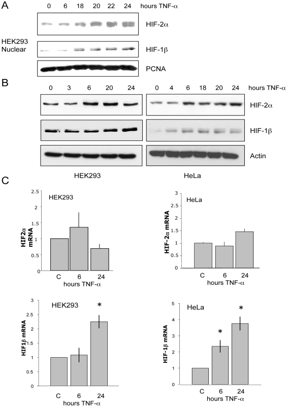
(A) HEK293 were treated with 20 ng/mL TNF-α for the indicated times prior to nuclear extraction. Levels of HIF-2α and HIF-1b were analysed by Western Blot. PCNA was used as a loading control. (B) HEK293 and HeLa cells were treated as in A, prior to lysis. Whole cell lysates were analysed by Western Blot. (C) HEK293 and HeLa cells were treated with 20 ng/mL TNF-α for the indicated periods of times prior to total RNA extraction. Following cDNA synthesis, qPCR was performed for the levels of HIF-2α and HIF-1β. TNF-α increases HIF-1β but not HIF-2α transcript levels
To investigate the mechanism behind HIF-1β and HIF-2α protein increases, the mRNA levels of these two genes following TNF-α exposure were analysed. qRT-PCR analysis indicated that in HEK293 cells or in HeLa cells, TNF-α treatment does not result in any significant increases of HIF-2α mRNA (Figure 1C). However, we found that HIF-1β transcript levels were induced in response to TNF-α in both cell types (Figure 1C).
IKK and NF-κB are required for TNF-α induction of HIF-1β mRNA
TNF-α activates the classical activation pathway that requires the IKK complex [17]. To test if TNF-α induced HIF-1β mRNA was due to IKK-dependent activation of NF-κB, cells were pre-treated with the IKK inhibitor Bay 11 7082 for 30 minutes, prior to TNF-α treatment. Total RNA was extracted and HIF-1β mRNA was measured using qRT-PCR. When IKK was inhibited TNF-α induced HIF-1β mRNA was greatly reduced (Figure 2A). Once again, we did not observe any effects of TNF-α on HIF-2α mRNA in the presence or absence of IKK activity. A significant reduction in the NF-κB subunit and RelA target, p100 mRNA in response to TNF-α, was also seen when IKK was inhibited (Figure 2A).
Fig. 2. TNF-α–induced HIF-1β mRNA is IKK- and RelA-dependent. 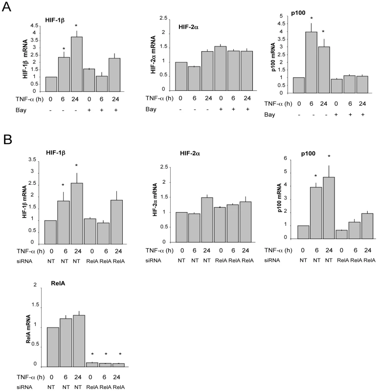
(A) HEK293 cells were pre-treated with the IKK inhibitor Bay 11 7082 prior to treatment with 20 ng/mL TNF-α for the indicated times. Total RNA was extracted, converted to cDNA, and qPCR was performed for the levels of HIF-1β, HIF-2α and p100. (B) HEK293 cells were transfected with control and RelA siRNA oligonucleotides for a total of 72 hours. Where indicated cells were treated with 20 ng/mL TNF-α prior to total RNA extraction. qPCR was performed for the levels of HIF-1β, HIF-2α, p100 and RelA. As IKK has NF-κB independent functions [22], we investigated the role of NF-κB in the control of HIF-1β. Using siRNA mediated depletion of RelA, TNF-α treatment was performed and the effects on the levels of HIF-1β and HIF-2α were analysed by qRT-PCR. The siRNA treatment was very effective at reducing the levels of RelA (Figure 2B). Importantly, depletion of RelA significantly impaired TNF-α mediated induction of HIF-1β and p100 mRNA (Figure 2B). HIF-2α mRNA levels were unaffected by RelA depletion in untreated and TNF-α treated cells (Figure 2B).
Since, RelA depletion only partially prevented TNF-α induction of HIF-1β, we assessed if other NF-κB subunits were also involved in this process. For this purpose, we targeted all NF-κB subunits concurrently, using a siRNA oligonucleotide directed against the Rel homology domain. This siRNA reduced the levels of all subunits of the NF-κB family (Figure S1A). Using RT-qPCR we analysed the levels of HIF-1β in both HEK293 and HeLa cells (Figure 3A). Importantly, depletion of all NF-κB subunits prevented TNF-α mediated induction of HIF-1β in both cell systems (Figure 3A). In addition it prevented the induction of HIF target genes such as GLUT3 and PGK1 (Figure 3B). As expected we did not observe any significant change in HIF-2α mRNA levels (Figure S1B). Consistent with our previous work, HIF-1α mRNA was also significantly decreased in the absence of NF-κB (Figure S1C) [14], [15]. These results support our finding with the IKK inhibitor and RelA depletion in addition to confirming an appreciable level of redundancy within the NF-κB pathway. Taken together these results suggest that IKK-dependent activation of NF-κB, mediates the induction of HIF-1β mRNA following TNF-α treatment.
Fig. 3. TNF-α–induced HIF mRNA levels and activity are NF-κB–dependent. 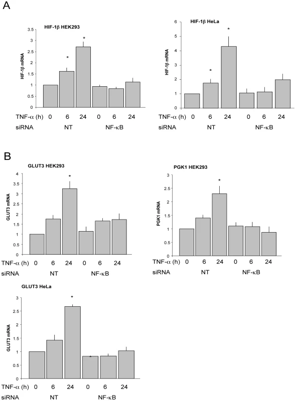
(A) HEK293 and HeLa cells were transfected with control and pan-NF-κB siRNA oligonucleotides for a total of 72 hours. Where indicated cells were treated with 20 ng/mL TNF-α prior to total RNA extraction. qPCR was performed for the levels of HIF-1β. (B) Cells were treated and processed as in A, and qPCR was performed for the indicated genes. HIF-1β is a novel NF-κB target
TNF-α treatment induced HIF-1β protein and mRNA in an IKK-NF-κB dependent manner (Figure 1, Figure 2, Figure 3). As NF-κB is a transcription factor, we wanted to determine if this control was direct. Therefore, we analysed the HIF-1β promoter for possible NF-κB binding sites, using MatInspector (www.genomatix.de). Our analysis revealed that there are three putative NF-κB binding sites, which are located at –36 bp, 59 bp, and 354 bp from the transcription start site. We designed primer sets encompassing these sites as well as a negative control region within the first intronic region of the gene. Chromatin Immunoprecipitation assays using antibodies specific for NF-κB subunits were used in extracts derived from cells untreated or treated with TNF-α for 24 h. PCRs were performed using two sets of primers and products were resolved on agarose gels. The results show that RelA is actively recruited to site 1 within the HIF-1β promoter following TNF-α treatment, while no binding was observed when the control region of the HIF-1β gene was analysed (Figure 4A). Furthermore, recruitment of an active marker of transcription, polymerase II, was also observed at the HIF-1β promoter regions where the NF-κB binding sites are located, following TNF-α treatment (Figure 4B).
Fig. 4. TNF-α induces RelA and RNA polymerase II recruitment to the HIF-1β promoter. 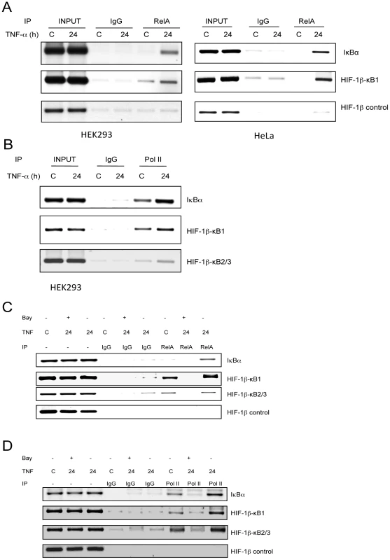
(A) HEK293 and HeLa cells were treated with 20 ng/mL TNF-α for 24 hours prior to crosslinking and lysis. ChIPs were performed using the indicated antibodies and purified DNA was amplified using PCR primers for the HIF-1β promoter and control regions. The IκB-α promoter was used as a positive control. (B) ChIPs were performed for RNA polymerase II and purified DNA processed as in A. (C) HeLa cells were pre-treated with the IKK inhibitor Bay 11 7082 for 30 minutes prior to TNF-α treatment. Cells were crosslinked and lysed 24 hours later. ChIPs were performed for the levels of RelA. (D) ChIPs were performed for RNA polymerase II and purified DNA processed as in A. IκB-α promoter was used as a positive control. As we had observed an active recruitment of NF-κB to the HIF-1β promoter following TNF-α treatment, we sought to formally demonstrate this occurred in an IKK-dependent manner. As such, cells were treated with the IKK inhibitor Bay 11 7082, prior to treatment with TNF-α and ChIPs were performed. TNF-α-dependent recruitment of RelA to the HIF-1β promoter was impaired in cells where IKK activity was reduced (Figure 4C). In addition, we could detect an IKK-dependent increase in binding of p50, c-Rel and p52 to the HIF-1β promoter (Figure S2A, S2B, S2C).
To determine the functional significance of the IKK-dependent recruitment of NF-κB to the HIF-1β promoter region, we examined if TNF-α induced polymerase loading was affected by IKK inhibition (Figure 4D). When the NF-κB pathway is inhibited, RNA polymerase II recruitment to the HIF-1β promoter is severely impaired (Figure 4D), indicating that NF-κB is a necessary factor for the formation of the RNA polymerase II transcriptional complex. These data show a direct regulation of the HIF-1β promoter by NF-κB.
IKK and NF-κB are required for TNF-α induced HIF-1β and HIF-2α protein
Our results indicate that changing NF-κB activity and levels results in altered HIF-1β mRNA. We wanted to determine if this was also seen at the protein level. HeLa cells were treated with IKK inhibitor prior to treatment with TNF-α. Analysis of the whole cell lysates demonstrated that in the absence of IKK activity, TNF-α treatment failed to induce HIF-1β expression (Figure 5A). These results support our mRNA analysis, where IKK inhibition resulted in impaired TNF-α induced HIF-1β (Figure 2A). We also analysed HIF-2α protein levels. Interestingly, inhibition of IKK also prevented TNF-α induced HIF-2α protein. These results suggest that IKK is controlling HIF-2α through a protein stabilisation mechanism and not as a result of increased HIF-2α mRNA production (Figure 2A and Figure 5A).
Fig. 5. TNF-α–induced HIF-1β and HIF-2α proteins are IKK- and NF-κB–dependent. 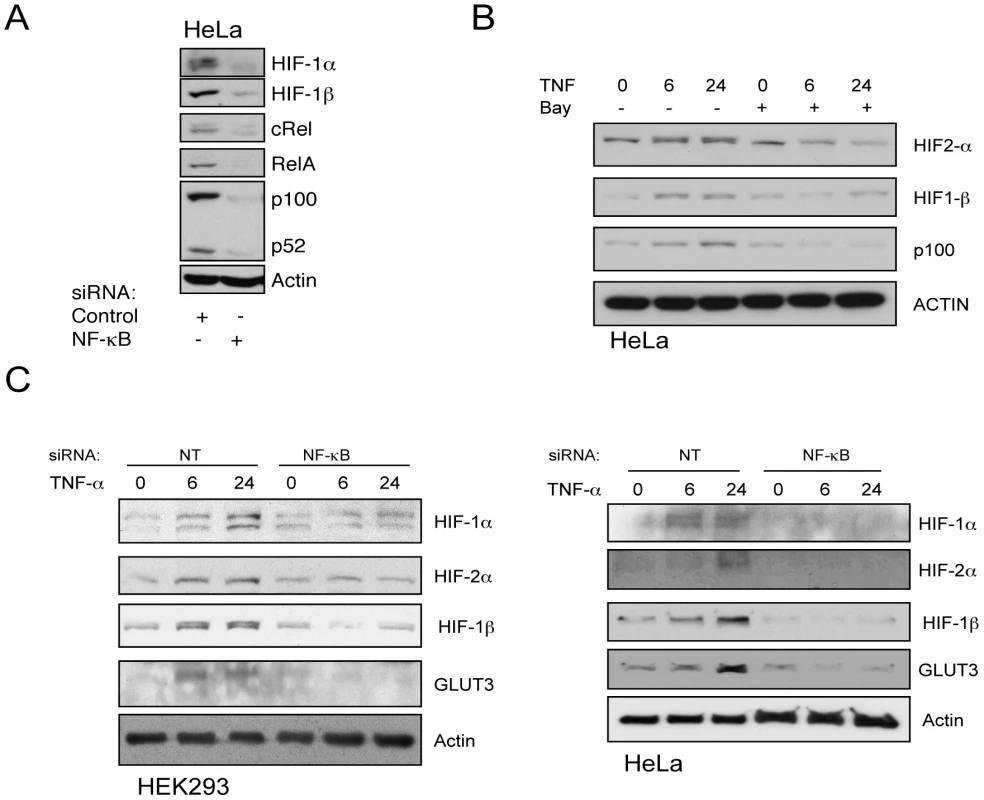
(A) HeLa cells were transfected with control or pan-NF-κB siRNA oligonucleotides and cultured for a total of 48 hours. Whole cell lysates were analysed by Western blot for the proteins mentioned. (B) HeLa cells were pre-treated with the IKK inhibitor Bay 11 7082 for 30 minutes prior to TNF-α treatment for the indicated periods of time prior to lysis. Whole cell lysates were analysed by Western blot for the proteins mentioned. (C) HEK293 and HeLa cells were transfected with control or pan-NF-κB siRNA oligonucleotides and cultured for a total of 72 hours. Where indicated, cells were treated with 20 ng/mL TNF-α prior to lysis. Whole cell lysates were analysed by Western blot for the proteins mentioned. To further demonstrate the involvement of NF-κB in the control of HIF-1β and HIF-2α proteins, NF-κB subunits were depleted by siRNA (Figure 5B). Under conditions of NF-κB impairment, TNF-α treatment did not increase HIF-1β, HIF-1α or, HIF-2α protein levels in HeLa or HEK293 cells (Figure 5B). Taken together these results indicate that IKK-NF-κB control HIF-1β, HIF-1α and HIF-2α proteins.
Changes in HIF-1β alter HIF-2α protein levels and result in AHR inhibition
TNF-α induces increases in HIF-1α [14], HIF-1β, and HIF-2α (Figure 1, Figure 5). We have demonstrated that HIF-1α and HIF-1β increases are both transcriptional ([14], Figure 2, Figure 3, Figure 4, Figure 5, Figure 6). However, we could not establish a direct link between NF-κB and HIF-2α. To investigate the mechanism behind HIF-2α stabilisation, we hypothesised that this was due to increased levels of its binding partner HIF-1β. HIF-1β increases could provide HIF-2α protection from proteolytic degradation. To test this hypothesis, we depleted HIF-1β directly, treated with TNF-α and analysed HIF-2α levels. In the absence of HIF-1β, TNF-α treatment did not result in any visible increases of HIF-2α protein in both HEK293 and HeLa cells (Figure 6A). These results suggest that HIF-1β levels can modulate HIF-2α subunit protein levels. Similar results were also obtained for HIF-1α, in U2OS cells exposed to hypoxia (Figure S3). To further investigate this mechanism, we performed gain of function experiments, where we overexpressed HIF-1β in the absence of any additional treatment. We could observe a dose dependent increase of HIF-1β as expected (Figure 6B). Importantly, this was also evident in the levels of HIF-2α protein (Figure 6B). Taken together these results indicate that HIF-1β is necessary and sufficient for the stabilisation of HIF-2α.
Fig. 6. HIF-1β is required for TNF-α–induced HIF-2α, which represses AHR function. 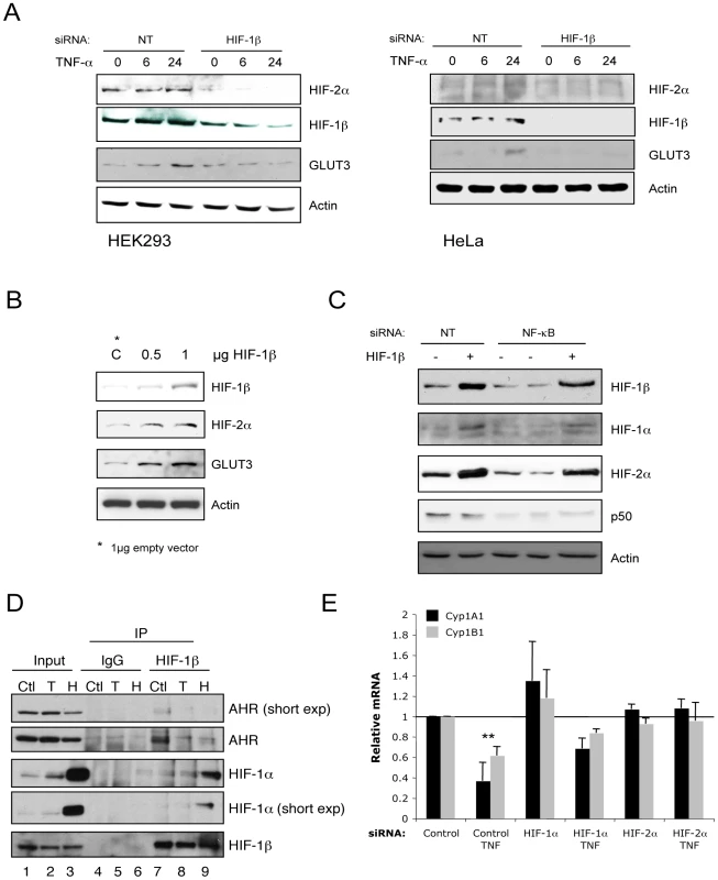
(A) HEK293 and HeLa cells were transfected with control or HIF-1β siRNA oligonucleotides and cultured for a total of 72 hours. Where indicated cells were treated with 20 ng/mL TNF-α prior to lysis. Whole cell lysates were analysed by Western blot for the proteins mentioned. (B) HEK293 cells were transfected with increasing amount of HIF-1β expression plasmid for 48 hours prior to lysates. Whole cell lysates were analysed by Western blot for the indicated proteins. (C) HEK293 cells were transfected with control or pan-NF-κB siRNA oligonucleotides and cultured for 72 hours. Where indicated cells were co-transfected with HIF-1β expression plasmids. Whole cell lysates were analysed by Western blot for the mentioned proteins. (D) HeLa cells were left untreated (Ctl) treated with 20 ng/mL TNF-α (T) or 1% O2 (H) for 24 hours prior to lysis. 200 µg of protein were used to immunoprecipitate HIF-1β. Levels of associated proteins were assessed by western blot using the indicated antibodies. Input corresponds to 10% of starting material. (E) HeLa cells were transfected with control, HIF-1α or HIF-2α siRNA oligonucleotides for a total of 48 hours. Where indicated cells were treated with 20 ng/mL TNF-α prior to total RNA extraction. qPCR was performed for the levels of the indicated genes. We have found that NF-κB mediates the TNF-α induction of HIF-1β mRNA and protein. In addition, gain and loss of function experiments, presented in Figure 6A and 6B, demonstrate that modulation of HIF-1β levels has a direct impact on HIF-2α protein levels. To specifically demonstrate that NF-κB induction of HIF-2α protein is dependent on HIF-1β levels, we performed NF-κB depletion by siRNA and simultaneously increased HIF-1β by exogenous overexpression. Cell extracts were analysed for both HIF-1β and HIF-2α levels by western blot (Figure 6C). Our analysis revealed that HIF-1β overexpression is sufficient to rescue the effects of NF-κB depletion on HIF-2α levels (Figure 6C).
Apart from binding HIF-α subunits, HIF-1β also binds the transcription factor Aryl Hydrocarbon Receptor (AHR) [23]. AHR is responsible for activating genes involved in detoxification and xenobiotic metabolism [23]. To determine if TNF-α induced HIF-1β and consequentially HIF-1α and HIF-2α had any functional significance in the modulation of AHR, we analysed if HIF-1β changed binding partners under these conditions. We co-immunoprecipitated HIF-1β and determined the levels of associated HIF-1α and AHR (Figure 6D). Under non-stimulated conditions, HIF-1β is significantly associated with AHR (Figure 6D, lane 7), however following TNF-α or hypoxia HIF-1β dissociates from AHR (Figure 6D, lane 8 and 9). This suggests that in response to TNF-α or hypoxia AHR function is reduced. To test this hypothesis, we assessed if TNF-α treatment had any effect on the levels of two AHR target genes, CYP1A1 and CYP1B1 (Figure S4A). Our analysis revealed that TNF-α treatment resulted in a time-dependent reduction in the levels of these two genes. Given that TNF-α induces HIF activity and a dissociation of HIF-1β from AHR, we assessed if the levels of CYP1A1 and CYP1B1 could be rescued by HIF-α depletion (Figure 6E). HIF-α depletion was effectively achieved (Figure S4B). Interestingly, the levels of CYP1A1 and CYP1B1 were not reduced by TNF-α treatment when HIF-2α was depleted. Knockdown of HIF-1α resulted in a partial recovery of these two genes (Figure 6E). Taken together these results indicate that TNF-α induced HIF-1β results in higher HIF-α activity and reduced AHR function, suggesting an impaired xenobiotic metabolism.
NF-κB mediated control of the HIF pathway is conserved in mouse and Drosophila
Alignment of the human and mouse HIF-1β promoter region revealed that these are well conserved (Figure S2C). Therefore, we hypothesised that in a murine model HIF-1β was likely to show a similar dependence on NF-κB. To test this hypothesis, Mouse Embryonic Fibroblasts (MEFs) were treated with TNF-α, and HIF-1β levels were analysed (Figure 7A). HIF-1β levels were increased after 24 hours of treatment. In addition, we treated MEFs with an IKK inhibitor (BAY11 7082) for 24 hours. Whole cell lysates were analysed by western blot and the results show that when NF-κB is inhibited, HIF-1β protein level is reduced (Figure 7B). In addition, analysis of IKKβ or IKKα/β null MEFs also revealed a reduction in HIF-1β levels when compared with wildtype MEFs (Figure 7C). Finally, we performed depletion of IKK subunits by siRNA in MEFs (Figure 7D). While depletion of IKKα and IKKβ did not alter the levels of HIF-1β, knockdown of IKKγ resulted in a visible decrease of the HIF-1β protein (Figure 7D). These results suggest that NF-κB dependent transcription of HIF-1β is conserved in mice.
Fig. 7. NF-κB–mediated control of the HIF system is conserved in mice. 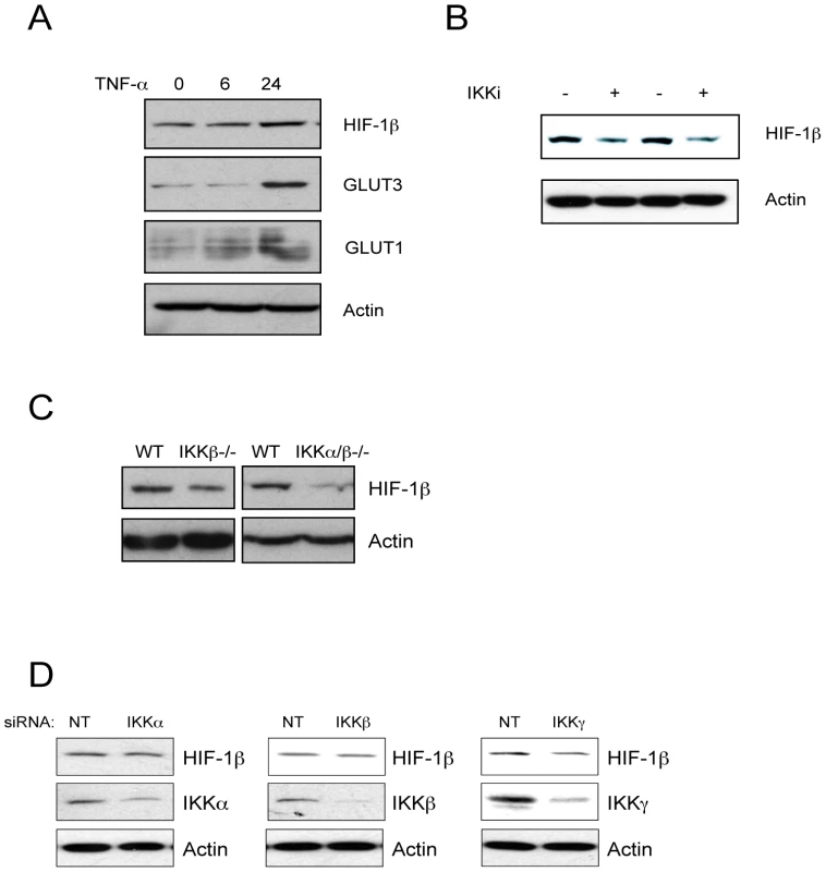
(A) Mouse embryo fibroblasts (MEFs) were treated with 20 ng/mL TNF-α for the indicated period of time prior to lysis. Whole cell lysates were analysed by Western blot. (B) MEFs were treated with the IKK inhibitor for 24 hours prior to lysis. Whole cell lysates were analysed by Western blot for the levels of HIF-1β. (C) Whole cell lysates were obtained from IKK wildtype (WT), IKKβ-/- or IKKα/β-/- MEFs and analysed by western blots. (D) MEFs were transfected with siRNA oligonucleotides for IKKα, IKKβ or IKKγ and the levels of HIF-1β were analysed by western blot. NF-κB and HIF are also conserved in insects [4], [20]. The Drosophila genome encodes three NF-κB family members, Dorsal, Dif and Relish [21], and HIF homologues are encoded by the genes similar (sima; HIF-1α) and tango (HIF-1β) [4]. To test if the NF-κB-mediated regulation of the HIF pathway is also conserved and hence important in Drosophila, we analysed the levels of HIF-1α and HIF-1β mRNA, as well as the HIF target PHD/Fatiga (encoded by Hph in Drosophila) levels in control and flies mutant for loss-of-function alleles of dorsal (Figure 8A). Our analysis revealed that in dorsal mutant flies, both sima and tango mRNA levels are significantly reduced compared to wildType controls (Figure 8A), while an unrelated gene, Iswi, is unaffected in dorsal mutants. Importantly, the levels of the HIF target and PHD2 homologue Hph/fatiga are also reduced (Figure 8A), indicating that the changes observed in mRNA are also functionality translated into lower target gene activation.
Fig. 8. NF-κB–mediated control of the HIF system is conserved in Drosophila. 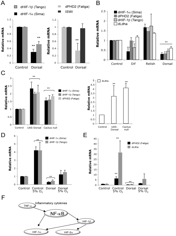
(A) Total RNA was extracted from control and Dorsal null flies, and qPCR was performed for the levels of Drosophila HIF-1α, HIF-1β, PHD and ISWI. (B) Total RNA was extracted from control Dif, Relish and Dorsal null flies, and qPCR was performed for the levels of the indicated genes. (C). Total RNA was extracted from control, UAS-Dorsal or Cactus null flies, and qPCR was performed for the levels of the indicated genes. (D) Total RNA was extracted from control and Dorsal null flies exposed or not to 5% O2 for 24 hours. qPCR was performed for the levels of Drosophila HIF-1α, HIF-1β. (E) As in D, but qPCR was performed for the levels of Drosophila PHD and Ldha. (F) Proposed model of NF-κB modulation of the HIF pathway. NF-κB directly regulates HIF-1α and HIF-1β genes. NF-κB induced HIF-1β mediates HIF-2α stability. Given that Drosophila possesses two additional NF-κB members (Dif and Relish), we next determined if these also contributed to the control of HIF levels and activity in the fly. While loss of dif, resulted in reduced levels of sima and tango, it had no effect in the levels of the target genes Hph/fatiga and ImpL3/ldha (Figure 8B). On the other hand, loss of relish resulted in higher levels of sima and tango and also Sima targets Hph/fatiga and ImpL3/ldha, suggesting that relish acts as a repressor of these genes.
To further analyse the role of NF-κB in the control of the HIF system in Drosophila, we performed gain of function experiments using ubiquitous overexpression of dorsal, and flies hemizygous for a loss of function allele of cactus, the fly homologue of IκB. In both of these genotypes, we observed an increase in the levels of sima and tango and their target genes Hph/fatiga and ImpL3/ldha (Figure 8C). These data suggest that activated Dorsal is able to induce the expression of HIF in the fly.
A recent study using Drosophila S2 cells has demonstrated that hypoxia induces increases in sima mRNA [24]. Given our findings, we next assessed if in adult flies, NF-κB was responsible for hypoxia induced sima mRNA and activity. For this purpose, we exposed adult flies to 5% O2 for 24 hours prior to mRNA extraction. We could confirm that like in S2 cells, hypoxia induces sima mRNA production in adult flies (Figure 8D). Interestingly, we also observed a significant increase in tango mRNA (Figure 8D). Importantly, these responses were abolished in dorsal loss of function flies (Figure 8D). Significantly, levels of the HIF target genes, Hph/fatiga and ImpL3/ldha, following hypoxia exposure, were significantly reduced in the absence of dorsal (Figure 8E). These results demonstrate that Dorsal is required for basal and hypoxia-induced HIF levels and activity in Drosophila.
All together our data demonstrate a novel control mechanism over the HIF system. By controlling HIF-1β, NF-κB is able to indirectly induce HIF-2α and thus has multiple levels of control over the HIF pathway (Figure 8F). Importantly, this is an evolutionary conserved mechanism, observed from flies to humans.
Discussion
In this report we demonstrate an evolutionary conserved and important mode of regulation of the HIF pathway by the transcription factor family NF-κB. We show that NF-κB can control the HIF pathway through several different mechanisms. Our results demonstrate that, in response to TNF-α, NF-κB controls the HIF-1β promoter directly (Figure 4). Depletion or inhibition of NF-κB activity results in low levels of HIF-1β and importantly, HIF-α proteins (Figure 6, Figure 7). Interestingly, we have identified a mechanism by which NF-κB can control HIF-2α levels and activity. We show that HIF-1β is necessary and sufficient for HIF-α stability in normoxia and hypoxia (Figure 6). Significantly, we show that NF-κB-mediated regulation of the HIF system is conserved in mice and Drosophila (Figure 7, Figure 8).
It is well established that HIF-α subunits are tightly regulated in the cell. However, very few studies have investigated how HIF-1β is controlled, or if it responds to stimulation. Yet, HIF-1β importance for development, homeostasis and disease is underscored by the numerous studies where HIF-1β is deleted, systemically or conditionally [25]-[30]. Systemic deletion of HIF-1β results in very early embryonic lethality with defects in placental, yolk sac and hemapoetic vascularization [30]. Conditional deletion of HIF-1β has demonstrated important roles in skin and liver [25], [26], [28].
HIF-1β is also known as Aryl Hydrocarbon Nuclear Translocator (ARNT), and apart from binding HIF-α subunits it is also an important binding partner for other transcription factors such as Aryl Hydrocarbon Receptor (AHR), a central regulator of xenobiotic metabolism [23]. As such, HIF-1β plays a role in both hypoxic and xenobiotic responses [31]. Importantly, several studies have demonstrated that ARNT has higher affinity for the HIF-α subunits, hence hypoxia induces a displacement of ARNT from AHR [32]. Furthermore, hypoxia or HIF-α stabilisation leads to inhibition of AHR activity [33], [34].
From our results, it would be predicted that NF-κB activation by TNF-α, apart from increasing HIF-1β and HIF-α subunits, would lead to the inhibition of AHR response. TNF-α and NF-kB have been shown previously to repress the expression of certain AHR targets such as CYP1A1, however, the mechanism behind such repression is not fully understood [35], [36]. We have thus investigated this possibility. Our results demonstrate that TNF-α induces the displacement of ARNT from AHR to HIF-α (Figure 6D). Furthermore, we show that as published previously [35], [36], TNF-α induces repression of AHR targets such as CYP1A1 and CYP1B1 (Figure 6E and Figure S4A). Importantly, this can be prevented by depletion of HIF-2α and, to some extent, by depletion of HIF-1α. These results indicate that hypoxia or inflammation will lead to a lowering of AHR activity and hence reduced xenobiotic metabolism.
Our results demonstrate a mechanism by which NF-κB can indirectly regulate HIF-2α protein levels. These events take place in normoxia, indicating a PHD independent mechanism. A few reports have analysed PHD-independent HIF-α degradation pathways. Amongst these, HSP90 and RACK1 compete for HIF-1α binding and hence modulate stability [37]–[39]. In these studies, HIF-1α degradation despite being PHD-VHL-independent is still proteasome-dependent [38], [39]. Similarly, GSK3 mediated degradation of HIF-1α is also VHL-independent but relies on the proteasome [40]. These are a few examples of direct regulation of the HIF-α subunits. Our analysis revealed that HIF-1β protects HIF-α subunits from degradation (Figure 6). HIF-1β disruption by both curcumin and EF24 results in proteasome independent degradation of HIF-1α even in hypoxia. How curcumin regulates HIF-1β is still a matter of debate. While some studies indicate the involvement of ubiquitin and the proteasome [41], other studies demonstrate that curcumin disrupts HIF-1β independent of the proteasome [42]. Of note, curcumin is known to inhibit NF-κB in many different cellular backgrounds [43], however, whether HIF-1β mRNA levels decrease in curcumin treated cells has not been investigated.
HIF-2α levels have been shown to be high in macrophages at normoxic levels and also in tumour areas, where near normal levels of oxygen are present [44]. The mechanism behind the increased HIF-2α levels in these areas has not been delineated thus far. While it is possible, that PHD inhibition might take place, our results demonstrate that NF-κB-mediated increases in HIF-1β, induce HIF-2α protein, provide an alternative explanation. However, the levels of HIF-1β under the conditions presented in the published studies, have not been investigated.
NF-κB and the HIF pathways are conserved in flies [4], [20]. While the VHL-PHD-dependent regulation has been shown to be conserved in Drosophila [4], the relevance and conservation of NF-κB-mediated control over HIF has not been previously established. Our analysis of dorsal mutant flies (c-Rel homologue), demonstrates that Dorsal controls the levels and activity of HIF also in Drosophila (Figure 8). We also found that dorsal, sima and tango are expressed in the head of the fly, a tissue we used for our analysis (Figure S5).
Loss of function of dif, although reducing the levels of sima and tango, does not alter Sima activity (Figure 8B), suggesting that the observed reduction in mRNA levels of the HIF subunits is not severe enough to have a functional effect at the protein level. Loss of Relish, an additional NF-κB family member in the fly, resulted in higher levels of sima, tango and its targets (Figure 8B). This suggests that Relish acts a repressor of these genes. In mammalian systems, it is well documented that p50 homodimers can act as repressor forms of NF-κB [45], [46], this is consistent with our analysis of Relish mutants.
Our results also demonstrate that activation of NF-κB, assessed by loss of function of Cactus and overexpression of Dorsal, will lead to increased levels and activity of the HIF system (Figure 8C). This raises the intriguing questions of whether in flies HIF is also a part of the immune system and whether NF-κB mutations alter the HIF response in hypoxia. These questions should thus be addressed in future studies.
Recently, a study in Drosophila S2 cells demonstrated that the levels of sima mRNA were hypoxia inducible [24]. Given these findings, we determined the role of NF-κB in regulating this event. Our results demonstrate that in adult flies, as observed in S2 cells, hypoxia induces sima mRNA. Interestingly, we also observed increases in tango mRNA levels. More importantly, we found that these increases were dependent on Dorsal (Figure 8D). Functionally, inhibition of Dorsal led to significant reduction in the levels of hypoxia-induced Sima-dependent genes (Figure 8E). This suggests that NF-κB also plays a role in the regulation of hypoxia induced HIF in Drosophila. Furthermore, it reveals that, as we had observed in mammalian cells [47], NF-κB is activated following hypoxia in the fly.
In summary, our results provide new evidence of the extensive control of the HIF pathway by the Rel homology domain containing transcription factor family, NF-κB, which is often found deregulated in a number of human pathologies, including cancer, autoimmune diseases and diabetes.
Materials and Methods
Cells
Human embryonic kidney cells HEK293 and human cervix carcinoma HeLa cells were obtained from the ATCC. Mouse Embryonic Fibroblasts were a kind gift from Prof. Ron Hay. All cells were maintained at 5% CO2 in Dulbecco's modified Eagle's medium (Lonza) supplemented with 10% fetal bovine serum (FBS) (Invitrogen), 1% penicillin-streptomycin (Lonza), and 1% L-glutamine (Lonza).
Treatments
TNF-α was obtained from Peprotech, dissolved in PBS and used at a final concentration of 20 ng/mL, subsequent to medium change. IKK inhibitor BAY 11-7082 and MG132 were purchased from Merck Biosciences, dissolved in DMSO and used at a final concentration of 20 µM.
Hypoxia treatments were performed in an InVIVO 300 hypoxia workstation (Ruskin, UK).
Plasmids
HIF-1β/ARNT expression plasmid was a kind gift from Prof. O. Hankinson (UCLA, USA) and was transfected into cells using the Calcium phosphate technique.
siRNA
siRNA oligonucleotides were purchased from MWG and used in a final concentration of 36 nM. siRNA oligonucleotides were transfected using the Calcium phosphate method (HEK293 and HeLa) or using interferin from Polyplus according to manufacturer's instructions.
siRNA sequences
Human
Control-5′ -aac agu cgc guu ugc gac ugg - 3′ [48]
RelA-5′ -gcu gau gug cac cga caa g - 3′ [48]
NF-κB-5′-AAG GUG CAG AAA GAG GAC A-3′
HIF-1β-5′-GGU CAG CAG UCU UCC AUG A-3′
HIF-1α-5′ - CUG AUG ACC AGC AAC UU-3′ [15], [49]
HIF-2α-5′ - CAG CAU CUU UGA CAG U-3′
Mouse
IKKα, GCAGAAGAUUAUUGAUCUA [50]
IKKβ, UGACGUGAAGCAUCUAGUA [50]
IKKγ, GGAUUCGAGCAGUUAGUGA [50]
Drosophila strains and RNA preparation
Fly culture and husbandry was performed after standard protocols. The amorphic dorsal alleles, dl1 and dl4 [51] were crossed to obtain transheterozygous flies lacking dorsal function. To generate cactus loss of function flies, the amorphic cact1 [52] allele was crossed to Df(2L)r10 and cact1 hemizygous adult flies were collected. For overexpression of dorsal, P[matα4-GAL-VP16] [53] was used to drive expression of P[UAS-dl.H]2. Adult flies homozygous for the loss of function alleles Dif1 and relE20 were viable and fertile and maintained as stock [54], [55]. As control an isogenized white1118 stock was used. Hypoxia treatment was performed for 24 hours at 25°C. For RNA preparations, groups of 70 adult flies were frozen in liquid nitrogen and heads were separated using a sieve. Total RNA was extracted using RNAeasy extraction kit (Qiagen, Germany).
Quantitative PCR analysis
Total RNA was extracted with the Nucleospin RNA II isolation system (Macherey Nagel; 740955) or Invisorb spin cell RNA (Invitek), according to the manufacturer's directions. RNA was converted to cDNA using Quantitect Reverse Transcription Kit (Qiagen). cDNA was used in semi-quantitative PCR. For quantitative PCR, Brilliant II Sybr green kit (Stratagene/Agilent), including specific MX3005P 96 well semi-skirted plates, were used to analyse samples on the Mx3005P QPCR platform (Stratagene/Agilent). Actin was used as a normalising gene in all experiments.
RT-qPCR sequences
Actin
For-CTGGGAGTGGGTGGAGGC
Rev-TCAACTGGTCTCAAGTCAGTG
GLUT3
For - CAA TGC TCC TGA GAA GAT CAT AA
Rev-AAA GCG GTT GAC GAA GAG T
HIF-1α
For - CAT AAA GTC TGC AAC ATG GAA GGT
Rev-ATT TGA TGG GTG AGG AAT GGG TT
HIF-2α
For - GCG CTA GAC TCC GAG AAC AT
Rev-TGG CCA CTT ACT ACC TGA CCC TT
HIF-1β
For-CAA GCC CCT TGA GAA GTC AG
Rev-GAG GGG CTA GGC CAC TAT TC
PGK1
For-AAG TGA AGC TCG GAA AGC TTC TAT
Rev-AGG GAA AAG ATG CTT CTG GG
RelA
For-CTGCCGGGATGGCTTCTAT
Rev-CCGCTTCTTCACACACTGGAT
p100
For-AGCCTGGTAGACACGTACCG
Rev-CCGTACGCACTGTCTTCCTT
CYP1A1
For-GAGGCCAGAAGAAACTCCGT
Rev-CCCAGCTCAGCTCAGTACCT
CYP1B1
For-CTGCACTCGAGTCTGCACAT
Rev-TATCACTGACATCTTCGGCG
Fly primers.
drActin
For-GCGTTTTGTACAATTCGTCAGCAACC
Rev-GCACGCGAAACTGCAGCCAA
drHIF-1α (Sima)
For-AGCCCAATCTGCCGCCAACC
Rev-TGGAGGCCAGGTGGTGGGAC
drHIF-1β (Tango)
For-CGGCTGCTCATACGCCCGAG
Rev-GGCCAGCATGTGCGTCTGGT
drPHD (Fatiga)
For-TGGCCCGCCGAGGTAGACAA
Rev-CAGGCCCGTCTCCATCCCCA
drISWI
For-ACAGGTTGCACACTC GATTTTCAGT
Rev-CGGATGAACTGGTGGCCGCA
dLhda
For - CAGTTCGCAACGAACGCGCA
Rev - CAGCTCGCCCTGCAGCTTGT
Whole-cell protein lysates
Briefly, medium was aspirated and cells were washed once in PBS and then resuspended in modified high salt whole cell extraction buffer (20 mM HEPES pH7.6, 400 mM NaCl, 1 mM EDTA, 25% glycerol, 0.1% NP-40, 1 mM dithiothreitol (DTT), 1 mM PMSF, 5 mM NAF, 500 µM Na3VO4, and 1 protease inhibitor cocktail tablet (Roche) per 10 mL buffer). The cells were lysed by incubating them at 4°C on a rotating wheel for 20 minutes before centrifugation at maximum speed for 15 minutes at 4°C.
Western blotting
Following SDS-PAGE, gels were transferred onto polyinylidine difluoride (PVDF) membrane using a semi-dry transfer system. Unless otherwise stated, western blots were incubated in primary antibody at 4°C overnight, followed by a one hour incubation in an HRP-linked secondary antibody solution and developed using Enhanced chemo-luminescence (ECL) solution (PIERCE).
Immunoprecipitations
200 µg of whole cell lysates were used per immunoprecipitation condition. Protein lystates were incubated overnight with 1 µg of antibody in a rotating platform at 4C. 20 µl of packed protein-G-sepharose beads were used to recover the immune-complexes, by incubation for 2 hours at 4C in a rotating platform. Following 3 washes with PBS, complexes were eluted from the beads using 30 µl of SDS-loading buffer and boiled for 5 minutes prior to being run in SDS-PAGE gels.
Antibodies
Antibodies used were: anti-HIF-1α (MAB1536, R&D Systems and sc-10790, Santa Cruz), anti-HIF-1β (3718, Cell Signalling and sc-5580, Santa Cruz), HIF-2α (NB100-122, Novus Biologicals), anti-100/p52 polyclonal (sc-848, Santa Cruz), anti-RelA (sc-372, Santa Cruz), anti-p105/p50 (sc-7178, Santa Cruz)), anti-RelB (sc-48366, Santa Cruz), anti-cRel (sc-71, Santa Cruz) anti-PCNA (P8825, Sigma), anti-Actin (A5441, Sigma), anti-Pol II (sc-47701, Santa Cruz), anti-Glut3 (53520, AnaSpec), anti-AHR (SA-210, Enzo Life Sciences).
Chromatin immunoprecipitation (ChIP)
Cells were grown in a 150 mm plate to 70% confluency and cross-linked with 1% formaldehyde at room temperature for 10 minutes. Glycine was added to a final concentration of 0.125M for 5 m at room temperature. Cells were washed twice with 10 mL of ice-cold phosphate-buffered saline (PBS) and then scraped into 2 mL ice cold harvest buffer (PBS, 1 mM phenylmethylsulfonyl fluoride (PMSF), 1 mg/mL leupeptin, 1 mg/mL aprotinin) before being centrifuged at 1000rpm in a Beckman & Coulter centrifuge at 4°C for 5 minutes. The supernatant was removed and the pellet was resuspended in 400 µL of lysis buffer (1% SDS, 10 mM EDTA, 50 mM Tris-HCl, pH 8.1, 1 mM PMSF, 1 mg/mL leupeptin, 1 mg/mL aprotinin) and left on ice for 10 minutes. Samples were then sonicated at 4°C eight times. Each sonication cycle lasted 15 seconds with a 30 second gap between each sonication using the 130W Sonics Vibracell. Supernatants were recovered by centrifugation at 12,000rpm in an eppendorf microfuge for 10 minutes at 4°C before being diluted 10 fold in dilution buffer (1% Triton X-100, 2 mM EDTA, 150 mM NaCl, 20 mM Tris-HCl, pH 8.1). Samples were then pre-cleared for 2 hours at 4°C with 2 mg of sheared salmon sperm DNA and 20 mL of protein A-Sepharose (50% slurry). At this stage, 10% of the material was kept and stored at –80°C as input material. Immunoprecipitations were performed overnight with specific antibodies (1–2 mg depending on antibody), with the addition of BRIJ-35 detergent to a final concentration of 0.1%. The immune complexes were captured by incubation with 30 mL of protein A-Sepharose (50% slurry) and 2 mg of single stranded sheared salmon sperm DNA (Sigma) for 1 hour at 4°C. The immunoprecipitates were washed sequentially for 5 m each at 4°C in Wash Buffer 1 (0.1% SDS, 1% Triton X-100, 2 mM EDTA, 20 mM Tris-HCl, pH 8.1, 150 mM NaCl), Wash Buffer 2 (0.1% SDS, 1% Triton X-100, 2 mM EDTA, 20 mM Tris-HCl, pH 8.1, 500 mM NaCl), and Wash Buffer 3 (0.25M LiCl, 1% Nonidet P-40, 1% deoxycholate, 1 mM EDTA, 10 mM Tris-HCl, pH 8.1). Beads were washed twice with Tris-EDTA (TE) buffer and eluted with 100 mL of Elution Buffer (1% SDS, 0.1M NaHCO3). Eluates were purified using a DNA purification kit (NBS Biologicals). Samples were then amplified using semi-quantitative PCR and analysed.
ChIP PCR sequences
HIF-1β Control
For - TTC CAC CCA TCC CCC TAT TTT
Rev - ATG CAA AGC TGT TCA ATA GCA TAA
HIF-1β κB1
For-CCA GCG GGC AGA GGG TTA
Rev-ATC CCG GGG ACT TGG GTA GA
HIF-1β κB2/3
For-CGC GGA ATC CAA GAT GGC
Rev-GCC CTC CCT TCA CTG GAC
IκB-α
For-CTT AGA AGT CTG GGG AAA GCA AAT
Rev-GTA ATC CTG TCC CTC TGC AAG T
Statistical analysis
ANOVA and Student's t-tests were performed on the means, and p values were calculated. *, p≤0.050; **, p≤0.010 and ***, p≤0.001.
Additional methods
Additional materials and methods can be found in Text S1.
Supporting Information
Zdroje
1. RochaS
2007
Gene regulation under low oxygen: holding your breath for transcription.
Trends Biochem Sci
32
389
397
2. KennethNS
RochaS
2008
Regulation of gene expression by hypoxia.
Biochem J
414
19
29
3. FandreyJ
GorrTA
GassmannM
2006
Regulating cellular oxygen sensing by hydroxylation.
Cardiovasc Res
71
642
651
4. GorrTA
GassmannM
WappnerP
2006
Sensing and responding to hypoxia via HIF in model invertebrates.
J Insect Physiol
52
349
364
5. EpsteinAC
GleadleJM
McNeillLA
HewitsonKS
O'RourkeJ
2001
C. elegans EGL-9 and mammalian homologs define a family of dioxygenases that regulate HIF by prolyl hydroxylation.
Cell
107
43
54
6. JiangH
GuoR
Powell-CoffmanJA
2001
The Caenorhabditis elegans hif-1 gene encodes a bHLH-PAS protein that is required for adaptation to hypoxia.
Proc Natl Acad Sci U S A
98
7916
7921
7. CentaninL
RatcliffePJ
WappnerP
2005
Reversion of lethality and growth defects in Fatiga oxygen-sensor mutant flies by loss of hypoxia-inducible factor-alpha/Sima.
EMBO Rep
6
1070
1075
8. Lavista-LlanosS
CentaninL
IrisarriM
RussoDM
GleadleJM
2002
Control of the hypoxic response in Drosophila melanogaster by the basic helix-loop-helix PAS protein similar.
Mol Cell Biol
22
6842
6853
9. MaE
HaddadGG
1999
Isolation and characterization of the hypoxia-inducible factor 1beta in Drosophila melanogaster.
Brain Res Mol Brain Res
73
11
16
10. TaylorPC
SivakumarB
2005
Hypoxia and angiogenesis in rheumatoid arthritis.
Curr Opin Rheumatol
17
293
298
11. CatrinaSB
OkamotoK
PereiraT
BrismarK
PoellingerL
2004
Hyperglycemia regulates hypoxia-inducible factor-1alpha protein stability and function.
Diabetes
53
3226
3232
12. BonelloS
ZahringerC
BelAibaRS
DjordjevicT
HessJ
2007
Reactive oxygen species activate the HIF-1alpha promoter via a functional NFkappaB site.
Arterioscler Thromb Vasc Biol
27
755
761
13. RiusJ
GumaM
SchachtrupC
AkassoglouK
ZinkernagelAS
2008
NF-kappaB links innate immunity to the hypoxic response through transcriptional regulation of HIF-1alpha.
Nature
453
807
811
14. van UdenP
KennethNS
RochaS
2008
Regulation of hypoxia-inducible factor-1alpha by NF-kappaB.
Biochem J
412
477
484
15. KennethNS
MudieS
van UdenP
RochaS
2009
SWI/SNF regulates the cellular response to hypoxia.
J Biol Chem
284
4123
4131
16. HaydenMS
GhoshS
2008
Shared principles in NF-kappaB signaling.
Cell
132
344
362
17. PerkinsND
2006
Post-translational modifications regulating the activity and function of the nuclear factor kappa B pathway.
Oncogene
25
6717
6730
18. PerkinsND
GilmoreTD
2006
Good cop, bad cop: the different faces of NF-kappaB.
Cell Death Differ
13
759
772
19. KarinM
YamamotoY
WangQM
2004
The IKK NF-kappa B system: a treasure trove for drug development.
Nat Rev Drug Discov
3
17
26
20. MinakhinaS
StewardR
2006
Nuclear factor-kappa B pathways in Drosophila.
Oncogene
25
6749
6757
21. HetruC
HoffmannJA
2009
NF-kappaB in the immune response of Drosophila.
Cold Spring Harb Perspect Biol
1
a000232
22. ChariotA
2009
The NF-kappaB-independent functions of IKK subunits in immunity and cancer.
Trends Cell Biol
19
404
413
23. BockKW
KohleC
2009
The mammalian aryl hydrocarbon (Ah) receptor: from mediator of dioxin toxicity toward physiological functions in skin and liver.
Biol Chem
390
1225
1235
24. DekantyA
RomeroNM
BertolinAP
ThomasMG
LeishmanCC
2010
Drosophila genome-wide RNAi screen identifies multiple regulators of HIF-dependent transcription in hypoxia.
PLoS Genet
6
e1000994
doi:10.1371/journal.pgen.1000994
25. WangXL
SuzukiR
LeeK
TranT
GuntonJE
2009
Ablation of ARNT/HIF1beta in liver alters gluconeogenesis, lipogenic gene expression, and serum ketones.
Cell Metab
9
428
439
26. YimSH
ShahY
TomitaS
MorrisHD
GavrilovaO
2006
Disruption of the Arnt gene in endothelial cells causes hepatic vascular defects and partial embryonic lethality in mice.
Hepatology
44
550
560
27. TomitaS
JiangHB
UenoT
TakagiS
TohiK
2003
T cell-specific disruption of arylhydrocarbon receptor nuclear translocator (Arnt) gene causes resistance to 2,3,7,8-tetrachlorodibenzo-p-dioxin-induced thymic involution.
J Immunol
171
4113
4120
28. GengS
MezentsevA
KalachikovS
RaithK
RoopDR
2006
Targeted ablation of Arnt in mouse epidermis results in profound defects in desquamation and epidermal barrier function.
J Cell Sci
119
4901
4912
29. TomitaS
SinalCJ
YimSH
GonzalezFJ
2000
Conditional disruption of the aryl hydrocarbon receptor nuclear translocator (Arnt) gene leads to loss of target gene induction by the aryl hydrocarbon receptor and hypoxia-inducible factor 1alpha.
Mol Endocrinol
14
1674
1681
30. AbbottBD
BuckalewAR
2000
Placental defects in ARNT-knockout conceptus correlate with localized decreases in VEGF-R2, Ang-1, and Tie-2.
Dev Dyn
219
526
538
31. SekineH
MimuraJ
YamamotoM
Fujii-KuriyamaY
2006
Unique and overlapping transcriptional roles of arylhydrocarbon receptor nuclear translocator (Arnt) and Arnt2 in xenobiotic and hypoxic responses.
J Biol Chem
281
37507
37516
32. GradinK
McGuireJ
WengerRH
KvietikovaI
fhitelawML
1996
Functional interference between hypoxia and dioxin signal transduction pathways: competition for recruitment of the Arnt transcription factor.
Mol Cell Biol
16
5221
5231
33. KallioPJ
PongratzI
GradinK
McGuireJ
PoellingerL
1997
Activation of hypoxia-inducible factor 1alpha: posttranscriptional regulation and conformational change by recruitment of the Arnt transcription factor.
Proc Natl Acad Sci U S A
94
5667
5672
34. FlemingCR
BilliardSM
Di GiulioRT
2009
Hypoxia inhibits induction of aryl hydrocarbon receptor activity in topminnow hepatocarcinoma cells in an ARNT-dependent manner.
Comp Biochem Physiol C Toxicol Pharmacol
150
383
389
35. KeS
RabsonAB
GerminoJF
GalloMA
TianY
2001
Mechanism of suppression of cytochrome P-450 1A1 expression by tumor necrosis factor-alpha and lipopolysaccharide.
J Biol Chem
276
39638
39644
36. ZordokyBN
El-KadiAO
2009
Role of NF-kappaB in the regulation of cytochrome P450 enzymes.
Curr Drug Metab
10
164
178
37. IsaacsJS
JungYJ
MimnaughEG
MartinezA
CuttittaF
2002
Hsp90 regulates a von Hippel Lindau-independent hypoxia-inducible factor-1 alpha-degradative pathway.
J Biol Chem
277
29936
29944
38. LiuYV
BaekJH
ZhangH
DiezR
ColeRN
2007
RACK1 competes with HSP90 for binding to HIF-1alpha and is required for O(2)-independent and HSP90 inhibitor-induced degradation of HIF-1alpha.
Mol Cell
25
207
217
39. LiuYV
HubbiME
PanF
McDonaldKR
MansharamaniM
2007
Calcineurin promotes hypoxia-inducible factor 1alpha expression by dephosphorylating RACK1 and blocking RACK1 dimerization.
J Biol Chem
282
37064
37073
40. FlugelD
GorlachA
MichielsC
KietzmannT
2007
Glycogen synthase kinase 3 phosphorylates hypoxia-inducible factor 1alpha and mediates its destabilization in a VHL-independent manner.
Mol Cell Biol
27
3253
3265
41. ChoiH
ChunYS
KimSW
KimMS
ParkJW
2006
Curcumin inhibits hypoxia-inducible factor-1 by degrading aryl hydrocarbon receptor nuclear translocator: a mechanism of tumor growth inhibition.
Mol Pharmacol
70
1664
1671
42. ThomasSL
ZhongD
ZhouW
MalikS
LiottaD
2008
EF24, a novel curcumin analog, disrupts the microtubule cytoskeleton and inhibits HIF-1.
Cell Cycle
7
2409
2417
43. LinJK
2007
Molecular targets of curcumin.
Adv Exp Med Biol
595
227
243
44. LofstedtT
FredlundE
Holmquist-MengelbierL
PietrasA
OvenbergerM
2007
Hypoxia inducible factor-2alpha in cancer.
Cell Cycle
6
919
926
45. TongX
YinL
WashingtonR
RosenbergDW
GiardinaC
2004
The p50-p50 NF-kappaB complex as a stimulus-specific repressor of gene activation.
Mol Cell Biochem
265
171
183
46. GuanH
HouS
RicciardiRP
2005
DNA binding of repressor nuclear factor-kappaB p50/p50 depends on phosphorylation of Ser337 by the protein kinase A catalytic subunit.
J Biol Chem
280
9957
9962
47. CulverC
SundqvistA
MudieS
MelvinA
XirodimasD
2010
Mechanism of hypoxia-induced NF-kappaB.
Mol Cell Biol
30
4901
4921
48. AndersonLA
PerkinsND
2003
Regulation of RelA (p65) function by the large subunit of replication factor C.
Mol Cell Biol
23
721
732
49. NewtonIP
KennethNS
AppletonPL
NathkeI
RochaS
2010
Adenomatous polyposis coli and hypoxia-inducible factor-1{alpha} have an antagonistic connection.
Mol Biol Cell
21
3630
3638
50. KennethNS
MudieS
RochaS
IKK and NF-kappaB-mediated regulation of Claspin impacts on ATR checkpoint function.
EMBO J
29
2966
2978
51. NUSSLEIN-VOLHARDC
1979
Maternal effect mutations that alter the spatial coordinates of the embryo of Drosophila melanogaster:
New York
Academic Press Inc
52. RothS
HiromiY
GodtD
Nusslein-VolhardC
1991
cactus, a maternal gene required for proper formation of the dorsoventral morphogen gradient in Drosophila embryos.
Development
112
371
388
53. BossingT
BarrosCS
BrandAH
2002
Rapid tissue-specific expression assay in living embryos.
Genesis
34
123
126
54. RutschmannS
JungAC
HetruC
ReichhartJM
HoffmannJA
2000
The Rel protein DIF mediates the antifungal but not the antibacterial host defense in Drosophila.
Immunity
12
569
580
55. HedengrenM
AslingB
DushayMS
AndoI
EkengrenS
1999
Relish, a central factor in the control of humoral but not cellular immunity in Drosophila.
Mol Cell
4
827
837
Štítky
Genetika Reprodukční medicína
Článek Composite Effects of Polymorphisms near Multiple Regulatory Elements Create a Major-Effect QTLČlánek Horizontal Transfer, Not Duplication, Drives the Expansion of Protein Families in ProkaryotesČlánek Segregating Variation in the Polycomb Group Gene Alters the Effect of Temperature on Multiple TraitsČlánek Global Analysis of the Impact of Environmental Perturbation on -Regulation of Gene ExpressionČlánek H3K9me-Independent Gene Silencing in Fission Yeast Heterochromatin by Clr5 and Histone DeacetylasesČlánek A Mutation in the Gene Encoding Mitochondrial Mg Channel MRS2 Results in Demyelination in the Rat
Článek vyšel v časopisePLOS Genetics
Nejčtenější tento týden
2011 Číslo 1
-
Všechny články tohoto čísla
- A Meta-Analysis of Genome-Wide Association Scans Identifies IL18RAP, PTPN2, TAGAP, and PUS10 As Shared Risk Loci for Crohn's Disease and Celiac Disease
- Composite Effects of Polymorphisms near Multiple Regulatory Elements Create a Major-Effect QTL
- Horizontal Transfer, Not Duplication, Drives the Expansion of Protein Families in Prokaryotes
- Genome-Wide Association Study SNPs in the Human Genome Diversity Project Populations: Does Selection Affect Unlinked SNPs with Shared Trait Associations?
- Friedreich's Ataxia (GAA)•(TTC) Repeats Strongly Stimulate Mitotic Crossovers in
- Zebrafish Mutation Leads to mRNA Splicing Defect and Pituitary Lineage Expansion
- Histone H4 Lysine 12 Acetylation Regulates Telomeric Heterochromatin Plasticity in
- Bub1-Mediated Adaptation of the Spindle Checkpoint
- Segregating Variation in the Polycomb Group Gene Alters the Effect of Temperature on Multiple Traits
- Signaling Role of Fructose Mediated by FINS1/FBP in
- RNF12 Activates and Is Essential for X Chromosome Inactivation
- Comparative Study between Transcriptionally- and Translationally-Acting Adenine Riboswitches Reveals Key Differences in Riboswitch Regulatory Mechanisms
- Global Analysis of the Impact of Environmental Perturbation on -Regulation of Gene Expression
- Application of a New Method for GWAS in a Related Case/Control Sample with Known Pedigree Structure: Identification of New Loci for Nephrolithiasis
- H3K9me-Independent Gene Silencing in Fission Yeast Heterochromatin by Clr5 and Histone Deacetylases
- A Mutation in the Gene Encoding Mitochondrial Mg Channel MRS2 Results in Demyelination in the Rat
- Transcription Initiation Patterns Indicate Divergent Strategies for Gene Regulation at the Chromatin Level
- The Transposon-Like Correia Elements Encode Numerous Strong Promoters and Provide a Potential New Mechanism for Phase Variation in the Meningococcus
- Proteins Encoded in Genomic Regions Associated with Immune-Mediated Disease Physically Interact and Suggest Underlying Biology
- A Novel RNA-Recognition-Motif Protein Is Required for Premeiotic G/S-Phase Transition in Rice ( L.)
- The Mucin-Like Protein OSM-8 Negatively Regulates Osmosensitive Physiology Via the Transmembrane Protein PTR-23
- Genome Sequencing and Comparative Transcriptomics of the Model Entomopathogenic Fungi and
- Rnf12—A Jack of All Trades in X Inactivation?
- Joint Genetic Analysis of Gene Expression Data with Inferred Cellular Phenotypes
- Evolutionary Conserved Regulation of HIF-1β by NF-κB
- Quaking Regulates Expression through Its 3′ UTR in Oligodendrocyte Precursor Cells
- PLOS Genetics
- Archiv čísel
- Aktuální číslo
- Informace o časopisu
Nejčtenější v tomto čísle- H3K9me-Independent Gene Silencing in Fission Yeast Heterochromatin by Clr5 and Histone Deacetylases
- Evolutionary Conserved Regulation of HIF-1β by NF-κB
- Rnf12—A Jack of All Trades in X Inactivation?
- Joint Genetic Analysis of Gene Expression Data with Inferred Cellular Phenotypes
Kurzy
Zvyšte si kvalifikaci online z pohodlí domova
Současné možnosti léčby obezity
nový kurzAutoři: MUDr. Martin Hrubý
Všechny kurzyPřihlášení#ADS_BOTTOM_SCRIPTS#Zapomenuté hesloZadejte e-mailovou adresu, se kterou jste vytvářel(a) účet, budou Vám na ni zaslány informace k nastavení nového hesla.
- Vzdělávání


