-
Články
Top novinky
Reklama- Vzdělávání
- Časopisy
Top články
Nové číslo
- Témata
Top novinky
Reklama- Videa
- Podcasty
Nové podcasty
Reklama- Kariéra
Doporučené pozice
Reklama- Praxe
Top novinky
ReklamaA Gatekeeper Chaperone Complex Directs Translocator Secretion during Type Three Secretion
Type Three Secretion Systems (T3SS) are essential virulence factors found in many pathogenic Gram-negative bacteria. These machines aid infection by delivering bacterial proteins into host cells where these proteins modulate host processes and help establish a niche for the bacteria. Protein delivery occurs in a highly regulated manner in which proteins involved in early steps in infection, or necessary to build the secretion conduit, are typically secreted before other substrates, a phenomenon termed secretion hierarchy. This study presents the structure of a molecular complex that physically links one class of early substrates, components of the secretion pore termed translocators, to a gatekeeper protein, a protein that has been implicated in the secretion hierarchy. Disruption of this interaction in Shigella disrupts the secretion of translocators, while supporting increased secretion of effectors, resulting in phenotypes indistinguishable from a gatekeeper deletion, and leading to the conclusion that a gatekeeper-chaperone-translocator complex is a critical component of the T3SS.
Published in the journal: . PLoS Pathog 10(11): e32767. doi:10.1371/journal.ppat.1004498
Category: Research Article
doi: https://doi.org/10.1371/journal.ppat.1004498Summary
Type Three Secretion Systems (T3SS) are essential virulence factors found in many pathogenic Gram-negative bacteria. These machines aid infection by delivering bacterial proteins into host cells where these proteins modulate host processes and help establish a niche for the bacteria. Protein delivery occurs in a highly regulated manner in which proteins involved in early steps in infection, or necessary to build the secretion conduit, are typically secreted before other substrates, a phenomenon termed secretion hierarchy. This study presents the structure of a molecular complex that physically links one class of early substrates, components of the secretion pore termed translocators, to a gatekeeper protein, a protein that has been implicated in the secretion hierarchy. Disruption of this interaction in Shigella disrupts the secretion of translocators, while supporting increased secretion of effectors, resulting in phenotypes indistinguishable from a gatekeeper deletion, and leading to the conclusion that a gatekeeper-chaperone-translocator complex is a critical component of the T3SS.
Introduction
Type Three Secretion Systems (T3SS) are conserved bacterial protein delivery machines used by many pathogenic Gram-negative bacteria to deliver a diverse group of protein molecules, termed effectors, into cells [1]–[4]. The type three secretion (T3S) apparatus is a conserved molecular machine that forms a protein-conducting channel from the bacterial cytosol to the target cell cytosol. Major structural components of the T3SS include: a cytosolic-ring complex, which includes the ATPase that catalyzes protein unfolding and secretion; a basal-body, which forms a pore across both the inner and outer bacterial membranes; a needle complex, which extends from the basal-body to the host cell; and translocators, which form a pore in the target cell membrane, termed a translocon [5], [6].
The translocon, through which effectors enter the host cell, is an oligomeric pore formed by bacterial proteins termed translocators that are themselves secretion substrates of the T3SS [2]. Translocator secretion is regulated such that it occurs prior to effector secretion, ensuring that effector secretion occurs after a functional conduit from the bacterial cytoplasm to the target cell has been formed [7], [8]. Efficient secretion is dependent on the interaction of specialized chaperones with cytosolic T3SS components [9]. Molecular structures have revealed two architectures for T3S chaperones: a mixed α/β homo or heterodimer and an all α-helical tetratricopeptide repeat (TPR) chaperone [10]–[14]. These chaperones, termed class I (α/β) and class II (TPR), are specific for effectors and translocators, respectively [9]. Our understanding of how the T3SS switches from translocator to effector secretion is limited, but in multiple systems this process is known to involve a conserved gatekeeper protein [15]–[20]. Gatekeepers are encoded as one of two molecular architectures, either as two separate proteins (YopN-TyeA family), or as a gene fusion (MxiC family) [20]. The importance of this architectural distinction is unknown, and a protein fusion resulting from ribosomal frameshifting has been reported without an evident functional change [21].
In Chlamydia, the gatekeeper, CopN, is known to directly bind Scc3, a translocator-specific chaperone [22]–[26]. Translocator-specific chaperones (class II chaperones) bind directly to translocators, prevent their degradation, and maintain the translocators in a secretion competent state [9], [13]. Structures from homologous class II chaperone/translocator pairs have revealed the chaperone to be a TPR protein with a conserved binding groove that binds an amino terminal chaperone-binding motif on the translocator [12]–[14], [27]. In addition to binding translocators, Scc3 also binds CopN, although the nature of this interaction is unknown [22], [23], [25], [26]. It is not known if CopN and the Chlamydial translocators (CopB/B2) compete for the TPR binding groove on Scc3, or if different binding determinants are responsible for the Scc3-CopN interaction. More fundamentally, it is not known how gatekeepers promote translocator secretion.
Gatekeepers and translocator chaperones have been observed in immunopurified complexes from other systems, but only as components of large complexes that also include other components of the T3SS [15], [17], [28]. Because such complexes are not readily accessible to structural study, we have focused our structural studies on the gatekeeper-translocator chaperone complex from Chlamydia. We reasoned that the CopN-Scc3 complex is likely to be involved in the ordered secretion of translocators prior to effectors, a conserved phenomenon termed the translocator-effector secretion hierarchy.
The origin of the translocator-effector secretion hierarchy is not understood, but has been proposed to arise from differential affinities and competition for binding sites either between chaperones and their effector or translocator cargo or between chaperone-effector/translocator complexes and cytosolic components of the T3SS [10], [11], [29]–[32]. To assess the importance of gatekeeper-translocator chaperone interactions in diverse pathogens, and because adequate tools and reagents for functional analysis of CopN mutants are not available in Chlamydia, we have extended our structural analysis with functional studies of MxiC and IpgC, the gatekeeper and translocator-specific chaperone from Shigella.
Results
Structure of Scc3-CopN complex
We determined the crystal structure of the Scc3-CopNΔ84 complex and refined the structure to 2.2 Å (PDB ID 4NRH). The amino terminal 84 residues of CopN were not included in the construct used for crystallization because they are unstructured [25]. Data collection and structure refinement statistics are given in Table 1, and representative electron density is shown in Supporting Figure S1. Two nearly identical Scc3-CopNΔ84 complexes (RMS deviation 0.34 Å for all CopNΔ84 mainchain atoms and 0.49 Å for all Scc3 mainchain atoms) are present in the asymmetric unit. CopNΔ84 forms a long cylindrical structure composed of three helical domains (Figure 1). A search for structurally similar proteins using the DALI software [33], indicates structural homology to the globin fold, which, aside from the use described here, is used in bacteria both as a light harvesting complex and as a stress response sigma factor [34]–[36]. In gatekeeper proteins multiple domains are concatenated through elongated connecting helices, whereas globin domains typically oligomerize through lateral contacts. CopNΔ84 is structurally similar to other gatekeeper proteins, both MxiC from Shigella and the YopN-TyeA complex from Yersinia. The most substantial differences among family members relate to the position of the carboxy-terminal domain or subunit (Figure 1 and [37], [38]). In the Scc3-CopNΔ84 complex, this domain is translated ∼9.5 Å and rotated ∼50° relative to the YopN-TyeA complex (Figure 1). Similarly, Scc3 is structurally similar to other translocator chaperones. The striking result from the Scc3-CopNΔ84 is the unexpected assembly of the complex and the role of the Scc3 amino terminus in binding CopNΔ84.
Fig. 1. Crystal structure of Scc3-CopNΔ84. 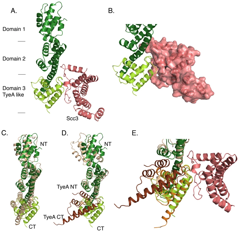
CopN is colored green, with the YopN homology region in dark green and the TyeA homology region in light green. A. A ribbon diagram of the Scc3-CopNΔ84 structure. Approximate domain boundaries are indicated. Scc3, salmon, binds across the domain 2-domain 3 domain interface. B. A close-up of the Scc3-CopNΔ84 interface, oriented as in A, with Scc3 shown as a molecular surface. Scc3 forms a relatively flat surface that bridges domains 2 and 3 of CopNΔ84. C., D. Comparisons of CopN and homologs. C. Comparison between MxiC and CopN. MxiC is colored tan and shown with thin helices. D. Comparison between CopN and the YopN/TyeA complex. YopN is tan and TyeA is brown. YopN and TyeA are and shown with thin helices. E. Overlay of TyeA in orientation shown in D. and when aligned as a rigid body to the carboxy terminal 91 residues of CopN (rmsd = 0.4 Å). Tab. 1. Data collection, phasing, and refinement statistics. 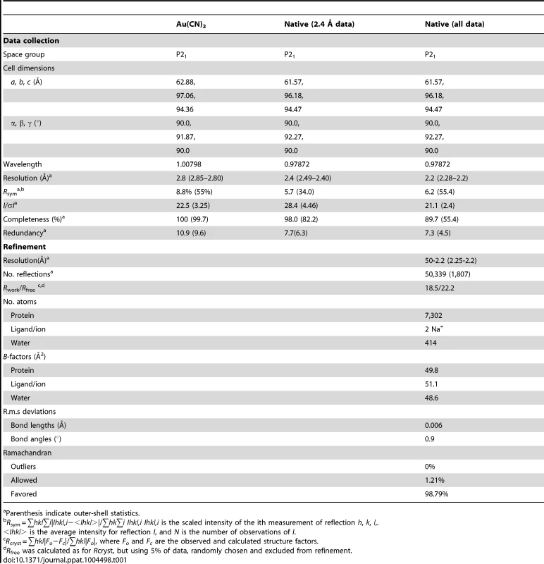
Parenthesis indicate outer-shell statistics. T3SS chaperones bind the amino terminus of effectors and translocators, and class II chaperones (those specific for translocators) use a conserved peptide-in-groove binding mode, utilizing the TPR binding groove, in which translocators bind in the concave face of the chaperone [9]–[14], [27]. The structure reveals that Scc3 does not engage CopNΔ84 using this conserved binding groove. Instead, the amino terminus, referred to here as a gatekeeper-binding region (GBR), forms a relatively flat surface, adjacent to the convex side of the TPR and binds across the interdomain interface of the last two domains of CopNΔ84 (Figure 1). The interface formed by this interaction results from burial of 980 Å2 of surface area. The Scc3 side of the interface is formed exclusively by residues from the GBR, consistent with separate functions of translocator and gatekeeper binding for the TPR and GBR regions of Scc3. Despite minimal sequence conservation, other translocator chaperones also have an amino terminal extension (GBR) prior to the TPR domain (Figure 2). In the homologs from Shigella and Pseudomonas this region mediates homo-dimerization, although translocator binding is known to disrupt these dimers such that in translocator-chaperone complexes this region (GBR) is no longer involved in homodimerization [13], [29], [39]. In the homologs from Yersinia and Pseudomonas crystallization and structure determination required removal of the GBR [12], [14], [27].
Fig. 2. Sequence conservation in CopN and Scc3. 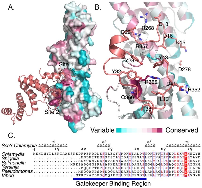
A., B. Sequence conservation displayed on the CopNΔ84 structure. Residues are colored by conservation (pink is conserved, blue is variable). A. Scc3 interacts with two conserved regions, site 1 and site 2, on CopN. B. An expanded view of the CopN-Scc3 interface. For clarity only the GBR region is shown. The interface is primarily composed of hydrophobic residues from Scc3 that surround a conserved arginine (R365) on CopN. C. Sequence conservation within GBR's is minimal. Scc3 homologs from multiple species are aligned based on conservation throughout their sequences, revealing that the GBR region is present, but not highly conserved in homologs. Sequences used are from C. pneumonia, S flexneri, S. enterica, B. pertussis, Y. enterocolitica, P. aeruginosa, V. parahaemolyticus. Multiple sequences from each genera used in C. were used in A. and B. (see methods). The Scc3-CopN interface is conserved in other bacteria
Scc3 engages CopNΔ84 with residues from the GBR, which bind a contiguous surface on CopNΔ84. This surface is formed by two distant patches of sequence conservation, site 1 and site 2, and spans the second and third domains of CopNΔ84, requiring these two domains to be appropriately oriented (Figures 1, 2). In the YopN-TyeA family, the third domain is encoded as a separate protein, such that in these homologs one would predict the interdomain interface recognized by the Scc3 homolog to be a dimer.
Residues 16–23 of Scc3 interact principally with site 1 of CopNΔ84, whereas residues 24–43 form a much larger interface in which Scc3 projects a ring of hydrophobic sidechains toward CopNΔ84 to surround a highly conserved arginine (R365) on CopNΔ84 (Figures 2, Supporting Figure S2). This interaction includes three tyrosines, one of which, Y43 is oriented to allow a π-cation interaction. Peripheral to this ring of hydrophobic residues are a collection of inter-molecular salt bridges (Figure 2). The circumscribed arginine (R365) is conserved across diverse species, including species with two polypeptide gatekeepers and is among the most conserved surface exposed residues in this protein family (this residue is an arginine in homologs from Shigella, Vibrio, Pseudomonas, Bordetella, and Yersinia and glutamine in Salmonella) (Supporting Figure S2). Residues on the CopN side of this interface are better conserved than those on the GBR, despite the fact that they span two proteins in the Yersinia architecture and are on the same protein in the architecture presented here (Figure 2, Supporting Figure S2).
To assess the importance of the two binding regions, we disrupted each interaction by mutagenesis. We made an amino terminal 24 amino acid deletion to Scc3 (Scc3Δ24), which eliminates the GBR-site one interaction. We also mutated the central arginine and two adjacent residues (A362R, R365D, and G369R) in site 2 of CopNΔ84 (CopNΔ84-RDR). A362, R365, and G369 are buried by Scc3 and likely solvent exposed in unliganded CopNΔ84. We introduced charged residues at these sites with the expectation that the solvent exposed charges would not disturb CopN, but would disrupt the CopNΔ84-Scc3 complex. CopNΔ84-RDR and Scc3Δ24, are well-folded, as judged by circular dichroism spectra similar to CopNΔ84 and Scc3 (Supporting Figure S3, Methods S1). As judged by the inability of Scc3Δ24 to bind CopNΔ84 and the inability of CopNΔ84-RDR to bind Scc3 (Supporting Figure S4), both regions are important for Scc3-CopNΔ84 complex formation.
The translocator-binding site in Scc3 is available in the Scc3-CopN complex
TPR family proteins are often unfolded when not bound to appropriate ligands and are considered to be somewhat flexible proteins [40], [41]. Scc3 has an appropriately organized but empty binding cleft when bound to CopN. Structural comparison with other class II chaperones, for which structures have been determined in complex with translocator-derived peptides, indicates that CopNΔ84 binding causes no significant reorganization of the translocator-binding site (Figure 3A). The Scc3-CopNΔ84 binding mode leaves the translocator-binding site on Scc3 unperturbed and available to bind translocators (Figure 3A). In support of this observation, the purified Scc3-CopNΔ84 complex is able to directly bind a translocator-derived peptide (presented as residues 158–177 from CopB fused to GST) and form a CopNΔ84-Scc3-CopB158-177 complex as judged by size exclusion chromatography (Figure 3B). Isothermal Titration Calorimetry (ITC) using a synthetic peptide (CopB residues 163–173) revealed Kd's of 79±16 µM for the Scc3-CopB peptide complex and 49±13 µM for the Scc3-CopNΔ84 peptide complex (Supporting Figure S5, Methods S1).
Fig. 3. The Scc3-CopNΔ84 complex binds directly to translocators. 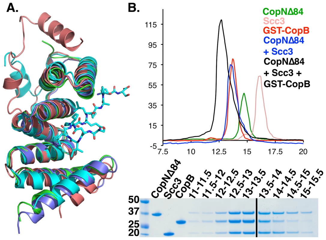
A. An overlay of class II T3S chaperones. All structures except the Scc3 structure were determined bound to translocator peptides. For clarity, only the IpaB peptide from Shigella is shown. Scc3 is shown in salmon, IpgC (Shigella) in teal, PcrH (Pseudomonas) in green, and SycD (Yersinia) in blue. The peptide-binding site is conserved and open in Scc3. B. CopN-Scc3 complex directly binds to the CopB translocator. Top: gel filtration traces reveal that the ternary complex is tight enough to survive gel filtration. Bottom: SDS PAGE confirming complex formation. Disruption of the Scc3-CopN interface alters secretion in Shigella
To determine the importance of the gatekeeper-chaperone interaction during T3S, we disrupted the homologous gatekeeper-translocator chaperone interface in Shigella. Shigella, unlike Chlamydia, are genetically tractable allowing disruption of the endogenous mxiC gene (the copN homolog) and rescue with a plasmid expressing mutant or wild-type MxiC. This is a well-established strategy that has been used to study other MxiC mutants [18], [28]. The CopN and Scc3 homologs in Shigella, MxiC and IpgC, form a complex that includes the T3SS ATPase [28]. The Scc3-CopNΔ84 structure was disrupted by mutation of A362R, R365D, and G369R on CopN (Supporting Figure S4), supporting the idea that the homologous mutations would disrupt IpgC-MxiC interface. We expressed the E331R/R334D/I338R MxiC mutant (MxiC-RDR) from a plasmid in a previously described mxiC null Shigella strain [18] and compared secretion profiles following Congo Red induction. MxiC-RDR is deficient in secretion of the translocators IpaB, IpaC, and IpaD, but efficiently secretes IpaA, an effector, and secretes elevated levels of the effectors OspC1-3 and IpgB (Figure 4A). IpaA is not secreted efficiently if wild type MxiC is present, but is secreted earlier and in greater quantities from ΔMxiC or MxiC-RDR strains (Figure 4B, compare IpaA secretion at 10 minutes and 60 minutes). The secretion profile of MxiC-RDR closely mimics that of the ΔMxiC strain (Figure 4A), highlighting the importance of gatekeeper-translocator chaperone complexes in translocator secretion. To further evaluate the importance of the conserved, central, arginine, we evaluated the secretion profile of a single point mutant, MxiC-R334D, revealing a phenotype similar to the triple mutant (Supporting Figure S6). Similar to CopN, MxiC is both the gatekeeper and a T3S substrate [18], [42], [43]. To verify that the mutations to MxiC did not prevent recognition and secretion of MxiC by the T3SS, we evaluated the secretion of MxiC-RDR, which is unaltered from wild-type MxiC (Figure 4A). To further confirm that the mutations didn't grossly alter the structure of MxiC, we compared circular dichroism specta of MxiCΔ74, MxiCΔ74-RDR, and MxiCΔ74-R334D, which indicated that all three proteins are similarly folded (Supporting Figure S3, Methods S1). In these constructs the first 74 residues of MxiC have been deleted to allow expression and purification from E. coli. MxiC-RDR is secreted in a similar manner to MxiC, indicating that the mutations do not disrupt its ability to interact with the T3SS, yet is unable to direct translocators for secretion, and is unable to prevent inappropriate secretion of the effectors, IpaA, OspC1-3, and IpgB.
Fig. 4. The gatekeeper-chaperone complex is needed for efficient translocator secretion. 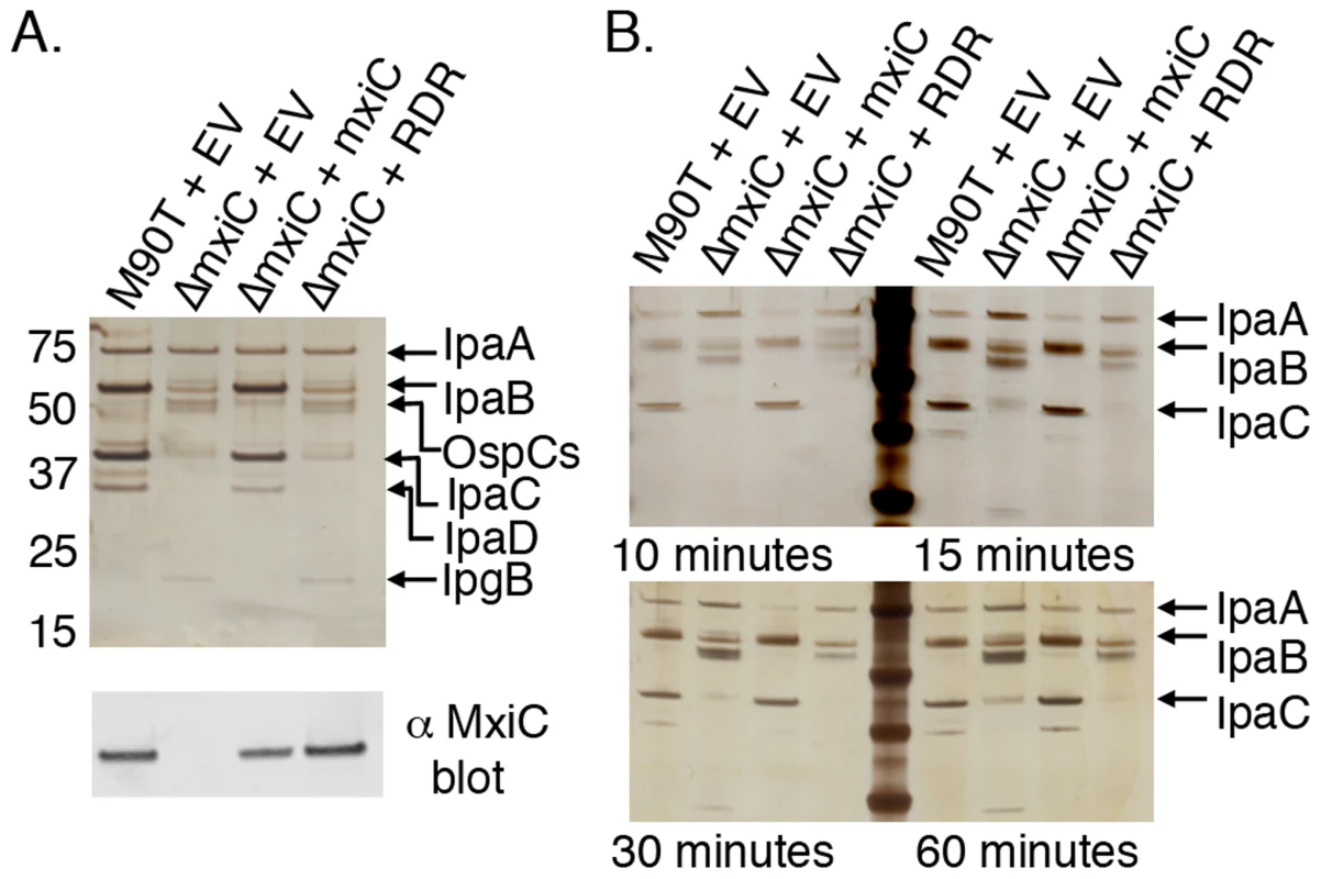
A. T3S was induced from wild type and mxiC mutant Shigella strains. M90T is a wild type strain. ΔmxiC is M90T derived, with mxiC deleted [18]. EV: empty vector. Strains were complemeted with mxiC, mxiC mutant (RDR), or empty vector (EV). Proteins were visualized by silver staining and identified by MS. IpaA, OspCs, and IpgB are effectors. IpaB, IpaC, and IpaD are translocators. Bottom: anti-MxiC blot of the same samples indicates that MxiC and MxiC-RDR are secreted normally. B. A secretion timecourse. The experiment shown in A. was repeated and samples taken a 10, 15, 30, and 60 minutes post induction. IpaA, an effector, is secreted at early time points in the absence of functional Mxic, but in the presence of MxiC it is not efficiently secreted until later time points. Discussion
In this study we have presented the structure of CopNΔ84 bound to Scc3. This structure provides the first description of a novel interaction for translocator-chaperones, which involves an amino-terminal extension, termed the GBR, binding across the YopN-TyeA-like domain interface in CopNΔ84. The translocator-chaperone-gatekeeper interaction involves a conserved arginine from the gatekeeper and the amino terminal GBR from the chaperone. Despite the lack of sequence conservation within the chaperone, the presence of a GBR in class II chaperones appears to be conserved (Figure 2). Scc3's GBR is ∼20 amino acids longer than others, implying that the extensive interactions made to the YopN-like domain are likely unique to the Scc3-CopNΔ84 complex. Although the unique α1 helix of Scc3 (amino acids 2–15) does not contact CopN, α1 does orient the α1–α2 loop, which contacts CopN. In organisms with shorter GBRs, other T3S apparatus components likely contribute to organizing this interface. This appears to be the case in Salmonella, where the gatekeeper interacts with the translocator-chaperone when it is bound to either translocator, but not to the empty chaperone [17]. In Shigella and Pseudomonas, the translocator-chaperones are known to homodimerize using the amino terminus, and translocator binding is necessary to disrupt this dimer [29], [39]. Although the monomeric chaperone is able to bind translocators, our data suggest a role for the chaperone amino terminus distinct from the role in homodimerization, namely a role in gatekeeper binding. We find that Scc3 is a monomer and the Scc3-CopNΔ84 complex is a heterodimer in solution (Supporting Figure S7). We suggest that disruption of chaperone homodimers, by translocator binding likely occurs prior to chaperone-gatekeeper complex formation, and that once the chaperone-translocator complex is formed, it is recruited to the T3S apparatus by the gatekeeper.
The binding mode observed in our structure leaves the canonical translocator-binding groove free and available to bind translocators. The Scc3-CopNΔ84 complex binds directly to CopB (Figure 3, Supporting Figure S5), a Chlamydial translocator, thus establishing a physical link between the gatekeeper and a translocator. By physically linking gatekeepers and translocators, the temporal order of secretion events in which translocators are secreted immediately after gatekeepers and prior to effectors is assured. The mechanism by which gatekeepers are recruited to the apparatus prior to effectors or translocators remains to be determined. The unusual CopN-Scc3 interface and relatively small buried-surface area (980 Å2) are consistent with the physiological role of this complex, in that the gatekeeper-translocator chaperone must assemble and disassemble each cycle of translocator secretion.
In Chlamydia the CopN-Scc3 interaction can be formed with purified proteins, suggesting that it is stronger than in homologous systems, likely because the GBR is longer and complex formation is not prevented by chaperone homodimerization. The precise advantage this affords Chlamydia is unclear, but T3S activation may represent an early “committed step” for Chlamydia infection. As obligate intracellular pathogens, the need for Chlamydia to enter their host subsequent to T3S activation seems absolute. Chlamydia are dependent on their hosts for ATP [44] and therefore a T3S event that doesn't result in entry is likely fatal for the bacterium. Consistent with this idea, Chlamydia also express a second translocator chaperone, Scc2, which is expressed during late stages of infection, after invasion, and does not bind CopN [22], [23].
Mutations shown to disrupt the CopN-Scc3 complex were evaluated in the MxiC-IpgC complex. The highly conserved central arginine and two additional charged residues, located at positions homologous to the sites mutated in CopN, were mutated (E331R/R334D/I338R) on a MxiC expressing plasmid and used to complement a mxiC deletion strain. This mutant MxiC (MxiC-RDR) was expressed and secreted normally, indicating that the mutations did not significantly disrupt MxiC. Strains harboring MxiC-RDR, however, mimicked deletion strains and were both deficient in translocator secretion and secreted elevated levels of effectors, resulting in significant effector secretion at early timepoints, prior to translocator secretion. These results support the conclusion that a key function of MxiC during secretion is to scaffold translocators and that the MxiC-IpgC complex is needed for this function. This arrangement, in which the gatekeeper directs translocators to the secretion apparatus, makes proper assembly of a gatekeeper-chaperone-translocator complex needed to promote translocator secretion. Gatekeepers are also needed to prevent effector secretion [15]–[20] indicating that the relevant “plug” that prevents premature effector secretion is likely the gatekeeper-chaperone-translocator complex. Consistent with this interpretation, disruption of IpaB (a Shigella translocator) or MxiC (the gatekeeper) both cause constitutive effector secretion [18]. In many systems, translocator chaperones have an additional role and act as transcription co-activators after translocator secretion thereby linking expression of some effector proteins to secretion of translocators [9].
Collectively, our results support a new mechanism for the translocator-effector hierarchy. We suggest that the translocators are recruited to the T3S pore as a molecular complex including the gatekeeper, translocator-chaperone, and translocator. There are likely multiple variations on this theme, one of which is evident in Pseudomonas aeruginosa, where translocators make multiple interactions with their chaperone, including regions outside the canonical chaperone binding region, at the extreme carboxy terminus [45]. The entire complex is needed both to promote translocator secretion and to prevent effector secretion. A triggering event from the tip of the T3S needle is known to induce gatekeeper release [18], which through the molecular complex described above is directly linked to translocator secretion. Secretion of the gatekeeper and translocator then allows effector secretion through a gatekeeper independent mechanism, similar to the efficient effector secretion seen in gatekeeper mutants.
Methods
Purification of His tagged proteins
CopNΔ84 and Scc3, from C. pneumoniae AR39, and MxiCΔ74 from S. flexneri were amplified and cloned into pET28 with an amino-terminal hexa-histadine tag. CopNΔ84 and MxiCΔ74 are amino terminal 84 and 74 amino acid deletions of CopN and MxiC, respectively. It was previously found that these were well-behaved variants [25], [37]. All CopN variants were made in pET28 using PCR based mutagenesis and verified by sequencing. CopN truncation mutants were designed from described limited proteolysis mass spectrometry analysis [25]. Proteins were expressed in BL21 (DE3) bacteria grown in Luria Broth at 37°C. Bacteria at an optical density (at 600 nm) of ∼0.6 were induced at 20°C with 0.1 mM isopropyl β-D-thiogalactopyranoside, and grown for ∼12 hours. Cultures were harvested by centrifugation and lysed with a French Press in phosphate buffered saline with ∼1 µg/mL chicken egg white lysozyme, ∼1 µg/mL bovine pancreatic deoxyribonuclease I, 10 µg/mL leupeptin, 1 mM PMSF, 0.7 µg/mL pepstatin. The lysate was clarified by centrifugation and proteins were purified by Co-NTA affinity using Talon resin. Eluted proteins were further purified by size exclusion chromatography, superdex 200, before being snap-frozen in liquid nitrogen and stored at −80°C until needed.
Expression and purification of GST-CopB
The Scc3 binding region of CopB, residues 158–178 based on sequence homology with IpaB from Shigella [13], was expressed as a GST fusion protein from PGEX-4T. Proteins were expressed and purified as for His tagged proteins with minor modifications; proteins were expressed from BL21 bacteria and purified with Glutathione Sepharose 4B (GE Healthcare).
Gel-filtration assays
Chaperone, and translocator peptide binding assays were performed by gel-filtration, using a 24 mL Superdex 200 10/300 GL (GE Healthcare), equilibrated in 10 mM Tris-HCl pH 7.5, 150 mM NaCl, run at 0.5 mL/min, and maintained at 4°C. Equivalent molar concentrations, determined from calculated extinction coefficients, of proteins were applied to the gel filtration column. Protein complexes were incubated for 15 minutes prior to analysis. Molecular weight determination was done under the same conditions using gel-filtration standards from BioRad.
Crystallization and preparation of heavy atom derivatives
Scc3-CopNΔ84 crystals were grown via vapor diffusion from a reservoir containing 0.2 M Na/K tartrate and 18–22% PEG 3350. Crystals were obtained from a 1∶1 mixture of reservoir and 15 mg/MmL Scc3-CopNΔ84. KAu(CN)2 derivatives were prepared by adding 100 mM KAu(CN)2 to the crystal drop to a final concentration of 2 mM KAu(CN)2. After two days of incubation derivative crystals were harvested. Native and derivative crystals were cryoprotected with 15% glycerol and flash cooled.
Data collection, structure determination, and analysis
Diffraction data were collected from single crystals on stations D and F at LS-CAT beam line at the Advanced Photon Source. Data were indexed, integrated and scaled with HKL2000 [46]. Three gold atoms were located and refined using Phenix [47]. The initial figure of merit for these sites was 0.46, which improved to 0.70 following density modification. The model was traced with a combination of automated and manual building in Phenix and COOT [47], [48]. Multiple rounds of refinement were done using Phenix. Refinement included simulated annealing, coordinate, individual B-factor, and TLS refinement as implemented in Phenix [47]. Non-crystallographic symmetry constraints were included in all rounds of positional refinement. Data collection and refinement statistics are given in Table S1. Figures were prepared using Pymol, ClustalW, ESPript, the DALI server, the PISA server, and the Consurf server [33], [49]–[52].
Alignments
Sequences used in the Consurf alignments of CopN homologs were chosen to represent the sequence diversity within genera shown in Figure 1 and included five Chlamydial sequences (NP_224529.1, NP_829326.1, YP_515466.1, 84785886, 332806765, YP_005809291.1), four Shigella sequences (YP_005712038, YP_313363.1, YP_001883209.1, YP_406185.1), three Salmonella sequences (1236849, 75349427, NP_461818.1), and three Bordetella sequences (NP_880900.1, WP_004568105.1, NP_884470.1). For the two-component gatekeepers, chimeric sequences were generated to agree with the spatial orientations in the YopN/TyeA structure (pdb accession code 1XL3). Yersinia YopN sequences used were NP_863522.1, NP_395173.1, and NP_052400.1. Because there is zero sequence diversity in TyeA from species evaluated here, we used YP_004210060.1 for all chimeras. An identical strategy was used for Pseudomonas and Vibrio. Pseudomonas sequences used were NP_250389.1, WP_003122865.1, and WP_010794024.1, WP_015648550.1, which were all matched with WP_009876220.1. Vibrio sequences used were NP_798046.1, YP_003285992.1, WP_005395115.1, WP_005377238.1, WP_005441804.1, WP_004745560.1, WP_005528936.1, which were all matched with WP_005395113.1.
Shigella secretion assay
Secretion assays were performed essentially as described [18], [53], with minor modifications. Shigella strain M90T was a gift from Marcia Goldberg. Shigella strain ΔmxiC as well as pUC19-mxiC have been previously described, and were gifts from Ariel Blocker [18]. The pmxiC-RDR and pmxiC-R334D were made by standard molecular biology methods and used to transform Shigella strain ΔmxiC. Strains were grown on tryptic soy broth (TSB) plates containing 100 µg/mL congo red, with appropriate antibiotics. Colonies were selected and grown overnight in liquid TSB broth at 37°C and harvested by centrifugation. Pellets were resuspended in 5 mLs of fresh liquid medium. A fraction, ∼1∶25 final dilution, of the resuspended cultures was added to 50 mL TSB cultures and grown to an optical density of 1.0 (600 nm). Cultures were harvested by centrifugation, washed with warm media, and resuspended to a final OD600 of 5.0 in PBS+100 mg/mL Congo Red at 37°C for 10 min and 30 min. Samples were analyzed by SDS-Page using both coomassie and silver staining, as well as western blotting. Western blotting was done with an α-MxiC antibody primary, which was a gift from Ariel Blocker, and goat anti-rabbit secondary antibody (LI-COR Biosciences). Blots were developed with an Odyssey fluorescent scanner. Protein bands were identified from Mass Spectrometry of trypsin-digested bands excised from coomassie-stained gels and was performed by the Vanderbilt University Proteomics Core.
Supporting Information
Zdroje
1. CornelisGR (2002) Yersinia type III secretion: send in the effectors. J Cell Biol 158 : 401–408.
2. CornelisGR (2006) The type III secretion injectisome. Nat Rev Microbiol 4 : 811–825.
3. GalanJE, Wolf-WatzH (2006) Protein delivery into eukaryotic cells by type III secretion machines. Nature 444 : 567–573.
4. GalanJE, CossartP (2005) Host-pathogen interactions: a diversity of themes, a variety of molecular machines. Curr Opin Microbiol 8 : 1–3.
5. HodgkinsonJL, HorsleyA, StabatD, SimonM, JohnsonS, et al. (2009) Three-dimensional reconstruction of the Shigella T3SS transmembrane regions reveals 12-fold symmetry and novel features throughout. Nat Struct Mol Biol 16 : 477–485.
6. AbrusciP, Vergara-IrigarayM, JohnsonS, BeebyMD, HendrixsonDR, et al. (2013) Architecture of the major component of the type III secretion system export apparatus. Nat Struct Mol Biol 20 : 99–104.
7. DeaneJE, AbrusciP, JohnsonS, LeaSM (2010) Timing is everything: the regulation of type III secretion. Cell Mol Life Sci 67 : 1065–1075.
8. StammLM, GoldbergMB (2011) Microbiology. Establishing the secretion hierarchy. Science 331 : 1147–1148.
9. ParsotC, HamiauxC, PageAL (2003) The various and varying roles of specific chaperones in type III secretion systems. Curr Opin Microbiol 6 : 7–14.
10. StebbinsCE, GalanJE (2001) Maintenance of an unfolded polypeptide by a cognate chaperone in bacterial type III secretion. Nature 414 : 77–81.
11. BirtalanSC, PhillipsRM, GhoshP (2002) Three-dimensional secretion signals in chaperone-effector complexes of bacterial pathogens. Mol Cell 9 : 971–980.
12. ButtnerCR, SorgI, CornelisGR, HeinzDW, NiemannHH (2008) Structure of the Yersinia enterocolitica type III secretion translocator chaperone SycD. J Mol Biol 375 : 997–1012.
13. LunelliM, LokareddyRK, ZychlinskyA, KolbeM (2009) IpaB-IpgC interaction defines binding motif for type III secretion translocator. Proc Natl Acad Sci U S A 106 : 9661–9666.
14. JobV, MatteiPJ, LemaireD, AttreeI, DessenA (2010) Structural basis of chaperone recognition of type III secretion system minor translocator proteins. J Biol Chem 285 : 23224–23232.
15. IriarteM, SoryMP, BolandA, BoydAP, MillsSD, et al. (1998) TyeA, a protein involved in control of Yop release and in translocation of Yersinia Yop effectors. EMBO J 17 : 1907–1918.
16. JosephSS, PlanoGV (2007) Identification of TyeA residues required to interact with YopN and to regulate Yop secretion. Adv Exp Med Biol 603 : 235–245.
17. KuboriT, GalanJE (2002) Salmonella type III secretion-associated protein InvE controls translocation of effector proteins into host cells. J Bacteriol 184 : 4699–4708.
18. Martinez-ArgudoI, BlockerAJ (2010) The Shigella T3SS needle transmits a signal for MxiC release, which controls secretion of effectors. Mol Microbiol 78 : 1365–1378.
19. O'ConnellCB, CreaseyEA, KnuttonS, ElliottS, CrowtherLJ, et al. (2004) SepL, a protein required for enteropathogenic Escherichia coli type III translocation, interacts with secretion component SepD. Mol Microbiol 52 : 1613–1625.
20. PallenMJ, BeatsonSA, BaileyCM (2005) Bioinformatics analysis of the locus for enterocyte effacement provides novel insights into type-III secretion. BMC Microbiol 5 : 9.
21. FerracciF, DayJB, EzelleHJ, PlanoGV (2004) Expression of a functional secreted YopN-TyeA hybrid protein in Yersinia pestis is the result of a +1 translational frameshift event. J Bacteriol 186 : 5160–5166.
22. SpaethKE, ChenYS, ValdiviaRH (2009) The Chlamydia type III secretion system C-ring engages a chaperone-effector protein complex. PLoS Pathog 5: e1000579.
23. SlepenkinA, de la MazaLM, PetersonEM (2005) Interaction between components of the type III secretion system of Chlamydiaceae. J Bacteriol 187 : 473–479.
24. FieldsKA, FischerER, MeadDJ, HackstadtT (2005) Analysis of putative Chlamydia trachomatis chaperones Scc2 and Scc3 and their use in the identification of type III secretion substrates. J Bacteriol 187 : 6466–6478.
25. ArchuletaTL, DuY, EnglishCA, LoryS, LesserC, et al. (2011) The Chlamydia effector, chlamydial outer protein N (CopN), sequesters tubulin and prevents microtubule assembly. J Biol Chem 286 : 33992–8.
26. Silva-HerzogE, JosephSS, AveryAK, CobaJA, WolfK, et al. (2011) Scc1 (CP0432) and Scc4 (CP0033) function as a type III secretion chaperone for CopN of Chlamydia pneumoniae. J Bacteriol 193 : 3490–3496.
27. SchreinerM, NiemannHH (2012) Crystal structure of the Yersinia enterocolitica type III secretion chaperone SycD in complex with a peptide of the minor translocator YopD. BMC Struct Biol 12 : 13.
28. CherradiY, SchiavolinL, MoussaS, MeghraouiA, MeksemA, et al. (2013) Interplay between predicted inner-rod and gatekeeper in controlling substrate specificity of the type III secretion system. Mol Microbiol 87 : 1183–99.
29. TomalkaAG, ZminaSE, StopfordCM, RietschA (2013) Dimerization of the P. aeruginosa translocator chaperone PcrH is required for stability not function. J Bacteriol 195 : 4836–43.
30. Lara-TejeroM, KatoJ, WagnerS, LiuX, GalanJE (2011) A sorting platform determines the order of protein secretion in bacterial type III systems. Science 331 : 1188–1191.
31. WinnenB, SchlumbergerMC, SturmA, SchupbachK, SiebenmannS, et al. (2008) Hierarchical effector protein transport by the Salmonella Typhimurium SPI-1 type III secretion system. PLoS ONE 3: e2178.
32. ThomasNA, DengW, BakerN, PuenteJ, FinlayBB (2007) Hierarchical delivery of an essential host colonization factor in enteropathogenic Escherichia coli. J Biol Chem 282 : 29634–29645.
33. HolmL, SanderC (1995) Dali: a network tool for protein structure comparison. Trends Biochem Sci 20 : 478–480.
34. MurrayJW, DelumeauO, LewisRJ (2005) Structure of a nonheme globin in environmental stress signaling. Proc Natl Acad Sci U S A 102 : 17320–17325.
35. QuinMB, BerrisfordJM, NewmanJA, BasleA, LewisRJ, et al. (2012) The bacterial stressosome: a modular system that has been adapted to control secondary messenger signaling. Structure 20 : 350–363.
36. ReuterW, WiegandG, HuberR, ThanME (1999) Structural analysis at 2.2 A of orthorhombic crystals presents the asymmetry of the allophycocyanin-linker complex, AP.LC7.8, from phycobilisomes of Mastigocladus laminosus. Proc Natl Acad Sci U S A 96 : 1363–1368.
37. DeaneJE, RoversiP, KingC, JohnsonS, LeaSM (2008) Structures of the Shigella flexneri type 3 secretion system protein MxiC reveal conformational variability amongst homologues. J Mol Biol 377 : 985–992.
38. SchubotFD, JacksonMW, PenroseKJ, CherryS, TropeaJE, et al. (2005) Three-dimensional structure of a macromolecular assembly that regulates type III secretion in Yersinia pestis. J Mol Biol 346 : 1147–1161.
39. AdamP, PatilM, DickensonN, ChoudhariS, BartaM, et al. (2012) Binding affects the tertiary and quaternary structures of the Shigella translocator protein IpaB and its chaperone IpgC. Biochemistry 51 : 4062–4071.
40. CliffMJ, WilliamsMA, Brooke-SmithJ, BarfordD, LadburyJE (2005) Molecular recognition via coupled folding and binding in a TPR domain. J Mol Biol 346 : 717–732.
41. LeNoue-NewtonM, WatkinsGR, ZouP, GermaneKL, McCorveyLR, et al. (2011) The E3 ubiquitin ligase - and protein phosphatase 2A (PP2A)-binding domains of the Alpha4 protein are both required for Alpha4 to inhibit PP2A degradation. J Biol Chem 286 : 17665–17671.
42. BotteauxA, SoryMP, BiskriL, ParsotC, AllaouiA (2009) MxiC is secreted by and controls the substrate specificity of the Shigella flexneri type III secretion apparatus. Mol Microbiol 71 : 449–460.
43. FieldsKA, HackstadtT (2000) Evidence for the secretion of Chlamydia trachomatis CopN by a type III secretion mechanism. Mol Microbiol 38 : 1048–1060.
44. SakaHA, ValdiviaRH (2009) Acquisition of nutrients by Chlamydiae: unique challenges of living in an intracellular compartment. Curr Opin Microbiol 13 : 4–10.
45. TomalkaAG, StopfordCM, LeePC, RietschA (2012) A translocator-specific export signal establishes the translocator-effector secretion hierarchy that is important for type III secretion system function. Mol Microbiol 86 : 1464–1481.
46. OtwinowskiZ, MinorW (1997) Processing of X-ray Diffraction Data Collected in Oscillation Mode. Methods in Enzymology 276A: 307–326.
47. AdamsPD, Grosse-KunstleveRW, HungLW, IoergerTR, McCoyAJ, et al. (2002) PHENIX: building new software for automated crystallographic structure determination. Acta Crystallogr D Biol Crystallogr 58 : 1948–1954.
48. EmsleyP, CowtanK (2004) Coot: model-building tools for molecular graphics. Acta Crystallogr D Biol Crystallogr 60 : 2126–2132.
49. DeLano WL (2002) The PyMOL Molecular Graphics System. San Carlos, CA: DeLano Scientific.
50. PanchenkoAR, BryantSH (2002) A comparison of position-specific score matrices based on sequence and structure alignments. Protein Sci 11 : 361–370.
51. GouetP, RobertX, CourcelleE (2003) ESPript/ENDscript: Extracting and rendering sequence and 3D information from atomic structures of proteins. Nucleic Acids Res 31 : 3320–3323.
52. AshkenazyH, ErezE, MartzE, PupkoT, Ben-TalN ConSurf 2010: calculating evolutionary conservation in sequence and structure of proteins and nucleic acids. Nucleic Acids Res 38 Suppl: W529–533.
53. DemersB, SansonettiPJ, ParsotC (1998) Induction of type III secretion in Shigella flexneri is associated with differential control of transcription of genes encoding secreted proteins. Embo J 17 : 2894–2903.
Štítky
Hygiena a epidemiologie Infekční lékařství Laboratoř
Článek Acidification Activates Motility and Egress by Enhancing Protein Secretion and Cytolytic ActivityČlánek Plasticity between MyoC- and MyoA-Glideosomes: An Example of Functional Compensation in InvasionČlánek Coronavirus Cell Entry Occurs through the Endo-/Lysosomal Pathway in a Proteolysis-Dependent MannerČlánek NK Cell Activation in Human Hantavirus Infection Explained by Virus-Induced IL-15/IL15Rα Expression
Článek vyšel v časopisePLOS Pathogens
Nejčtenější tento týden
2014 Číslo 11- Jak souvisí postcovidový syndrom s poškozením mozku?
- Měli bychom postcovidový syndrom léčit antidepresivy?
- Farmakovigilanční studie perorálních antivirotik indikovaných v léčbě COVID-19
- 10 bodů k očkování proti COVID-19: stanovisko České společnosti alergologie a klinické imunologie ČLS JEP
-
Všechny články tohoto čísla
- Peculiarities of Prion Diseases
- Inhibitors of Peptidyl Proline Isomerases As Antivirals in Hepatitis C and Other Viruses
- War and Infectious Diseases: Challenges of the Syrian Civil War
- Microbial Contamination in Next Generation Sequencing: Implications for Sequence-Based Analysis of Clinical Samples
- Acidification Activates Motility and Egress by Enhancing Protein Secretion and Cytolytic Activity
- Co-dependence of HTLV-1 p12 and p8 Functions in Virus Persistence
- Shed GP of Ebola Virus Triggers Immune Activation and Increased Vascular Permeability
- Plasticity between MyoC- and MyoA-Glideosomes: An Example of Functional Compensation in Invasion
- The Type III Translocon Is Required for Biofilm Formation at the Epithelial Barrier
- Retromer Regulates HIV-1 Envelope Glycoprotein Trafficking and Incorporation into Virions
- IFI16 Restricts HSV-1 Replication by Accumulating on the HSV-1 Genome, Repressing HSV-1 Gene Expression, and Directly or Indirectly Modulating Histone Modifications
- Coronavirus Cell Entry Occurs through the Endo-/Lysosomal Pathway in a Proteolysis-Dependent Manner
- Silencing by H-NS Potentiated the Evolution of
- Crystal Structure of Cytomegalovirus IE1 Protein Reveals Targeting of TRIM Family Member PML via Coiled-Coil Interactions
- GAPDH-A Recruits a Plant Virus Movement Protein to Cortical Virus Replication Complexes to Facilitate Viral Cell-to-Cell Movement
- Genomic Insights into the Fungal Pathogens of the Genus : Obligate Biotrophs of Humans and Other Mammals
- Unravelling Human Trypanotolerance: IL8 is Associated with Infection Control whereas IL10 and TNFα Are Associated with Subsequent Disease Development
- The Skin Microbiome: A Focus on Pathogens and Their Association with Skin Disease
- Human Cytomegalovirus Vaccine Based on the Envelope gH/gL Pentamer Complex
- IL-37 Inhibits Inflammasome Activation and Disease Severity in Murine Aspergillosis
- Host-Specific Parvovirus Evolution in Nature Is Recapitulated by Adaptation to Different Carnivore Species
- Activation of HIV Transcription with Short-Course Vorinostat in HIV-Infected Patients on Suppressive Antiretroviral Therapy
- PUL21a-Cyclin A2 Interaction is Required to Protect Human Cytomegalovirus-Infected Cells from the Deleterious Consequences of Mitotic Entry
- Programmed Ribosomal Frameshift Alters Expression of West Nile Virus Genes and Facilitates Virus Replication in Birds and Mosquitoes
- Aminoterminal Amphipathic α-Helix AH1 of Hepatitis C Virus Nonstructural Protein 4B Possesses a Dual Role in RNA Replication and Virus Production
- NK Cell Activation in Human Hantavirus Infection Explained by Virus-Induced IL-15/IL15Rα Expression
- Structure and Specificity of the Bacterial Cysteine Methyltransferase Effector NleE Suggests a Novel Substrate in Human DNA Repair Pathway
- Genetics, Receptor Binding Property, and Transmissibility in Mammals of Naturally Isolated H9N2 Avian Influenza Viruses
- A Gatekeeper Chaperone Complex Directs Translocator Secretion during Type Three Secretion
- A Conserved Peptide Pattern from a Widespread Microbial Virulence Factor Triggers Pattern-Induced Immunity in
- Succinate Dehydrogenase is the Regulator of Respiration in
- The Plasmodesmal Protein PDLP1 Localises to Haustoria-Associated Membranes during Downy Mildew Infection and Regulates Callose Deposition
- Dysregulated B Cell Expression of RANKL and OPG Correlates with Loss of Bone Mineral Density in HIV Infection
- Restriction of Genetic Diversity during Infection of the Vector Midgut
- The Epithelial αvβ3-Integrin Boosts the MYD88-Dependent TLR2 Signaling in Response to Viral and Bacterial Components
- The Relationship between Host Lifespan and Pathogen Reservoir Potential: An Analysis in the System
- Multiple Roles of the Cytoskeleton in Bacterial Autophagy
- The Evolution and Genetics of Virus Host Shifts
- ChIP-seq and In Vivo Transcriptome Analyses of the SREBP SrbA Reveals a New Regulator of the Fungal Hypoxia Response and Virulence
- PLOS Pathogens
- Archiv čísel
- Aktuální číslo
- Informace o časopisu
Nejčtenější v tomto čísle- Coronavirus Cell Entry Occurs through the Endo-/Lysosomal Pathway in a Proteolysis-Dependent Manner
- Peculiarities of Prion Diseases
- Host-Specific Parvovirus Evolution in Nature Is Recapitulated by Adaptation to Different Carnivore Species
- War and Infectious Diseases: Challenges of the Syrian Civil War
Kurzy
Zvyšte si kvalifikaci online z pohodlí domova
Současné možnosti léčby obezity
nový kurzAutoři: MUDr. Martin Hrubý
Všechny kurzyPřihlášení#ADS_BOTTOM_SCRIPTS#Zapomenuté hesloZadejte e-mailovou adresu, se kterou jste vytvářel(a) účet, budou Vám na ni zaslány informace k nastavení nového hesla.
- Vzdělávání


