-
Články
Top novinky
Reklama- Vzdělávání
- Časopisy
Top články
Nové číslo
- Témata
Top novinky
Reklama- Videa
- Podcasty
Nové podcasty
Reklama- Kariéra
Doporučené pozice
Reklama- Praxe
Top novinky
ReklamaSequence Divergent RXLR Effectors Share a Structural Fold Conserved across Plant Pathogenic Oomycete Species
article has not abstract
Published in the journal: . PLoS Pathog 8(1): e32767. doi:10.1371/journal.ppat.1002400
Category: Pearls
doi: https://doi.org/10.1371/journal.ppat.1002400Summary
article has not abstract
The availability of genome sequences for some of the most devastating eukaryotic plant pathogens has led a revolution in our understanding of how these parasites cause disease, and how their hosts respond to invasion [1]–[7]. One of the most significant discoveries from the genome sequences of plant pathogenic oomycetes is the plethora of putative translocated effector proteins these organisms encode. Many effector genes display signatures of rapid evolution and tend to reside in dynamic regions of the pathogen genomes. Once inside the host, effector proteins modulate cellular processes, mainly suppressing plant immunity [8]–[12]. Effectors can also be recognized directly or indirectly by the plant immune system through the action of disease resistance (R) proteins [13], [14].
Plant Pathogenic Oomycetes Express RXLR Effector Proteins
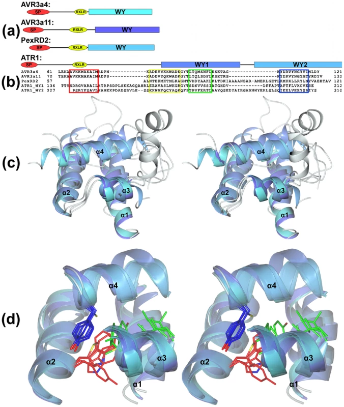
One expanded family of effector proteins is defined by the sequence RXLR (Arg-X-Leu-Arg, where X is any amino acid), which in some cases is followed by an acidic-rich dEER motif (Asp-Glu-Glu-Arg) (Figure 1). The RXLR motif was originally identified by comparing sequences of effectors from Hyaloperonospora arabidopsidis, Phytophthora infestans, and Phytophthora sojae [15]. It has since been shown that the RXLR motif is important for translocation of oomycete effectors into plant cells [16], [17]. It is widely accepted that RXLR effectors are modular proteins comprising an N-terminal secretion signal, followed by the RXLR region, and a C-terminal “effector” domain that encodes the biochemical activity of the protein when expressed directly in plant cells [18], [19]. A large family of Phytophthora RXLR effectors contain conserved sequence motifs (W, Y, and L) in their C-terminal domains that often form tandem repeats [2], [20].
Fig. 1. Structural conservation of the WY-domain fold in RXLR effectors from P. infestans, P. capsici, and H. arabidopsidis. 
(a) Schematic representation (to scale) of the domain architectures of AVR3a4, AVR3a11, PexRD2, and ATR1 showing the positions of the WY-domains used in the structural overlays. SP = signal peptide region, RXLR = RXLR/dEER region, WY = WY-domain regions (schematics are aligned at the end of the RXLR/dEER region). (b) Structure-based sequence alignment showing the positions of the conserved helices in each WY-domain. (c) Stereo view of an overlay comprising the WY-domains from AVR3a11, ATR1-WY1, ATR1-WY2, PexRD2, and AVR3a4 (α2–α4 span the WY-domain, α1 is the N-terminal helix present in all but PexRD2). The helices of the structures are colored in grades of blue through to cyan. Connecting regions are in gray (all structures). (d) Stereo view (orientation as in (c)) showing the positions of important residues within the hydrophobic core of the WY-domain (red = the W position, blue = the Y position, in green are two positions contributed from α3). Only the conserved helices are shown in cartoon representation (connecting regions removed for clarity). Structural Biology Uncovers an Effector Fold Conserved across Oomycete Species
Our laboratories have employed structural biology to investigate the molecular basis of RXLR effector function. A total of four structures have recently been published, those of AVR3a4 and AVR3a11 (paralogues from Phytophthora capsici), PexRD2 (from P. infestans), and ATR1 (from H. arabidopsidis) [21]–[23]. Each publication focused on a different aspect of structure/function analysis including phospholipid binding, protein folding, and effector recognition by the host.
The studies of Boutemy et al. and Chou et al. independently described the structural homology of AVR3a11 and a domain of ATR1, respectively, to the cyanobacterial four-helix bundle protein KaiA [24]. This strongly implied they would also be structurally related to each other. This is unexpected, as these Phytophthora and H. arabidopsidis effectors do not share any significant sequence similarity: the conservation was only apparent after the structures were determined and compared. Further, the structural conservation across different oomycete species was particularly intriguing, as studies with the Phytophthora proteins AVR3a11 and PexRD2 [21] had suggested a three-helix bundle fold could be the basic structural unit adopted by the repeating W-Y motifs found in >520 Phytophthora RXLR effectors (44% of annotated RXLR effectors in P. infestans, Phytophthora ramorum, and P. sojae). Using Hidden Markov Model (HMM)-based sequence searches, these motifs had also been detected in H. arabidopsidis RXLR effector proteins, with 35 out of 134 (26%) containing this fold (HMM score>0), including ATR1 with a low confidence score [21]. Boutemy et al. named this conserved structural unit the “WY-domain” and it comprises three α-helices connected by variable loop regions. The minimal three-helix WY-domain is found in PexRD2, but Avr3a4, Avr3a11, and ATR1 all have an N-terminal helix as an extension to this unit that forms a four-helix bundle. Further analysis of the ATR1 structure revealed that not only residues 139–210 (the domain originally identified as having a KaiA-like fold), but also 226–308 comprised a WY-domain four-helix bundle (ATR1 also has a fifth helix that creates a five-helix repeat) [22]. This tandem repeat could not be detected from amino acid sequence comparison, and was only discovered after the ATR1 structure was determined [22]. The structure of ATR1 shows how tandem WY-domains encoded by very divergent amino acid sequences are linked in three-dimensional space. This is significant, as it provides insight into how WY-domains may be arranged in other WY motif repeat RXLR effectors.
The Conserved Fold Is Based on a “Flexible” Hydrophobic Core
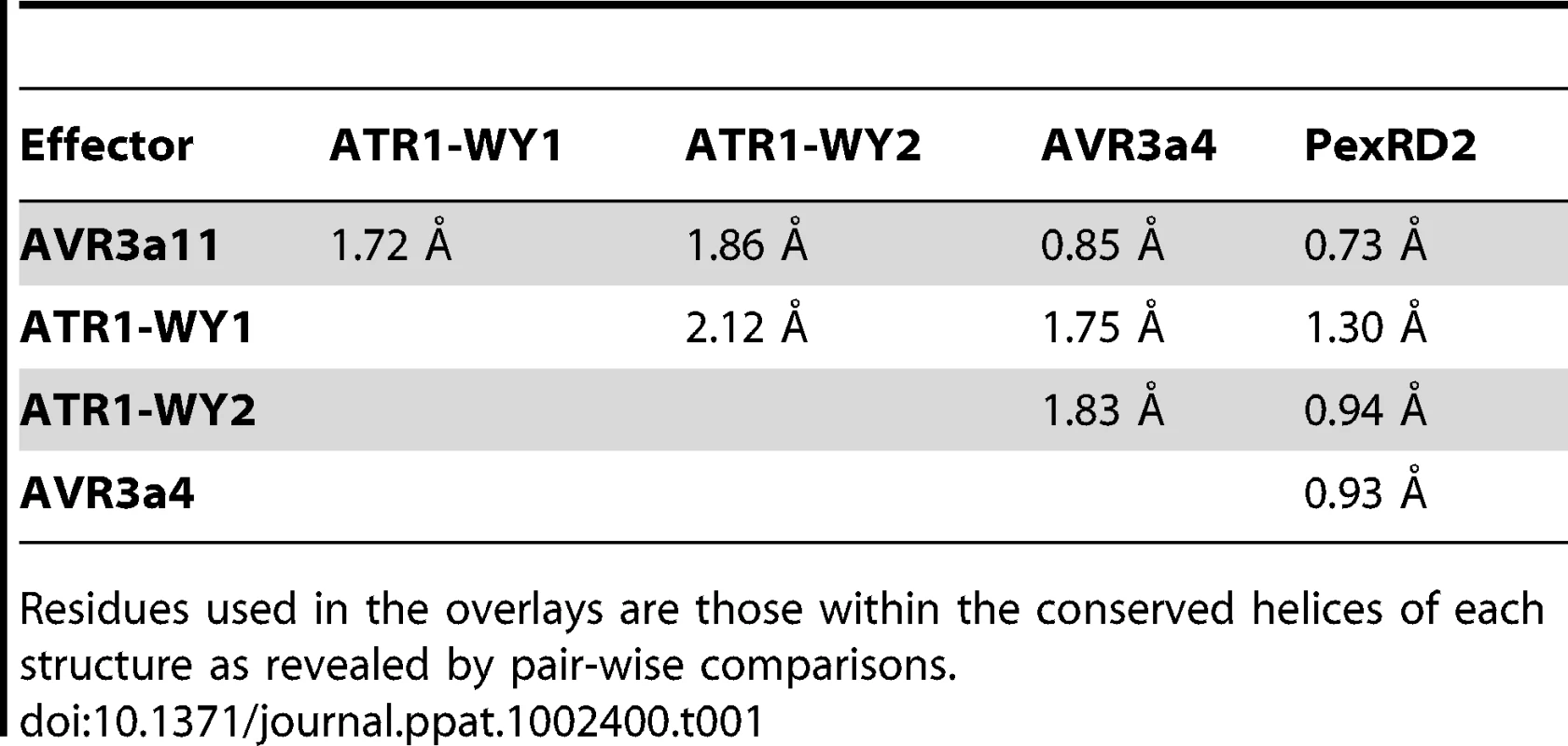
The availability of these four oomycete RXLR effector domain structures, from three different pathogens, allows us to present a detailed analysis of the WY-domain fold. Structural overlays of the conserved WY-domains from each of the effectors are shown in Figure 1, and the root mean square deviations derived from the overlays are given in Table 1 (obtained using Secondary Structure Matching (SSM) algorithms [25]). What are the features of this fold that allow structural conservation with little, if any, identifiable pair-wise sequence identity? In the HMM models, the conserved motif is largely defined by residues such as the W and Y (for Trp and Tyr) that, in each structure, are buried in the hydrophobic core of the helical bundle (other hydrophobic residues that contribute to the core are also prevalent in the HMM). Critically, the identity of these residues can change, without affecting the fold, as long as their hydrophobic potential is maintained. For example, in the WY-domain structures available, the W and Y positions are Trp-Tyr (AVR3a4 and AVR3a11), Met-Tyr (PexRD2), and Trp-Cys/Tyr-Tyr (for the two WY-domains of ATR1). Further, there is evidence from the existing structures that solvent remains excluded from the hydrophobic core when mutations from bulkier to smaller side chains occurs through complementary mutations at other positions that fill the available space. The ability of this structural fold to accommodate the rapid evolution of protein sequence explains why the WY-domain was not identifiable in pair-wise sequence comparisons.
Tab. 1. Root mean square deviations (based on Cα atoms) for overlays of the published RXLR effector structures (only the A-chains of ATR1 and PexRD2 were considered). 
Residues used in the overlays are those within the conserved helices of each structure as revealed by pair-wise comparisons. The WY-Domain Fold Is Restricted to the Peronosporales
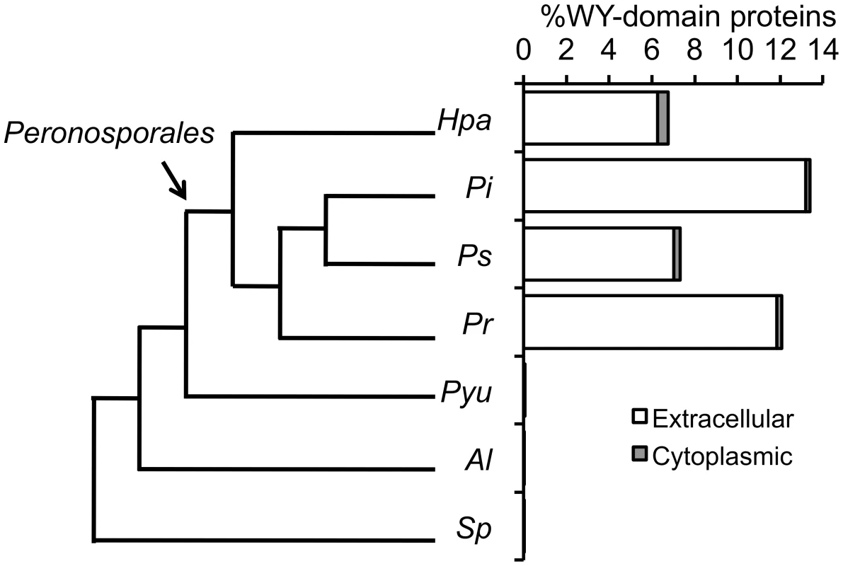
We believe that the conservation of this “flexible” hydrophobic core fold indicates that a large family of plant pathogenic oomycete effectors may have been derived from a common ancestor. Intriguingly, this effector family has rapidly diverged to gain new and/or adapt existing virulence functions and/or evade detection by plant immune systems. New or modified effector functions may be derived from surface point mutations or indels in the connecting regions between helices (including domain duplication). To test the argument for a common ancestor, we extended previous analyses and searched the proteomes of various organisms using the HMM for the WY-domain, as described in [21]. Firstly, we searched the proteomes of 47 different eukaryotes [26]. These searches included diverse species, from fungi through plants and animals. We found no evidence for the presence of the WY-domain signature beyond the level of our previously described false-positive rate [21]. We then narrowed our search and screened the available proteomes of phylogenetically diverse oomycetes: Saprolegnia parasitica [27], Albugo labachii [28], and Pythium ultimum [29], adding to the Phytophthora and Hyaloperonospora proteomes searched previously [21] (Figure 2). These searches show that, with the data available, the WY-domain is limited to a single clade within the oomycetes, the Peronosporales, that are exclusively plant pathogens [30]. This suggests that the WY-domain may be an innovation within the Peronosporales. The WY-domain is also correlated with the emergence of the RXLR motif in the Peronosporales, and is linked with the evolution of haustoria as a possible interface for effector delivery in this lineage [19], [29]. Of the seven oomycetes whose genome sequences are available, all of the Peronosporales (four species) have RXLRs and WY-domains (Figure 2); the non-Peronosporales (three species) encode essentially no RXLR or WY-domain proteins, and those few identified may be false positives given that they are not enriched in the secretome (as in the Peronosporales). It is also notable that oomycetes with expansions of their RXLR effector repertoire (P. infestans>P. sojae>P. ramorum>H. arabidopsidis) also encode a significantly higher percentage of WY-domains in their secretomes (P. infestans>P. ramorum>P. sojae>H. arabidopsidis, Figure 2). Further, as WY-domains are found in both Phytophthora (hemibiotrophs) and Hyaloperonospora (obligate biotrophs), but not P. ultimum, it appears that the WY-domain emerged with biotrophy in this lineage along with the evolution of haustoria and RXLR effectors [19], [31]. Whilst arguing in favour of a common ancestor of the WY-domain within the Peronosporales, we acknowledge that alternative interpretations (including convergent evolution to a fold adapted for stability in the plant cell and/or well-suited to a particular function, such as secretion and/or translocation) remain possible.
Fig. 2. Phylogenetic relationship and presence of the WY-domain HMM signature in sequenced oomycete genomes. 
Schematic representation of the phylogenetic relationship among seven oomycetes for which genome sequences are available and the distribution of WY-domain in their extracellular and cytoplasmic proteomes (as a percentage of the total). Hpa = H. arabidopsidis; Pi = P. infestans; Ps = P. sojae; Pr = P. ramorum; Pyu = P. ultimum; Al = A. labachii; Sp = S. parasitica. Some of the most notorious and agriculturally important pathogenic oomycetes contain RXLR:WY-domain effectors, suggesting that this structure has been critical for the success of these pathogens. This raises questions such as, why has this fold been preserved and what can it tell us about the function of these proteins? How can we use this knowledge to design novel disease management strategies? Future studies will help define the roles of the WY-domain fold in the virulence mechanisms of these pathogens, in particular how it engages with plant cell targets, and will help to unravel the extent of structural diversity in RXLR effectors. Our initial studies have laid the foundation for new, exciting discoveries addressing the function of oomycete effectors.
Zdroje
1. BaxterLTripathySIshaqueNBootNCabralA 2010 Signatures of adaptation to obligate biotrophy in the Hyaloperonospora arabidopsidis genome. Science 330 1549 1551
2. HaasBJKamounSZodyMCJiangRHHandsakerRE 2009 Genome sequence and analysis of the Irish potato famine pathogen Phytophthora infestans. Nature 461 393 398
3. The Genome Portal of the Department of Energy Joint Genome Institute 2011 Phytophthora capsici genome project. Available: http://genome.jgi-psf.org/Phyca11/Phyca11.home.html. Accessed 12 December 2011
4. RaffaeleSFarrerRACanoLMStudholmeDJMacLeanD 2010 Genome evolution following host jumps in the Irish potato famine pathogen lineage. Science 330 1540 1543
5. SpanuPDAbbottJCAmselemJBurgisTASoanesDM 2010 Genome expansion and gene loss in powdery mildew fungi reveal tradeoffs in extreme parasitism. Science 330 1543 1546
6. TylerBMTripathySZhangXDehalPJiangRH 2006 Phytophthora genome sequences uncover evolutionary origins and mechanisms of pathogenesis. Science 313 1261 1266
7. SchirawskiJMannhauptGMunchKBrefortTSchipperK 2010 Pathogenicity determinants in smut fungi revealed by genome comparison. Science 330 1546 1548
8. DoddsPNRathjenJP 2010 Plant immunity: towards an integrated view of plant-pathogen interactions. Nat Rev Genet 11 539 548
9. BosJIArmstrongMRGilroyEMBoevinkPCHeinI 2010 Phytophthora infestans effector AVR3a is essential for virulence and manipulates plant immunity by stabilizing host E3 ligase CMPG1. Proc Natl Acad Sci U S A 107 9909 9914
10. KearneyBStaskawiczBJ 1990 Widespread distribution and fitness contribution of Xanthomonas campestris avirulence gene avrBs2. Nature 346 385 386
11. KimMGda CunhaLMcFallAJBelkhadirYDebRoyS 2005 Two Pseudomonas syringae type III effectors inhibit RIN4-regulated basal defense in Arabidopsis. Cell 121 749 759
12. NomuraKDebroySLeeYHPumplinNJonesJ 2006 A bacterial virulence protein suppresses host innate immunity to cause plant disease. Science 313 220 223
13. JonesJDDanglJL 2006 The plant immune system. Nature 444 323 329
14. ChisholmSTCoakerGDayBStaskawiczBJ 2006 Host-microbe interactions: shaping the evolution of the plant immune response. Cell 124 803 814
15. RehmanyAPGordonARoseLEAllenRLArmstrongMR 2005 Differential recognition of highly divergent downy mildew avirulence gene alleles by RPP1 resistance genes from two Arabidopsis lines. Plant Cell 17 1839 1850
16. WhissonSCBoevinkPCMolelekiLAvrovaAOMoralesJG 2007 A translocation signal for delivery of oomycete effector proteins into host plant cells. Nature 450 115 118
17. DouDKaleSDWangXChenYWangQ 2008 Conserved C-terminal motifs required for avirulence and suppression of cell death by Phytophthora sojae effector Avr1b. Plant Cell 20 1118 1133
18. OlivaRWinJRaffaeleSBoutemyLBozkurtTO 2010 Recent developments in effector biology of filamentous plant pathogens. Cell Microbiol 12 705 715
19. SchornackSvan DammeMBozkurtTOCanoLMSmokerM 2010 Ancient class of translocated oomycete effectors targets the host nucleus. Proc Natl Acad Sci U S A 107 17421 17426
20. JiangRHTripathySGoversFTylerBM 2008 RXLR effector reservoir in two Phytophthora species is dominated by a single rapidly evolving superfamily with more than 700 members. Proc Natl Acad Sci U S A 105 4874 4879
21. BoutemyLSKingSRWinJHughesRKClarkeTA 2011 Structures of Phytophthora RXLR effector proteins: a conserved but adaptable fold underpins functional diversity. J Biol Chem 286 35834 35842
22. ChouSKrasilevaKVHoltonJMSteinbrennerADAlberT 2011 Hyaloperonospora arabidopsidis ATR1 effector is a repeat protein with distributed recognition surfaces. Proc Natl Acad Sci U S A 108 13323 13328
23. YaenoTLiHChaparro-GarciaASchornackSKoshibaS 2011 Phosphatidylinositol monophosphate-binding interface in the oomycete RXLR effector AVR3a is required for its stability in host cells to modulate plant immunity. Proc Natl Acad Sci U S A 108 14682 14687
24. MarksonJSO'SheaEK 2009 The molecular clockwork of a protein-based circadian oscillator. FEBS Lett 583 3938 3947
25. KrissinelEHenrickK 2004 Secondary-structure matching (SSM), a new tool for fast protein structure alignment in three dimensions. Acta Crystallogr D Biol Crystallogr 60 2256 2268
26. WinJMorganWBosJKrasilevaKVCanoLM 2007 Adaptive evolution has targeted the C-terminal domain of the RXLR effectors of plant pathogenic oomycetes. Plant Cell 19 2349 2369
27. Broad Institute of Harvard and MIT 2011 Saprolegnia parasitica sequencing project. Availalble: http://www.broadinstitute.org/annotation/genome/Saprolegnia_parasitica/MultiHome.html. Accessed 12 December 2011
28. KemenEGardinerASchultz-LarsenTKemenACBalmuthAL 2011 Gene gain and loss during evolution of obligate parasitism in the white rust pathogen of Arabidopsis thaliana. PLoS Biol 9 e1001094 doi:10.1371/journal.pbio.1001094
29. LévesqueCABrouwerHCanoLHamiltonJPHoltC 2010 Genome sequence of the necrotrophic plant pathogen Pythium ultimum reveals original pathogenicity mechanisms and effector repertoire. Genome Biol 11 R73
30. BeakesGWGlocklingSLSekimotoS 2011 The evolutionary phylogeny of the oomycete “fungi”. Protoplasma. E-pub ahead of print 20 March 2011 doi:10.1007/s00709-011-0269-2
31. ThinesMKamounS 2010 Oomycete-plant coevolution: recent advances and future prospects. Curr Opin Plant Biol 13 427 433
Štítky
Hygiena a epidemiologie Infekční lékařství Laboratoř
Článek vyšel v časopisePLOS Pathogens
Nejčtenější tento týden
2012 Číslo 1- Jak souvisí postcovidový syndrom s poškozením mozku?
- Měli bychom postcovidový syndrom léčit antidepresivy?
- Farmakovigilanční studie perorálních antivirotik indikovaných v léčbě COVID-19
- 10 bodů k očkování proti COVID-19: stanovisko České společnosti alergologie a klinické imunologie ČLS JEP
-
Všechny články tohoto čísla
- How “Humane” Is Your Endpoint?—Refining the Science-Driven Approach for Termination of Animal Studies of Chronic Infection
- Sexual Development in : Lessons from Functional Analyses
- Type 1 Interferons and Antiviral CD8 T-Cell Responses
- Historical Contingencies Modulate the Adaptability of
- Human-like PB2 627K Influenza Virus Polymerase Activity Is Regulated by Importin-α1 and -α7
- -Sialidase in Complex with a Neutralizing Antibody: Structure/Function Studies towards the Rational Design of Inhibitors
- Zebrafish: A See-Through Host and a Fluorescent Toolbox to Probe Host–Pathogen Interaction
- How Do Bacteria Know They Are on a Surface and Regulate Their Response to an Adhering State?
- Sequence Divergent RXLR Effectors Share a Structural Fold Conserved across Plant Pathogenic Oomycete Species
- The Murine Coronavirus Hemagglutinin-esterase Receptor-binding Site: A Major Shift in Ligand Specificity through Modest Changes in Architecture
- Temporal Expression of Bacterial Proteins Instructs Host CD4 T Cell Expansion and Th17 Development
- PLOS Pathogens
- Archiv čísel
- Aktuální číslo
- Informace o časopisu
Nejčtenější v tomto čísle- Type 1 Interferons and Antiviral CD8 T-Cell Responses
- Sequence Divergent RXLR Effectors Share a Structural Fold Conserved across Plant Pathogenic Oomycete Species
- Temporal Expression of Bacterial Proteins Instructs Host CD4 T Cell Expansion and Th17 Development
- Sexual Development in : Lessons from Functional Analyses
Kurzy
Zvyšte si kvalifikaci online z pohodlí domova
Současné možnosti léčby obezity
nový kurzAutoři: MUDr. Martin Hrubý
Všechny kurzyPřihlášení#ADS_BOTTOM_SCRIPTS#Zapomenuté hesloZadejte e-mailovou adresu, se kterou jste vytvářel(a) účet, budou Vám na ni zaslány informace k nastavení nového hesla.
- Vzdělávání


