-
Články
Top novinky
Reklama- Vzdělávání
- Časopisy
Top články
Nové číslo
- Témata
Top novinky
Reklama- Videa
- Podcasty
Nové podcasty
Reklama- Kariéra
Doporučené pozice
Reklama- Praxe
Top novinky
ReklamaClpP1 and ClpP2 Function Together in Protein Degradation and Are Required for Viability and During Infection
In most bacteria, Clp protease is a conserved, non-essential serine protease that regulates the response to various stresses. Mycobacteria, including Mycobacterium tuberculosis (Mtb) and Mycobacterium smegmatis, unlike most well studied prokaryotes, encode two ClpP homologs, ClpP1 and ClpP2, in a single operon. Here we demonstrate that the two proteins form a mixed complex (ClpP1P2) in mycobacteria. Using two different approaches, promoter replacement, and a novel system of inducible protein degradation, leading to inducible expression of clpP1 and clpP2, we demonstrate that both genes are essential for growth and that a marked depletion of either one results in rapid bacterial death. ClpP1P2 protease appears important in degrading missense and prematurely terminated peptides, as partial depletion of ClpP2 reduced growth specifically in the presence of antibiotics that increase errors in translation. We further show that the ClpP1P2 protease is required for the degradation of proteins tagged with the SsrA motif, a tag co-translationally added to incomplete protein products. Using active site mutants of ClpP1 and ClpP2, we show that the activity of each subunit is required for proteolysis, for normal growth of Mtb in vitro and during infection of mice. These observations suggest that the Clp protease plays an unusual and essential role in Mtb and may serve as an ideal target for antimycobacterial therapy.
Published in the journal: . PLoS Pathog 8(2): e32767. doi:10.1371/journal.ppat.1002511
Category: Research Article
doi: https://doi.org/10.1371/journal.ppat.1002511Summary
In most bacteria, Clp protease is a conserved, non-essential serine protease that regulates the response to various stresses. Mycobacteria, including Mycobacterium tuberculosis (Mtb) and Mycobacterium smegmatis, unlike most well studied prokaryotes, encode two ClpP homologs, ClpP1 and ClpP2, in a single operon. Here we demonstrate that the two proteins form a mixed complex (ClpP1P2) in mycobacteria. Using two different approaches, promoter replacement, and a novel system of inducible protein degradation, leading to inducible expression of clpP1 and clpP2, we demonstrate that both genes are essential for growth and that a marked depletion of either one results in rapid bacterial death. ClpP1P2 protease appears important in degrading missense and prematurely terminated peptides, as partial depletion of ClpP2 reduced growth specifically in the presence of antibiotics that increase errors in translation. We further show that the ClpP1P2 protease is required for the degradation of proteins tagged with the SsrA motif, a tag co-translationally added to incomplete protein products. Using active site mutants of ClpP1 and ClpP2, we show that the activity of each subunit is required for proteolysis, for normal growth of Mtb in vitro and during infection of mice. These observations suggest that the Clp protease plays an unusual and essential role in Mtb and may serve as an ideal target for antimycobacterial therapy.
Introduction
Intracellular protein degradation is critical for maintaining cellular homeostasis through protein quality control and regulation of numerous biological pathways [1], [2]. In eukaryotes, the ubiquitin-proteasome pathway constitutes the predominant degradation pathway [3]. Most prokaryotes, however, possess a variety of ATP-dependent serine protease complexes, such as Lon and Clp protease [4], and some actinomycetes and archaea contain proteasomes, which are threonine proteases. Interestingly, Mycobacterium tuberculosis (Mtb) encodes both a proteasome and Clp protease. While recent work has explored the role of the Mtb proteasome [5]–[7], little is known about mycobacterial Clp protease. This serine protease was first discovered and is best characterized in Escherichia coli [8], [9]. The Clp proteolytic complex is formed by the association of proteolytic subunits, ClpP, with ATPase adapters, ClpX or ClpA in Gram-negative organisms and ClpX or ClpC in Gram-positive organisms. E. coli ClpP is a tetradecamer composed of two stacked heptameric rings of identical ClpP subunits that form an internal proteolytic chamber [10]. This core associates with distinct hexameric ATPase adapters, ClpX and ClpC1 in mycobacteria, which provide substrate specificity and catalyze ATP-dependent unfolding of globular proteins [11], [12]. In E. coli, the ClpXP protease is involved in the regulation of the DNA damage response and degradation of SsrA-tagged peptides stalled on the ribosome [13], [14]. Clp proteolytic enzymes are also required for full virulence in several pathogenic organisms, including Listeria monocytogenes where the protease is required for the production of α-listeriolysin [15], [16]. In most bacteria including E. coli, Clp protease is dispensable for normal growth, and in fact, until recently, the only organism in which clpP has been found to be essential is Caulobacter crescentus, where Clp degrades CtrA, an inhibitor of cell cycle progression [17].
Unlike most bacteria, which have a single ClpP subunit, the genome of Mtb encodes two closely related ClpP homologs, clpP1 and clpP2, in a single operon. A transposon-based mutagenesis screen for essential genes in Mtb predicted that ClpP2 and the ATPase adapters ClpC1 and ClpX, were required for normal growth [18] while a recent publication has shown that ClpP1 is essential [19]. Here, we show that both ClpP1 and ClpP2 are required for growth, and that their activity is important for the removal of abnormal proteins. Our data suggest that ClpP1 and ClpP2 assemble to form a single proteolytic complex, referred to as ClpP1P2, that is required for normal growth in vitro and during infection. In related studies, we have found that although pure ClpP1 and ClpP2 by themselves form tetradecamers, they are inactive. However, in the presence of low molecular weight activators they reassociate to form a mixed tetradecamer, ClpP1P2, which is capable of proteolysis (Akopian et. al., manuscript submitted). The unusual properties of this heteromeric complex, the absence of such an enzyme in the eukaryotic cytoplasm, and the essentiality of both subunits make ClpP1P2 protease an attractive target for novel therapeutic development for the treatment of tuberculosis.
Results
ClpP1 and ClpP2 subunits interact to form a single proteolytic complex
Mycobacterial genomes contain two homologous ClpP protease genes, clpP1 and clpP2, arranged in a putative operon. To investigate whether the two proteins may function together in a complex, we co-expressed Mtb clpP1 and clpP2, each containing a different C terminal epitope tag, in Mycobacterium smegmatis (Msm). We used affinity chromatography with nickel resin to isolate 6×-His tagged Mtb ClpP2 together with associated proteins from the Msm cell lysate. As shown in Figure 1A, a fraction of the c-myc tagged ClpP1 bound to the Ni column and co-eluted with ClpP2. To verify that ClpP1 and ClpP2 co-eluted from the Ni column may be associated in a complex, we applied the fraction from the Ni column containing both proteins to an anti-c-myc agarose column and analyzed by SDS PAGE. Figure 1B shows that a large fraction of the ClpP2 was associated with ClpP1. Incidentally, expression of the Mtb proteins in Msm also led to the co-isolation of Msm ClpP1 and ClpP2, as shown by tandem mass spectrometry of the purified complex. In each case, peptides present uniquely in Mtb or Msm ClpP1 and ClpP2 were detected (Figure 1C).
Fig. 1. Mtb ClpP1 and ClpP2 interact, forming a multi-component protease, and share substantial similarity with ClpP1 and ClpP2 homologs in Msm. 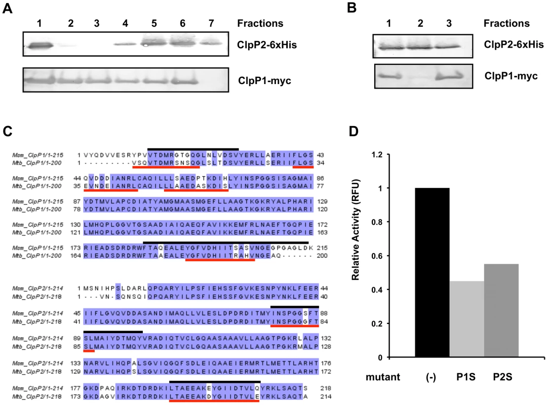
(A) C-terminally myc-tagged Mtb ClpP1 and 6×His-tagged Mtb ClpP2 were expressed in Msm. Lysate (lane 1) was prepared and loaded onto a Ni-column. After washing with PBS (lanes 2,3), Ni-bound material was eluted with 50 mM (lane 4), 100 mM (lane 5), 250 mM (lane 6, 7) of imidazole in PBS, and analyzed by immunoblotting using anti α-myc and α-6×His antibodies. (B) Fraction 6 from (A) was applied to an anti-myc column (lane 1). The flow through (lane 2), and bound material (lane 3) were analyzed by immunoblot with α-myc and α-His antibodies. Bound material was released from the anti-myc agarose beads by boiling in Laemmli buffer after washing with PBS. (C) Bands representing ClpP1 and ClpP2 from (B) were sequenced by MS/MS revealing the presence of both Mtb and Msm homologs. Msm specific peptides are indicated by black lines, those specific to Mtb are indicated by red lines. (D) Cleavage of fluorescent peptide Z-Gly-Gly-Leu-AMC was measured in the presence of 1 µg ClpP1, 1 µg Clp2, and the activating peptide Z-Leu-Leu (see accompanying paper). Addition of 5 µg of catalytically inactive mutants of either ClpP1 (ClpP1S) or ClpP2 (ClpP2S) markedly inhibited cleavage by the ClpP1P2 protease. Results graphed are a representative sample of results obtained. If ClpP1 and ClpP2 do in fact associate to form a single proteolytic core, we reasoned that mutations blocking the catalytic activity of one subunit might reduce that activity of the enzyme. We identified likely active site residues of ClpP1 and ClpP2 by mapping the Mtb proteins onto E. coli ClpP and locating the catalytic triad of Asp-His-Ser, which is characteristic of serine proteases. In both cases, the serine likely to be responsible for nucleophillic attack was replaced by an alanine (ClpP1 S98A and ClpP2 S110A). To analyze the effects of these mutations, we expressed and purified 6×His-tagged forms of each protein,and assayed their effect on the enzymatic activity of the wild type ClpP1P2 in an in vitro peptidase assay (Akopian et. al., manuscript submitted). Enzyme activity of the reconstituted ClpP1P2 complex was quantified using cleavage of the fluorescence reporter, Z-Gly-Gly-Leu-AMC. As seen in Figure 1D, addition of an excess of mutated ClpP1 or ClpP2 to the active wild type ClpP1P2 complex inhibited proteolytic cleavage of a fluorescent peptide substrate, presumably by replacing the wild type subunits. These results suggest that the ClpP1 and ClpP2 subunits interact to form a single proteolytic complex in vitro, that each active site is important for activity, and that these mutations can be used as dominant negative inhibitors.
Both ClpP1 and ClpP2 are required for normal growth in vitro
We employed three complementary strategies to determine if ClpP1 and ClpP2 are required for normal growth in mycobacteria. First, using mycobacterial recombineering [20], we replaced the endogenous promoter of clpP1 and clpP2 in Msm with a tetracycline-inducible promoter (Figure 2A, Figure S1). Introduction of a tetracycline repressor resulted in a strain (ptet_clpP1P2) that could only be maintained in the presence of the inducer anhydrotetracycline (ATc) (Figure 2B). In the absence of this compound, growth did not occur, but could be restored by the presence of an episomal plasmid containing both clpP1 and clpP2. Plasmids expressing only clpP1 or clpP2 alone could not rescue growth and depletion of either subunit resulted in bacterial death (Figure 2C). Since complementation was conducted with Mtb homologs and subunits from different species associate into a functional tetradecamer, the ClpP1P2 complex is likely very similar in Msm and Mtb. Furthermore, active site mutants of either ClpP1 or ClpP2 were unable to complement ptet_clpP1P2 in the absence of ATc, suggesting that the activity of both subunits were required for normal growth (data not shown).
Fig. 2. Both ClpP1 and ClpP2 are essential for normal growth in mycobacteria. 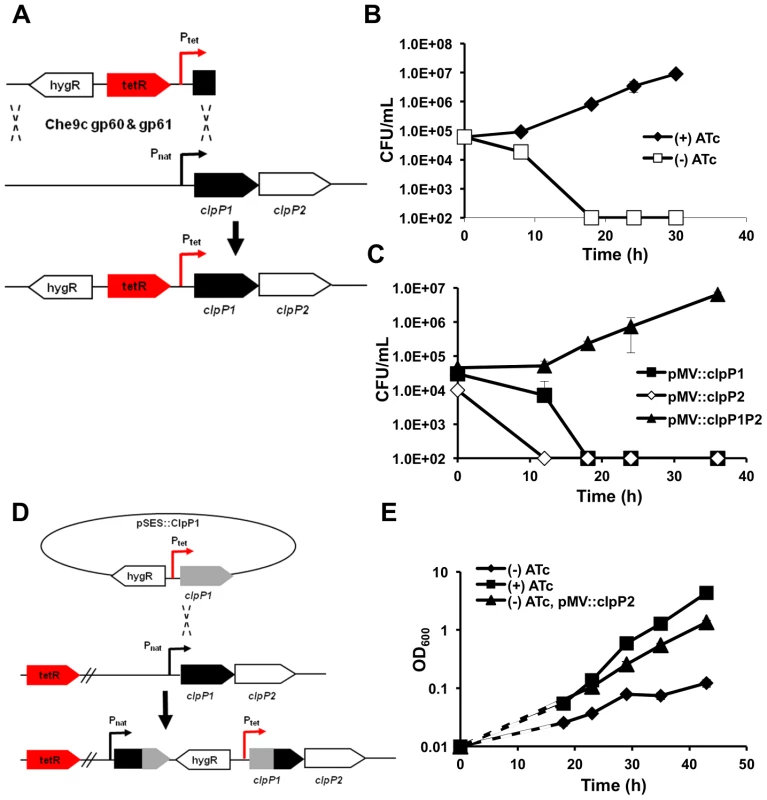
(A) Schematic representation of mycobacterial recombineering, employed to replace the endogenous promoter of the clpP1P2 operon with a ATc-inducible promoter (Msm strain ptet_clpP1P2). (B) Growth curves of Msm ptet_clpP1P2 in the presence (50 ng/mL) or absence of inducer ATc. Data are represented as mean CFU/mL +/− standard deviation. (C) Growth curves of Msm ptet_clpP1P2 complemented with clpP1, clpP2 or both clpP1 and clpP2 in the absence of inducer ATc. Data are represented as mean CFU/mL +/− standard deviation. (D) Schematic representation of genetic strategy used to create a tetracycline inducible conditional Msm ClpP2 mutant (Msm strain ptet_ClpP2) (E) Growth curves of Msm ptet_clpP2 in the presence (50 ng/mL) or absence of inducer ATc. Msm ptet_clpP2 was also complemented with clpP2 in the absence of ATc. Data are represented as mean OD600 +/− standard deviation. Dashed lines represent assumed growth rates until first measured growth point. Second, we inserted a tetracycline inducible promoter upstream of the clpP1P2 operon via homologous recombination in Msm creating a strain in which clpP2 was inducibly expressed (Figure 2D), and clpP1 was under the control of its native promoter (ptet_clpP2). In accord with the previous findings, the growth of this strain was dramatically inhibited in the absence of ATc (Figure 2E).
Third, we used a system of inducible protein degradation recently developed in Msm (Figure 3A) [21]. Briefly, we employed mycobacterial recombineering to add an inducible degradation (ID) tag to the C-terminus of ClpP2 (clpP2_ID). Upon cleavage of the tag by a tetracycline inducible HIV-2 protease, an SsrA sequence is revealed on the substrate that directs degradation of the protein. By inserting epitope tags C-terminally to the HIV-2 protease recognition motif (FLAG) and N-terminally to the SsrA tag (c-myc), we were able to monitor the amount of ClpP2 by immunoblotting. As shown in Figure 3B, induction of HIV-2 protease resulted in degradation of the majority of ClpP2 and inhibited bacterial growth (Figure 3C). Using this system, we did not observe cell death, perhaps due to incomplete inhibition, as would be expected for a system where the protease targets itself. Loss of ClpP2, as measured by immunoblotting, was rapid and reached near completion within hours. Furthermore, the growth defect was complemented by expression of Mtb clpP2 using a constitutively active promoter. A similar approach with ClpP1 was unsuccessful as extended C-terminal tagging was not tolerated, and the ID tag was indiscriminately cleaved. Collectively, these results confirm that both ClpP1 and ClpP2 are required for normal growth in mycobacteria, presumably because they function together in the ClpP1P2 complex.
Fig. 3. Inducible protein degradation demonstrates requirement of ClpP2 for normal growth. 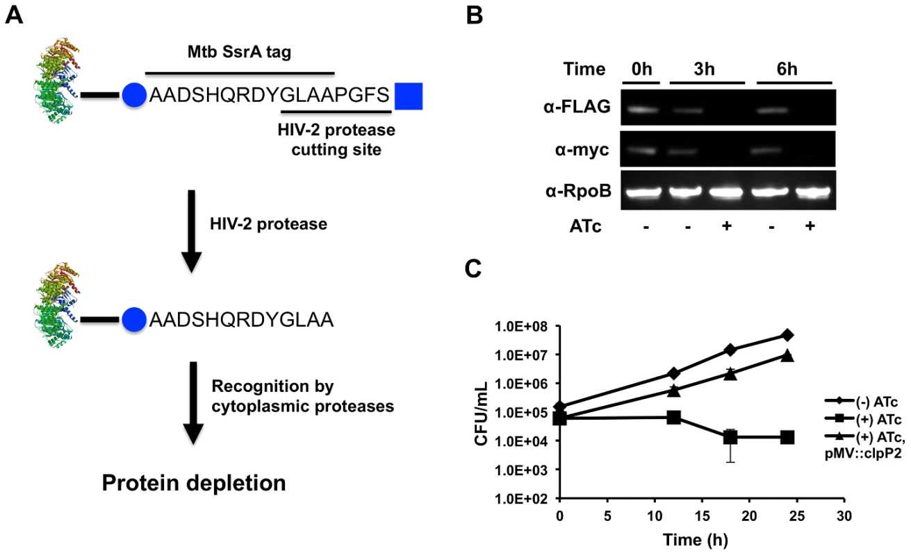
(A) Schematic representation of the inducible degradation system used to inducibly deplete ClpP2 (Msm strain clpP2_ID). Induction of HIV-2 protease with ATc leads to cleavage of the HIV-2 protease cutting site and exposure of a SsrA tag on the tagged protein. Cleavage by HIV-2 protease and subsequent degradation can be tracked via the FLAG (square) and c-myc (circle) epitope tags, respectively, included on the inducible degradation tag. (B) Degradation of ClpP2 in clpP2_ID was tracked by Western in the absence or presence of inducer ATc. Blots were probed α-FLAG (loss indicates HIV-2 protease cleavage), α-myc (loss indicates target degradation), and α-RpoB (loading control). (C) Growth curves of Msm clpP2_ID in the absence or presence (50 ng/mL) of inducer ATc. Msm clpP2_ID was also complemented with clpP2 in the presence of ATc. Data are represented as mean CFU/mL +/− standard deviation. Mycobacterial Clp protease plays a role in protein quality control
In other bacteria, Clp plays a role in degrading abnormal proteins such as SsrA-tagged peptides that stall on the ribosome [22]. To determine the importance of ClpP1P2 protease in the degradation of misfolded proteins, we used antibiotics that alter protein synthesis in distinct ways including chloramphenicol, which blocks protein elongation without increasing mistranslation rates [23], and streptomycin and amikacin, which induce translational errors resulting in missesnse or prematurely-terminated polypeptides [24]. We found that the strain ptet_ClpP2, in which clpP2 expression is regulated by anhydrotetracycline, grows well in low or high concentrations of ATc, 1 to 100 ng/mL (Figure 4A). Treatment with sublethal concentrations of chloramphenicol resulted in no difference in viability between bacteria maintained on low or high concentrations of ATc (Figure 4A, bottom). In contrast, sub-MIC concentrations of the aminoglycosides streptomycin and amikacin significantly inhibited the growth of strains incubated in low concentrations of ATc, while they had no effect on growth of the strain maintained in high concentrations of ATc (Figure 4A, top). Together, these results suggest that ClpP1P2 protease protects against error-prone translation by catalyzing the degradation of misfolded proteins.
Fig. 4. Clp protease is required for degradation of abnormal proteins and SsrA-tagged proteins in mycobacteria. 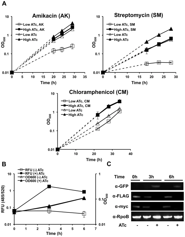
(A) Growth curves of Msm ptet_clpP2 in growth medium containing low (1 ng/mL) or high (100 ng/mL) concentrations of inducer ATc, in the presence of either no drug, or amikacin (top left, 0.03 µg/mL), streptomycin (top right, 0.125 µg/mL), and chloramphenicol (bottom, 7.5 µg/mL). Data are represented as mean OD600 +/− standard deviation. Dashed lines represent assumed growth rates until first measured growth point. (B) Increase in fluorescence (RFU, 485/520) and initial growth curve (OD600) of Msm clpP2_ID expressing the fusion construct GFP-SsrA on a constitutively expressing plasmid, in the presence and absence of inducer, ATc. Data are represented as mean RFU or OD600 +/− standard deviation. (C) Depletion of ClpP2 and increase in GFP-SsrA in Msm clpP2_ID expressing the fusion construct GFP-SsrA on a constitutively expressing plasmid was tracked by immunoblot. Blots were probed α-GFP, α-myc, α-FLAG, and α-RpoB (loading control). To specifically assess whether ClpP1P2 is responsible for the removal of SsrA-tagged proteins in mycobacteria, we fused the mycobacterial SsrA-tag to the C-terminus of GFP (GFP-SsrA) and expressed the construct constitutively on an episomal plasmid. This construct was introduced into the strain clpP2_ID, in which ClpP2 degradation was regulated. In the presence of ClpP2 (and in wild type cells), there was no detectable GFP-SsrA. However, upon depletion of ClpP2, there was substantial accumulation of GFP-SsrA, as measured by both fluorescence and immunoblot analysis after four hours (Figure 4B, 4C). Quantitative PCR showed that the rise of GFP-SsrA was not due to transcriptional activation of the gene (Figure S2). GFP lacking the SsrA tag is present at similar levels in all strains (data not shown). Because we cannot detect GFP-SsrA in the presence of Clp activity, we were unable to accurately measure changes in protein stability. However, the rate of accumulation of GFP-SsrA was consistent with the time course of ClpP2 depletion, which occurred over six hours, as shown by immunoblotting. Thus, functional ClpP1P2 protease is vital for the rapid clearance of SsrA-tagged substrates in mycobacteria.
Functional Clp protease is required for growth of Mtb in vitro and during infection
As shown above, catalytically inactive forms of ClpP1 and ClpP2 inhibit proteolysis by the wild type enzyme, possibly by replacement of wild type subunits with inactive ones. To assess whether ClpP1P2 activity is required for the growth of Mtb, we expressed a catalytically inactive form of Mtb clpP1, clpP1 S98A, on a tetracycline-inducible plasmid in wild type Mtb. Addition of ATc led to expression of the catalytically inactive mutant protein and resulted in a significant inhibition of growth (Figure 5A) while overexpression of wild type Mtb clpP1 had no effect.
Fig. 5. A catalytically inactive ClpP allele inhibit Mtb growth in vitro and during infection. 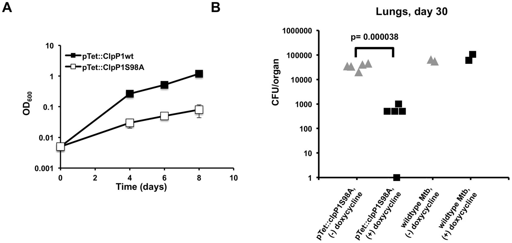
(A) Growth curves for Mtb overexpressing wild type ClpP1 or ClpP1 S98A via an ATc-inducible expression vector. Data are represented as mean OD600 +/− standard deviation. Dashed lines represent assumed growth rates until first measured growth point. (B) Growth of Mtb containing a doxycycline-inducible plasmid expressing the mutant allele ClpP1 S98A in lungs of C57BL/6 mice 30 days post aerosol infection. Mice were infected via aerosol with a 3∶1 mixture of mutant and wild type bacteria. Mice were fed either with chow containing (dark squares, N = 5 mice) or lacking (gray triangles, N = 5 mice) the inducer doxycycline. As a control, wild type Mtb was co-infected, and representative CFU/organs for the control are represented (right). Each point represents calculated total CFU/organ for each mouse. Not all mice received enough wild type bacteria to quantitate. To determine if the dominant negative mutant of ClpP1 affected ClpP1P2 function during infection, we infected mice with a 3∶1 mixture of Mtb expressing clpP1 S98A on a hygromycin-resistant doxycycline inducible plasmid and wild type Mtb (containing a kanamycin-resistant control vector). Mice were fed either normal chow or chow infused with the inducer doxycycline. Growth of Mtb was monitored by assessing CFU in lung tissue at day 30 post-infection. While there were no differences in the growth of wild type Mtb between treated and untreated mice, expression of the active site mutant significantly inhibited growth (Figure 5B). Our results suggest that functional ClpP1P2 protease is required for the growth of Mtb both in vitro and during infection.
Discussion
We find that the mycobacterial ClpP1P2 protease has two unusual properties that distinguishes it from other members of the prokaryotic ClpP family. First, the protease consists of distinct types of subunits, each of which is required for full activity of a single proteolytic complex. While other species do encode multiple ClpP subunits, two different proteolytic subunits forming a single protease has not been documented. Second, unlike in most bacteria that have been studied, ClpP1P2 activity is absolutely required for normal growth. This requirement is particularly striking as mycobacteria contain several cytoplasmic ATP-dependent proteolytic complexes, including FtsH, and the proteasome [7], [25], [26]. Clearly, the mycobacterial ClpP1P2 proteolytic core has unique roles that are important for viability.
The ClpP proteases that have been characterized biochemically in other bacteria and mitochondria are tetradecameric complexes containing a single type of proteolytic subunit. In mycobacteria, however, two different protein species contribute to protease activity. Although Mtb ClpP1 forms a tetradecameric complex, a crystal structure of Mtb ClpP1 lacks appropriate active site geometry to support proteolysis [27]. The presence of two ClpP subunits with distinct substrate preferences may facilitate an expansion of the peptide specificity of the complex, much like the eukaryotic proteasome. Interestingly, the Mtb proteasome is composed of a single type of subunit, and the presence of distinct subunits comprising a single proteolytic core is rare among prokaryotes.
There is at least one example of an essential role for ClpP. In Caulobacter crescentus, ClpXP degrades CtrA, a protein that normally inhibits cell cycle progression during cellular replication [28]. In this case, a single protein target is responsible for the essentiality of the enzyme. ClpP1P2 protease might play a similar role in mycobacteria. While this may be true, screens for essential proteins in mycobacteria suggest that, in addition to the clpP1 and clpP2, multiple Clp-associated ATPase adapters (clpX and clpC1) are also essential [18]. The requirement of multiple adapters makes it possible that accumulation of multiple protein substrates contribute to the poor growth phenotype observed on depleting the ClpP1 and ClpP2 subunits in mycobacteria.
ClpP1P2 might be important for other reasons. As shown here, ClpP1P2 protease is required for the clearance of SsrA-tagged proteins. These tagged polypeptides are generated under conditions when protein synthesis is stalled and are required for ribosome release. In the absence of ClpP1P2-mediated proteolysis, protein synthesis might eventually be inhibited. In addition, ClpP1P2 protease is necessary for degrading abnormal proteins, such as those produced in the presence of certain antibiotics. Accumulation of such non-functional misfolded proteins might result in cellular stress in the absence of an effective system for their removal [29]. Clearance of damaged proteins might be particularly important in Mtb during infection, when cells are exposed to multiple oxidative and nitrosoative radicals that can induce protein damage. In fact, a transcriptional activator of the clpP1P2 operon, clgR, is critically activated upon reaeration of hypoxic Mtb and during Mtb growth within the macrophage [30], [31]. Degradation of pre-existing proteins during such stressful transitions may be the initial event that triggers adaptation and facilitates the bacterium's ability to handle a wide array of environmental challenges. Using a dominant negative overexpression mutant in Mtb, we have confirmed that optimal Clp proteolytic activity is required for growth during infection.
The essential nature of ClpP1P2 protease makes it an attractive target for antibiotic development, particularly because the proteases as a class are druggable enzymes and have already been validated as therapeutic targets in the treatment of HIV, hepatitis, and cancer [32]. In organisms where ClpP is not essential, uncontrolled activation of ClpP activity can be toxic. For example, in E. coli, acyldepsipeptide compounds reorganize the ClpP proteolytic core, causing dissociation from ATPase adapters, and indiscriminate protein degradation [33]. Compounds that produce a similar effect should result in toxicity in a broad range of organisms. In fact, it was recently discovered that the natural product cyclomarin kills Mtb by targeting the ClpC1 ATPase and presumably increasing Clp-mediated proteolysis, as demonstrated in a whole cell fluorescence-based assay [34]. In mycobacteria, where ClpP1P2 protease activity is required and depletion of either subunit is bactericidal, either non-specific activation or inhibition could effectively limit bacterial growth. An example of a ClpP inhibitor with potential therapeutic activity already exists. In S. aureus, beta-lactones have been found to inhibit Clp protease activity and decrease the virulence of the organism [35]. Additionally, the synergistic nature of ClpP1P2 protease depletion with aminoglycosides, a class of drugs already used to treat tuberculosis, points to a potential combination therapy against Mtb. As ClpP1P2 protease is most likely involved in preventing the accumulation of misfolded proteins and the degradation of critical endogenous regulatory proteins, small molecule modulators of ClpP1P2 activity would target a critical aspect of Mtb physiology, and might prove useful in the face of growing multi-drug resistance in one of the world's most successful pathogens.
Materials and Methods
Ethics statement
The animal experiments were preformed with protocols approved by the Harvard Medical School Animal Management Program, which is accredited by the Association for Assessment and Accreditation of Laboratory Animal Care, International (AAALAC) and meets National Institute of Health standards as set forth in the Guide for the Care and Use of Laboratory Animals (Revised, 2010). The institution also accepts as mandatory the PHS Policy on Humane Care and Use of Laboratory Animals by Awardee Institutions and NIH Principles for the Utilization and Care of Vertebrate Animals Testing, Research, and Training. An Animal Welfare Assurance of Compliance is on file with the Office of Laboratory Animal Welfare (OLAW) (#A3431-01).
Bacterial strains and plasmids
Msm mc2155 (Msm) or Mtb H37Rv were grown at 37°C in Middlebrook 7H9 broth with 0.05% Tween 80 and ADC (0.5% BSA, 0.2% dextrose, 0.085% NaCl, 0.003 g catalase/1L media). Mtb was additionally supplemented with oleic acid (0.006%). For growth curves, overnight cultures were diluted into the appropriate media and growth was either measured by OD600 or colony forming units per mL. A summary of all strains, plasmids, and primers used in this study as well as a summary of the construction of conditional mutants can be found in the supporting information (Text S1).
Protein purification and in vitro peptidase assay
The C-terminally 6× His-tagged wild type clpP1, wild type clpP2, clpP1Ser98Ala, and clpP2Ser110Ala subunits were overexpressed in Msm using an anhydrotetracycline (ATc) inducible expression system. After overnight induction with ATc (100 ng/mL), cells were lysed by French press, and lysates were centrifuged for 1 h at 100,000 g. The subunits were purified from the supernatant by Ni-NTA affinity chromatography (Qiagen). Eluted fractions containing ClpP proteins were pooled and further purified by size exclusion chromatography on Sephacryl S-300 column. Equal amounts of ClpP1 and ClpP2 (1 µg each) were mixed in the reaction buffer (50 mM K-phosphate buffer pH 7,5, 100 mM KCl, 5% glycerol, 2 mM BME, 5 mM Z-Leu-Leu) and peptidase activity was measured by a rise in fluorescence at 460 nm (Ex at 340 nm) with 0.1 mM Z-Gly-Gly-Leu-AMC as a substrate. To measure dominant negative effect of active site mutants, same reaction was carried out in the presence of 5 µg of the mutant proteins.
Animal infections
Six to eight week old C57BL/6 mice (Jackson Laboratory) were used for animal infections. Mice were infected via aerosolization with 5×10⁶ CFU each of a 3∶1 mixture of Mtb pTet::ClpP1 S98A and Mtb pTet::GFP (wild type Mtb transformed with a control pTet plasmid containing GFP ). Mice were fed with chow with or without inducer doxycycline. At 30 days after infection, mice were sacrificed, lungs were homogenized and appropriate dilutions were plated on 7H10 plates containing hygromycin or kanamycin to select for the Clp mutant or the control respectively.
Supporting Information
Zdroje
1. IngmerHBrøndstedL 2009 Proteases in bacterial pathogenesis. Res Microbiol 160 704 710 doi:10.1016/j.resmic.2009.08.017
2. GoldbergAL 2003 Protein degradation and protection against misfolded or damaged proteins. Nature 426 895 899 doi:10.1038/nature02263
3. GlickmanMHCiechanoverA 2002 The ubiquitin-proteasome proteolytic pathway: destruction for the sake of construction. Physiol Rev 82 373 428 doi:10.1152/physrev.00027.2001
4. FreesDSavijokiKVarmanenPIngmerH 2007 Clp ATPases and ClpP proteolytic complexes regulate vital biological processes in low GC, Gram-positive bacteria. Mol Microbiol 63 1285 1295 doi:10.1111/j.1365-2958.2007.05598.x
5. BurnsKEPearceMJDarwinKH 2010 Prokaryotic ubiquitin-like protein provides a two-part degron to Mycobacterium proteasome substrates. J Bacteriol 192 2933 2935 doi:10.1128/JB.01639-09
6. Cerda-MairaFAPearceMJFuortesMBishaiWRHubbardSR 2010 Molecular analysis of the prokaryotic ubiquitin-like protein (Pup) conjugation pathway in Mycobacterium tuberculosis. Mol Microbiol 77 1123 1135 doi:10.1111/j.1365-2958.2010.07276.x
7. DarwinKH 2003 The Proteasome of Mycobacterium tuberculosis Is Required for Resistance to Nitric Oxide. Science 302 1963 1966 doi:10.1126/science.1091176
8. Katayama-FujimuraYGottesmanSMauriziMR 1987 A multiple-component, ATP-dependent protease from Escherichia coli. J Biol Chem 262 4477 4485
9. HwangBJParkWJChungCHGoldbergAL 1987 Escherichia coli contains a soluble ATP-dependent protease (Ti) distinct from protease La. Proc Natl Acad Sci U S A 84 5550 5554
10. WangJHartlingJAFlanaganJM 1997 The structure of ClpP at 2.3 A resolution suggests a model for ATP-dependent proteolysis. Cell 91 447 456
11. MogkADouganDWeibezahnJSchliekerCTurgayK 2004 Broad yet high substrate specificity: the challenge of AAA+ proteins. J Struct Biol 146 90 98 doi:10.1016/j.jsb.2003.10.009
12. KennistonJABakerTAFernandezJMSauerRT 2003 Linkage between ATP Consumption and Mechanical Unfolding during the Protein Processing Reactions of an AAA+ Degradation Machine. Cell 114 511 520 doi:10.1016/S0092-8674(03)00612-3
13. FarrellCMGrossmanADSauerRT 2005 Cytoplasmic degradation of ssrA-tagged proteins. Mol Microbiol 57 1750 1761 doi:10.1111/j.1365-2958.2005.04798.x
14. PruteanuMBakerTA 2009 Controlled degradation by ClpXP protease tunes the levels of the excision repair protein UvrA to the extent of DNA damage. Mol Microbiol 71 912 924 doi:10.1111/j.1365-2958.2008.06574.x
15. GaillotOBregenholtSJaubertFDi SantoJPBercheP 2001 Stress-Induced ClpP Serine Protease of Listeria monocytogenes Is Essential for Induction of Listeriolysin O-Dependent Protective Immunity. Infect Immun 69 4938 4943 doi:10.1128/IAI.69.8.4938-4943.2001
16. GaillotOPellegriniEBregenholtSNairSBercheP 2000 The ClpP serine protease is essential for the intracellular parasitism and virulence of Listeria monocytogenes. Mol Microbiol 35 1286 1294
17. JenalUFuchsT 1998 An essential protease involved in bacterial cell-cycle control. EMBO J 17 5658 5669 doi:10.1093/emboj/17.19.5658
18. SassettiCMBoydDHRubinEJ 2003 Genes required for mycobacterial growth defined by high density mutagenesis. Mol Microbiol 48 77 84
19. CarrollPFaray-KeleM-CParishT 2011 Identifying vulnerable pathways in Mycobacterium tuberculosis using a knock-down approach. Appl Environ Microbiol: 77 5040 5043 doi:10.1128/AEM.02880-10
20. van KesselJCHatfullGF 2008 Mycobacterial recombineering. Methods Mol Biol 435 203 215 doi:10.1007/978-1-59745-232-8_15
21. WeiJ-RKrishnamoorthyVMurphyKKimJ-HSchnappingerD 2011 Depletion of antibiotic targets has widely varying effects on growth. Proc Natl Acad Sci USA 108 4176 4181 doi:10.1073/pnas.1018301108
22. FreesDIngmerH 1999 ClpP participates in the degradation of misfolded protein in Lactococcus lactis. Mol Microbiol 31 79 87
23. HAHNFEWISSEMANCLHOPPSHE 1955 Mode of action of chloramphenicol. III. Action of chloramphenicol on bacterial energy metabolism. J Bacteriol 69 215 223
24. WykaMASt JohnAC 1990 Effects of production of abnormal proteins on the rate of killing of Escherichia coli by streptomycin. Antimicrob Agents Chemother 34 534 538
25. SmithCKBakerTASauerRT 1999 Lon and Clp family proteases and chaperones share homologous substrate-recognition domains. Proc Natl Acad Sci U S A 96 6678 6682
26. KiranMChauhanADziedzicRMaloneyEMukherjiSK 2009 Mycobacterium tuberculosis ftsH expression in response to stress and viability. Tuberculosis (Edinb) 89 S70 S73 doi:10.1016/S1472-9792(09)70016-2
27. IngvarssonHMatéMJHögbomMPortnoïDBenaroudjN 2007 Insights into the inter-ring plasticity of caseinolytic proteases from the X-ray structure of Mycobacterium tuberculosis ClpP1. Acta Crystallogr D Biol Crystallogr 63 249 259 doi:10.1107/S0907444906050530
28. QuonKCMarczynskiGTShapiroL 1996 Cell cycle control by an essential bacterial two-component signal transduction protein. Cell 84 83 93
29. GoldbergAL 1972 Degradation of abnormal proteins in Escherichia coli (protein breakdown-protein structure-mistranslation-amino acid analogs-puromycin). Proc Natl Acad Sci U S A 69 422 426
30. EstorninhoMSmithHTholeJHarders-WesterveenJKierzekA 2010 ClgR regulation of chaperone and protease systems is essential for Mycobacterium tuberculosis parasitism of the macrophage. Microbiology 156 3445 3455 doi:10.1099/mic.0.042275-0
31. SherridAMRustadTRCangelosiGAShermanDR 2010 Characterization of a Clp Protease Gene Regulator and the Reaeration Response in Mycobacterium tuberculosis. PLoS ONE 5 e11622 doi:10.1371/journal.pone.0011622
32. DragMSalvesenGS 2010 Emerging principles in protease-based drug discovery. Nat Rev Drug Discov 9 690 701 doi:10.1038/nrd3053
33. KirsteinJHoffmannALilieHSchmidtRRübsamen-WaigmannH 2009 The antibiotic ADEP reprogrammes ClpP, switching it from a regulated to an uncontrolled protease. EMBO Mol Med 1 37 49 doi:10.1002/emmm.200900002
34. SchmittEKRiwantoMSambandamurthyVRoggoSMiaultC 2011 The Natural Product Cyclomarin Kills Mycobacterium Tuberculosis by Targeting the ClpC1 Subunit of the Caseinolytic Protease. Angew Chem Int Ed Engl 50 5889 5891 doi:10.1002/anie.201101740
35. BöttcherTSieberSA 2008 Beta-lactones as specific inhibitors of ClpP attenuate the production of extracellular virulence factors of Staphylococcus aureus. J Am Chem Soc 130 14400 14401 doi:10.1021/ja8051365
Štítky
Hygiena a epidemiologie Infekční lékařství Laboratoř
Článek vyšel v časopisePLOS Pathogens
Nejčtenější tento týden
2012 Číslo 2- Jak souvisí postcovidový syndrom s poškozením mozku?
- Měli bychom postcovidový syndrom léčit antidepresivy?
- Farmakovigilanční studie perorálních antivirotik indikovaných v léčbě COVID-19
- 10 bodů k očkování proti COVID-19: stanovisko České společnosti alergologie a klinické imunologie ČLS JEP
-
Všechny články tohoto čísla
- Mechanisms of Pathogenesis, Infective Dose and Virulence in Human Parasites
- Structural and Functional Insights into the Pilotin-Secretin Complex of the Type II Secretion System
- Characterising the Mucosal and Systemic Immune Responses to Experimental Human Hookworm Infection
- Population Genetic Analyses Reveal the African Origin and Strain Variation of var.
- How Do Microbial Pathogens Make s?
- Substance P Causes Seizures in Neurocysticercosis
- Phagosomal Rupture by Results in Toxicity and Host Cell Death
- Absence of HIV-1 Evolution in the Gut-Associated Lymphoid Tissue from Patients on Combination Antiviral Therapy Initiated during Primary Infection
- Five Questions about Viral Trafficking in Neurons
- Selecting an Invertebrate Model Host for the Study of Fungal Pathogenesis
- A Co-Opted DEAD-Box RNA Helicase Enhances Tombusvirus Plus-Strand Synthesis
- Biochemical Properties of Highly Neuroinvasive Prion Strains
- ClpP1 and ClpP2 Function Together in Protein Degradation and Are Required for Viability and During Infection
- Discrete Cyclic di-GMP-Dependent Control of Bacterial Predation versus Axenic Growth in
- PLOS Pathogens
- Archiv čísel
- Aktuální číslo
- Informace o časopisu
Nejčtenější v tomto čísle- Discrete Cyclic di-GMP-Dependent Control of Bacterial Predation versus Axenic Growth in
- Characterising the Mucosal and Systemic Immune Responses to Experimental Human Hookworm Infection
- How Do Microbial Pathogens Make s?
- Substance P Causes Seizures in Neurocysticercosis
Kurzy
Zvyšte si kvalifikaci online z pohodlí domova
Současné možnosti léčby obezity
nový kurzAutoři: MUDr. Martin Hrubý
Všechny kurzyPřihlášení#ADS_BOTTOM_SCRIPTS#Zapomenuté hesloZadejte e-mailovou adresu, se kterou jste vytvářel(a) účet, budou Vám na ni zaslány informace k nastavení nového hesla.
- Vzdělávání


