-
Články
Top novinky
Reklama- Vzdělávání
- Časopisy
Top články
Nové číslo
- Témata
Top novinky
Reklama- Videa
- Podcasty
Nové podcasty
Reklama- Kariéra
Doporučené pozice
Reklama- Praxe
Top novinky
ReklamaFungal Disease and the Developing Story of Bat White-nose Syndrome
article has not abstract
Published in the journal: . PLoS Pathog 8(7): e32767. doi:10.1371/journal.ppat.1002779
Category: Pearls
doi: https://doi.org/10.1371/journal.ppat.1002779Summary
article has not abstract
Two recently emerged cutaneous fungal diseases of wildlife, bat white-nose syndrome (WNS) [1] and amphibian chytridiomycosis [2], have devastated affected populations. Fungal diseases are gaining recognition as significant causes of morbidity and mortality to plants, animals, and humans [3], yet fewer than 10% of fungal species are known [4]. Furthermore, limited antifungal therapeutic drugs are available, antifungal therapeutics often have associated toxicity, and there are no approved antifungal vaccines. The unexpected emergence of WNS, the rapidity with which it has spread, and its unprecedented severity demonstrate both the impacts of novel fungal disease upon naïve host populations and challenges to effective management of such diseases.
When Was WNS Discovered and What Is the Cause?
The first evidence suggestive of WNS in North America is a photograph of hibernating bats with an unusual white substance on their muzzles taken in February 2006, at a cave in east central New York. Since then, WNS has spread to 19 US states and four Canadian provinces (see http://www.fws.gov/whitenosesyndrome/maps/WNSMAP04-27-12_300dpi.jpg) and is estimated to have killed over 5 million insectivorous bats (see http://www.fws.gov/whitenosesyndrome/pdf/WNS_Mortality_2012_NR_FINAL.pdf). A recent study predicted the little brown bat (Myotis lucifugus), a species particularly hard-hit by WNS and once the most common bat species in the northeastern US, may be regionally extirpated by the year 2026 as a result of this disease [5]. Epizootic disease on the order of WNS is unprecedented among wild mammals.
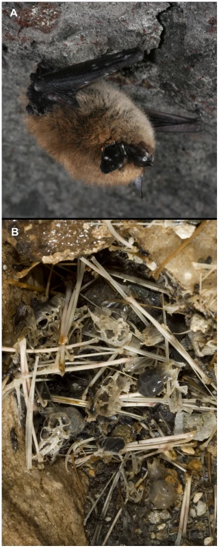
Biologists first observed unusual mortality and other clinical signs suggestive of WNS (Figure 1) in March 2007, at five underground bat hibernation sites (hibernacula) near Albany, New York. A multi-institutional disease investigation was initiated in January, 2008. The gross clinical presentation of WNS, a white substance on bats' muzzles, ears, and wings, suggested a fungal cause. A key finding was made when microscopy revealed that bats were colonized by a fungus with unique curve-shaped conidia. In parallel, skin samples from infected bats cultured at 7°C, a temperature consistent with the body temperature of hibernating bats, consistently yielded isolates of this same fungus [1], [6], a previously unknown species that was subsequently named Geomyces destructans [7].
Fig. 1. Images documenting clinical signs of white-nose syndrome (WNS) and devastating effects of the disease upon a wild bat population. 
(A) Eastern small-footed bat (Myotis leibii) with WNS photographed in April 2009, in Graphite Mine, New York, USA. Note characteristic white and gray fungal growth on the bat's muzzle, ear, and wing. (B) Bat skeletal remains on the floor of Aeolus Cave, Vermont, USA, photographed in 2010, approximately two years after white-nose syndrome was first identified at this site. Photo credit: Alan Hicks. Infection trials have since demonstrated that G. destructans is the sole causative agent of WNS [8], [9]. As a psychrophile, growth of G. destructans is restricted to temperatures below approximately 20°C [1], [7], making WNS an unusual disease among “warm blooded” mammals. Bats infected by G. destructans only develop WNS during hibernation when they dramatically reduce their core body temperature to a level conducive to proliferation of the fungus. Because of this temperature restriction, G. destructans is not known to be a pathogen of humans or domestic animals, and WNS is widely perceived strictly as a wildlife conservation problem, not as a threat to public health or agricultural production. However, while insectivorous bats lack the readily definable market value of domestic animals, they have great value both as critical components of ecosystems and for the ecological services they provide. A recent analysis estimated that insect suppression services provided by bats to commercial agriculture in the continental US has an average value of $22.9 billion per year [10].
Is G. destructans Related to Other Fungal Pathogens?
Ribosomal RNA gene region sequence analyses place the WNS pathogen in the anamorphic genus Geomyces, which consists predominately of cold-tolerant environmental saprobes [11]. Within the phylum Ascomycota, Geomyces belongs to the class Leotiomycetes, a group of fungi that includes several important plant pathogens (e.g., Botrytis cinerea and Schlerotinia sclerotiorum) but that is not recognized to include major pathogens of animals [12]. With the exception of a few reports of G. pannorum acting as a dermatophyte [13], Geomyces spp. are not commonly recognized as animal pathogens. Work is ongoing to identify characteristics that differentiate G. destructans from its non-pathogenic relatives.
Has G. destructans Been Found Beyond the Boundaries of North America?
Following the initial description of G. destructans in North America, investigators found the same fungus on hibernating bats in at least 12 European countries but without associated unusual mortality among infected animals [14]. It was then shown that isolates of G. destructans from North America were of a single clonal genotype [15], and an experimental study demonstrated that a European isolate of G. destructans was lethal to little brown bats, a North American bat species that does not exist in Europe [9]. Together, these findings support the hypothesis that the fungus was introduced from a foreign source to North America where it is acting as an emergent pathogen among a naïve population of hosts. Although differences in WNS lethality among bats of North America and Europe are still not understood, they likely stem from intercontinental differences in bat population dynamics, host species, and/or environmental conditions within hibernacula. Planned laboratory infectivity studies with a European bat species may yield additional insights.
Why Is G. destructans Pathogenic to Hibernating Bats?
While pathogenesis of G. destructans is not yet fully elucidated, an unrelated fungal skin infection of wildlife, amphibian chytridiomycosis caused by the fungus Batrachochytrium dendrobatidis [16], may provide important clues toward developing a more complete understanding of WNS. Skin of amphibians plays a vital role in maintaining osmotic homeostasis, and infection by B. dendrobatidis inhibits electrolyte transport across the epidermis of infected animals, disrupting plasma sodium and potassium concentrations and causing cardiac arrest and death [17]. Unlike B. dendrobatidis and other fungal dermatophytes that superficially colonize epidermis, nails, and hair, G. destructans is an aggressive fungal pathogen that penetrates the epidermis and invades underlying connective tissues [18]. Pathology analyses have shown the primary site for infection by G. destructans is the skin of bats' wings [18], [19]. In addition to its unique role in mammalian flight, bat wing skin also serves a critical role in maintaining physiological homeostasis. Intact bat wing skin prevents evaporative water loss, helps to release carbon dioxide through passive cutaneous exchange, aids in regulation of blood pressure, and supports thermoregulatory processes [19]. The importance of these physiological functions may be further elevated during hibernation, the exclusive time when bats are vulnerable to infection by G. destructans and a physiological state that may render them immunologically or otherwise unable to counteract both fungal infection and its sequelae. Additionally, infection of bats by G. destructans has been shown to cause increased arousal from torpor during hibernation, which likely contributes to depletion of energy reserves and death [9], [20]. A comprehensive understanding of disruptions to the delicate physiology of hibernating bats, either caused directly by the invasive skin infection that characterizes WNS and/or as mediated by yet uncharacterized fungal effector proteins, will likely be key to understanding this deadly disease.
What Can We Learn from the Discovery of WNS?
Recent applications of culture-independent microbial discovery techniques have yielded extraordinary advances in pathogen discovery [21]. However, the story of the discovery of G. destructans demonstrates that traditional culture - and histopathology-based laboratory methods remain highly relevant in disease investigation. For a cutaneous fungal disease such as WNS, the high diversity of fungi found in environments occupied by hibernating bats [22] and the non-sterile nature of skin makes the identification of a single clinically relevant microorganism difficult. Additionally, classification of the agent causative of WNS among Leotiomycetes, fungi not previously recognized as primary pathogens of animals [12], further complicates differentiation of a fungal pathogen from common environmental microflora. In the case of WNS, morphological characterization of the infectious agent in laboratory culture and on the skin of infected bats by both direct microscopy and histopathology readily provided the ability to make the original connection between G. destructans and WNS [1], [6], [18]. Definitive identification of G. destructans as the causative agent of WNS was ultimately determined through fulfillment of Koch's Postulates using a model system involving hibernating little brown bats [8]. Careful field observations, detailed pathology analyses, and controlled experimental confirmations bringing together scientists from multiple disciplines including microbiology, pathology, and ecology will continue to remain important in determining etiologies of novel fungal diseases.
Conclusions
Infectious diseases occur when a pathogen is introduced into a population of susceptible hosts under appropriate environmental conditions. In the case of WNS, the pathogen is G. destructans [8], the hosts are hibernating bats [1], and the environments that promote development of disease are cold underground hibernacula that bats occupy during winter. Unlike pathogenic microbes such as viruses that require host species for their survival, fungal pathogens can survive in the environment in the absence of hosts, providing them with the unique potential to extirpate host populations [3]. Thus, WNS presents a dire threat to populations of insectivorous hibernating bats in North America. However, much is known about the physiology of bat host species [19], and since the first published description of WNS [1], we have isolated and identified the pathogen [7] and developed model systems to study host–pathogen aspects of WNS in the laboratory [8], affording opportunities to fully define mechanisms of WNS pathogenesis. To date, efforts to manage the spread of WNS have focused on implementation of universal precautions, including restricting access of humans to sensitive bat hibernation sites and decontaminating equipment and clothing when sites are accessed for disease surveillance, research, or recreational purposes. Despite the considerable challenge of managing infectious disease in free-ranging wildlife while avoiding unintended adverse consequences, additional research aimed at increasing our understanding of the ecology of WNS in bats and their environment continues to offer the greatest potential for identifying novel strategies to mitigate the effects of this unprecedented disease.
Zdroje
1. BlehertDSHicksACBehrMJMeteyerCUBerlowski-ZierBM 2009 Bat white-nose syndrome: An emerging fungal pathogen? Science 323 227
2. SkerratLFBergerLSpeareRCashinsSMcDonaldKR 2007 Spread of chytridiomycosis has caused the rapid global decline and extinction of frogs. Ecohealth 4 125 134
3. FisherMCHenkDABriggsCJBrownsteinLCMadoffLC 2012 Emerging fungal threats to animal, plant and ecosystem health. Nature 484 186 194
4. HibbettDSOhmanAGlotzerDNuhnMKirkP 2011 Progress in molecular and morphological taxon discovery in Fungi and options for formal classification of environmental sequences. Fungal Biol Rev 25 38 47
5. FrickWFPollockJFHicksACLangwigKEReynoldsDS 2010 An emerging disease causes regional population collapse of a common North American bat species. Science 329 679 682
6. ChaturvediVSpringerDJBehrMJRamaniRLiX 2010 Morphological and molecular characterizations of psychrophilic fungus Geomyces destructans from New York bats with white-nose syndrome (WNS). PLoS ONE 5 e10783 doi:10.1371/journal.pone.0010783
7. GargasATrestMTChristensenMVolkTJBlehertDS 2009 Geomyces destructans sp. nov. associated with bat white-nose syndrome. Mycotaxon 108 147 154
8. LorchJMMeteyerCUBehrMJBoylesJGCryanPM 2011 Experimental infection of bats with Geomyces destructans causes white-nose syndrome. Nature 480 376 378
9. WarneckeLTurnerJMBollingerTKLorchJMMisraV 2012 Inoculation of bats with European Geomyces destructans supports the novel pathogen hypothesis for the origin of white-nose syndrome. Proc Natl Acad Sci U S A 109 6999 7003 doi:10.1073/pnas.1200374109
10. BoylesJGCryanPMMcCrackenGFKunzTH 2011 Economic importance of bats in agriculture. Science 332 41 42
11. RiceABCurrahRS 2006 Two new species of Pseudogymnoascus with Geomcyes anamorphs and their phylogenetic relationship with Gymnostellatospora. Mycologia 98 307 318
12. BerbeeML 2001 The phylogeny of plant and animal pathogens in the Ascomycota. Physiol Mol Plant Pathol 59 165 187
13. GianniCCarettaGRomanoC 2003 Skin infection due to Geomyces pannorum var. pannorum. Mycoses 46 430 432
14. PuechmailleSJWibbeltGKornVFullerHForgetF 2011 Pan-European distribution of white-nose syndrome fungus (Geomyces destructans) not associated with mass mortality. PLoS ONE 6 e19167 doi:10.1371/journal.pone.0019167
15. RajkumarSSLiXRuddRJOkoniewskiJCXuJ 2011 Clonal genotype of Geomyces destructans among bats with white-nose syndrome, New York, USA. Emerg Infect Dis 17 1273 1276
16. LongcoreJEPessierAPNicholsDK 1999 Batrachochytrium dendrobatidis gen. et sp. nov., a chytrid pathogenic to amphibians. Mycologia 91 219 227
17. VoylesJYoungSBergerLCampbellCVoylesWF 2009 Pathogenesis of chytridiomycosis, a cause of catastrophic amphibian declines. Science 326 582 585
18. MeteyerCUBucklesELBlehertDSHicksACGreenDE 2009 Pathology criteria for confirming white-nose syndrome in bats. J Vet Diagn Invest 21 411 414
19. CryanPMMeteyerCUBoylesJGBlehertDS 2010 Wing pathology of white-nose syndrome in bats suggests life-threatening disruption of physiology. BMC Biol 8 135 doi:10.1186/1741-7007-1188-1135
20. ReederDMFrankCLTurnerGGMeteyerCUKurtaA 2012 Frequent arousal from hibernation linked to severity of infection and mortality in bats with white-nose syndrome. PLoS ONE 7 e38920 doi:10.1371/journal.pone.0038920
21. LipkinWI 2010 Microbe hunting. Microbiol Mol Biol Rev 74 363 377
22. LindnerDLGargasALorchJMBanikMTGlaeserJ 2011 DNA-based detection of the fungal pathogen Geomyces destructans in soil from bat hibernation sites. Mycologia 103 241 246
Štítky
Hygiena a epidemiologie Infekční lékařství Laboratoř
Článek vyšel v časopisePLOS Pathogens
Nejčtenější tento týden
2012 Číslo 7- Jak souvisí postcovidový syndrom s poškozením mozku?
- Měli bychom postcovidový syndrom léčit antidepresivy?
- Farmakovigilanční studie perorálních antivirotik indikovaných v léčbě COVID-19
- 10 bodů k očkování proti COVID-19: stanovisko České společnosti alergologie a klinické imunologie ČLS JEP
-
Všechny články tohoto čísla
- Fungal Disease and the Developing Story of Bat White-nose Syndrome
- The Lectin Pathway of Complement Activation Is a Critical Component of the Innate Immune Response to Pneumococcal Infection
- Toll-Like Receptors Promote Mutually Beneficial Commensal-Host Interactions
- Identifying Host Factors That Regulate Viral Infection
- Gastrointestinal Cell Mediated Immunity and the Microsporidia
- Polydnaviruses as Symbionts and Gene Delivery Systems
- The Pore-Forming Toxin β hemolysin/cytolysin Triggers p38 MAPK-Dependent IL-10 Production in Macrophages and Inhibits Innate Immunity
- Evidence for Antigenic Seniority in Influenza A (H3N2) Antibody Responses in Southern China
- PLOS Pathogens
- Archiv čísel
- Aktuální číslo
- Informace o časopisu
Nejčtenější v tomto čísle- Polydnaviruses as Symbionts and Gene Delivery Systems
- The Lectin Pathway of Complement Activation Is a Critical Component of the Innate Immune Response to Pneumococcal Infection
- Gastrointestinal Cell Mediated Immunity and the Microsporidia
- Evidence for Antigenic Seniority in Influenza A (H3N2) Antibody Responses in Southern China
Kurzy
Zvyšte si kvalifikaci online z pohodlí domova
Současné možnosti léčby obezity
nový kurzAutoři: MUDr. Martin Hrubý
Všechny kurzyPřihlášení#ADS_BOTTOM_SCRIPTS#Zapomenuté hesloZadejte e-mailovou adresu, se kterou jste vytvářel(a) účet, budou Vám na ni zaslány informace k nastavení nového hesla.
- Vzdělávání


