-
Články
Top novinky
Reklama- Vzdělávání
- Časopisy
Top články
Nové číslo
- Témata
Top novinky
Reklama- Videa
- Podcasty
Nové podcasty
Reklama- Kariéra
Doporučené pozice
Reklama- Praxe
Top novinky
ReklamaLarge-Scale Population Study of Human Cell Lines Indicates that Dosage Compensation Is Virtually Complete
X chromosome inactivation in female mammals results in dosage compensation of X-linked gene products between the sexes. In humans there is evidence that a substantial proportion of genes escape from silencing. We have carried out a large-scale analysis of gene expression in lymphoblastoid cell lines from four human populations to determine the extent to which escape from X chromosome inactivation disrupts dosage compensation. We conclude that dosage compensation is virtually complete. Overall expression from the X chromosome is only slightly higher in females and can largely be accounted for by elevated female expression of approximately 5% of X-linked genes. We suggest that the potential contribution of escape from X chromosome inactivation to phenotypic differences between the sexes is more limited than previously believed.
Published in the journal: . PLoS Genet 4(1): e9. doi:10.1371/journal.pgen.0040009
Category: Research Article
doi: https://doi.org/10.1371/journal.pgen.0040009Summary
X chromosome inactivation in female mammals results in dosage compensation of X-linked gene products between the sexes. In humans there is evidence that a substantial proportion of genes escape from silencing. We have carried out a large-scale analysis of gene expression in lymphoblastoid cell lines from four human populations to determine the extent to which escape from X chromosome inactivation disrupts dosage compensation. We conclude that dosage compensation is virtually complete. Overall expression from the X chromosome is only slightly higher in females and can largely be accounted for by elevated female expression of approximately 5% of X-linked genes. We suggest that the potential contribution of escape from X chromosome inactivation to phenotypic differences between the sexes is more limited than previously believed.
Introduction
Dosage compensation is a regulatory process that alters gene expression along entire X chromosomes resulting in equivalent levels of X-linked gene products in males and females. Dosage compensation has evolved independently several times and is achieved in various ways. In Drosophila melanogaster, for example, the male X chromosome is hypertranscribed, doubling the output of X-linked genes (reviewed in [1] and [2]). This balances gene expression between the sexes and also satisfies the second requirement of a dosage compensation system, which is to balance X chromosome gene expression with that of the autosomes. The situation in mammals is more complex. Inactivation of one of the X chromosomes in female mammals [3] balances X-linked gene expression between males and females. However, if this were the only component of the mammalian system, the single active X chromosome of females and males would effectively make both sexes aneuploid with respect to autosome gene expression. Ohno hypothesized therefore that balance would be achieved by doubling the output from the male X (and active female X) [4]. The hypothesis was confirmed recently when 2-fold upregulation of the X chromosome was demonstrated for both human [5] and mouse [6].
The molecular mechanism for this X chromosome upregulation in mammals is unknown. By contrast X chromosome inactivation (XCI) has been known about for almost fifty years and is extensively characterized. XCI is a complex and tightly regulated process unique to mammals that results in heterochromatization and transcriptional silencing of one of the female X chromosomes (for a review see reference [7]). In eutherian mammals, the maternal or paternal X chromosome is inactivated randomly early in embryogenesis, and once established the pattern is mitotically stable. XCI was first suggested in 1961 to explain mosaic phenotypes seen in female mice heterozygous for sex-linked mutations in coat colour genes [3]. The theory was supported by the observation that cloned fibroblasts from human females heterozygous for an electrophoretic protein variant from the X-linked gene G6PD expressed either the paternal or the maternal allele, but not both [8]. Similar observations were reported for HPRT [9], PGK [10,11] and a number of other X-linked genes.
This pattern of complete silencing of one allele in females is seen for the majority of X-linked genes tested. However, the finding that the steroid sulphatase gene (STS) was always expressed in female fibroblast clones with one STS-deficient allele, regardless of which X was inactivated, suggested that some genes are not subject to XCI [12]. This “escape” from XCI results in differential expression of STS loci on the active X (Xa) and inactive X (Xi) chromosomes: clones expressing STS from the Xi have approximately half the level of STS enzyme activity of clones expressing from the Xa [13].
The use of rodent-human hybrid cell lines retaining an inactive human X chromosome has contributed greatly to our knowledge of escape from X inactivation [14–16]. This approach has the advantage of being able to assay gene expression from the Xi without the interference of the active copy. The largest study of this type has estimated that minimally 16% of human genes escape from XCI [16]. In a complementary approach, the same authors compared expression from maternal and paternal alleles of 94 genes in a panel of cell lines with skewed XCI and found that 15% of these were consistently expressed from both X chromosomes [16].
These approaches can detect low levels of expression from Xi. However, measuring the effect on dosage compensation requires a method to compare gene expression between the sexes. Expression microarrays, designed using the annotated X chromosome sequence [17], are suitable for such comparisons. Microarrays have previously been used to identify human genes escaping XCI by comparing gene expression in cell lines with supernumerary X chromosomes [18], in male and female lymphocytes [19], and in a range of male and female tissues [20]. A genome-wide survey of sex-differences in gene expression in lymphoblastoid cells also yielded several examples of X chromosome genes with elevated female expression levels [21].
In the light of this reported widespread escape from X inactivation, we sought to determine its effect on dosage compensation in the largest comparison to date of X chromosome gene expression in females and males. Here we report analysis of a microarray expression dataset obtained using lymphoblastoid cell lines from 210 individuals in four populations. We show that the proportion of X-linked genes with significantly higher expression in females is around 5%, and that dosage compensation in these cell lines is virtually complete.
Results
Expression from the X Chromosome in Male Cell Lines Is Upregulated 2-Fold
Microarray data from the Gene Expression Variation project (GENEVAR [22–24]) were analysed for 210 unrelated individuals from the four HapMap populations [25], designated CEU, CHB, YRI and JPT (see Methods).
First, we identified 371 genes on the X chromosome and 11,952 genes on autosomes that are expressed in the cell lines (see Methods). We then compared gene expression from autosomes and the male X chromosome. The median expression value of the 11,952 autosomal genes was plotted against the median of the 371 X chromosome genes for 105 unrelated male individuals from four populations (Figure 1A). There is a clear linear relationship between autosomal and X gene expression in all populations. The majority of data points fall along a diagonal close to that where the autosomal median is equal to the X median. We compared mean expression of the 371 X genes with randomly selected sets of 371 autosome genes (n = 100) for 30 YRI males using Student's t-test and saw no significant difference (data not shown). Average expression from the single X chromosome in males is, therefore, similar to expression from an autosome pair.
Fig. 1. Gene Expression from the Single Male X Chromosome Is Similar to Expression from Autosome Pairs 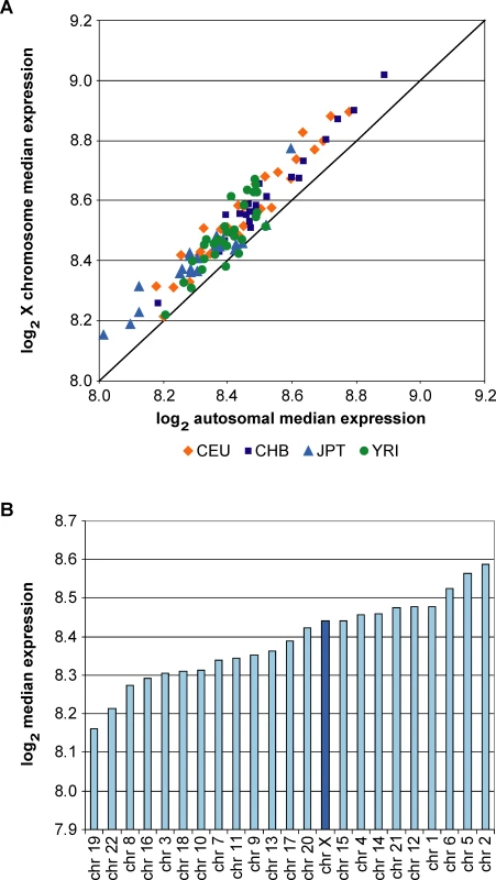
(A) Scatter graph of median expression of 11,952 autosomal genes against median expression of 371 X chromosome genes for 105 males. The diagonal indicates the position at which median expression levels from X and autosomes are the same. Next, we compared the male X chromosome with each autosome pair separately. Results for the YRI population are shown in Figure 1B, and very similar results were obtained for the other populations (data not shown). Median expression from the single X chromosome falls within the normal range seen for autosome pairs and is slightly above average. The latter accounts for the observation that most data points in Figure 1A lie above the diagonal.
We conclude that expression from the single X in male cell lines is upregulated 2-fold relative to autosomes, thus achieving dosage parity between the X and autosomes. Upregulation occurs precisely and consistently in 105 individual male samples. This supports and extends the findings of Nguyen and Disteche [5].
The Contribution of the Inactive X Chromosome in Females Is Small
The two X chromosomes in females are not equivalent as the majority of genes on one are subject to X inactivation; however, it is well established that a number of genes escape the silencing process. Therefore, we compared expression of X chromosome genes in females and males, reasoning that escape from XCI should produce a substantially higher level of expression in females.
First we compared expression of the 11,952 expressed autosome genes and 371 expressed X genes in 30 males and 30 females from the YRI population. Figure 2A shows median male expression plotted against female expression for each gene. The autosome genes lie on a diagonal with the vast majority showing very similar expression in males and females. Most X genes lie on the same diagonal, indicating that X chromosome gene expression too is similar in males and females and is not proportional to the number of X chromosomes. These data suggest that for most X chromosome genes, dosage compensation is achieved between males and females.
Fig. 2. Limited Contribution of the Xi to Female Gene Expression 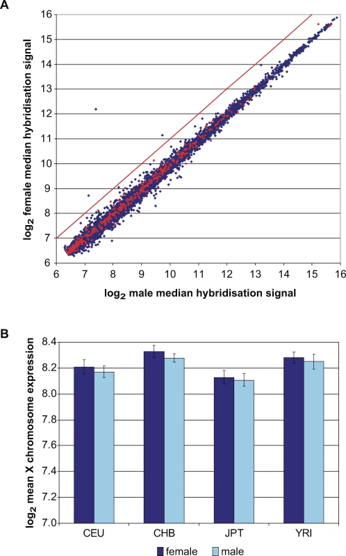
(A) Median expression of 11,952 autosome genes (blue) and 371 X genes (red) for 30 YRI males plotted against median expression for 30 females. The red diagonal indicates the expected position of X-linked genes if expression were proportional to copy number. We then normalised expression of the 371 X chromosome genes to the median of the 11,952 autosome genes for each individual, and calculated the mean of the normalised X chromosome genes for 105 males and 105 females. In each population mean expression of X chromosome genes was higher in females than males (Figure 2B). However, these differences are small, representing increased expression in females of just 2.6%, 3.4%, 1.5%, and 2.2% for CEU, CHB, JPT and YRI, respectively. Genes escaping X inactivation do not, therefore, have a great effect on the overall level of X-linked gene expression in the female cell lines. This indicates that dosage compensation is occurring effectively.
Identification and Analysis of Genes with Significantly Higher Female Expression
Although the difference is small, there is a measurable and consistent increase in X chromosome gene expression in females compared to males. In order to identify the genes that contribute to this difference, we looked for X chromosome genes with significantly higher expression in females. We used a Student's t-test to assess differences in expression levels between females and males within each population separately for the 371 X chromosome genes and 11,952 autosome genes (Table S1).
Figure 3 compares the proportion of X chromosome and autosome genes expressed more highly in females or males at three different levels of significance: p < 0.05, p < 0.01 and p < 0.001. The proportion of X chromosome genes expressed more highly in females is greater than that from autosomes in each population. The difference between X and autosomes is consistent across the three levels of significance and at p < 0.001 ranges from 3.5% to 4.9% of genes in different populations. In contrast, the proportion of X chromosome genes with higher expression in males remains similar to that of autosome genes at all levels of significance (Figure 3).
Fig. 3. The Proportion of Genes with Higher Female Expression Is Greater from X Than Autosomes 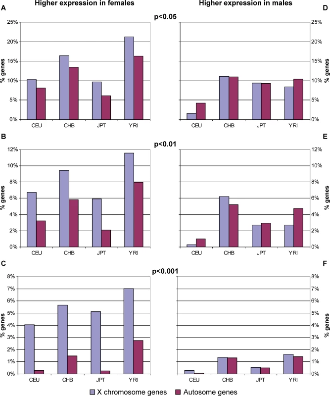
Percentage of X chromosome and autosome genes showing higher expression in females (A–C) or males (D–F) at three levels of significance: p < 0.05, p < 0.01, and p < 0.001. Note that the y-axis scale changes for different levels of significance. The most likely explanation for the observations above is that a proportion of X chromosome genes escape from X inactivation to a measurable level in these female cell lines. We supposed that X-linked genes able to escape the silencing process would be expressed more highly in females in all populations. We also reasoned that other X-linked or autosomal genes reaching a significance threshold may not do so in all populations. We therefore assessed the population commonality of autosomal and X chromosome genes with higher female expression at different levels of significance (Table 1). At p < 0.05 and p < 0.01, the proportion of genes with significantly higher female expression is almost identical for autosomes and the X chromosome. However, the distribution of genes among populations is strikingly different, with a far greater percentage of X chromosome genes achieving significance in all four populations. When the significance threshold is raised to p < 0.001, the proportion of genes retained is now lower for autosomes than for the X chromosome, and no autosome gene is common to three or four populations (Table 1). By contrast, approximately 3% of X chromosome genes are significantly elevated in the females of all populations at p < 0.001 (Table 1). A single gene (CD99) is expressed more highly in the males of all four populations (p < 0.001). This is the only notable difference between X and autosomes in respect of higher male expression (Table 1).
Tab. 1. 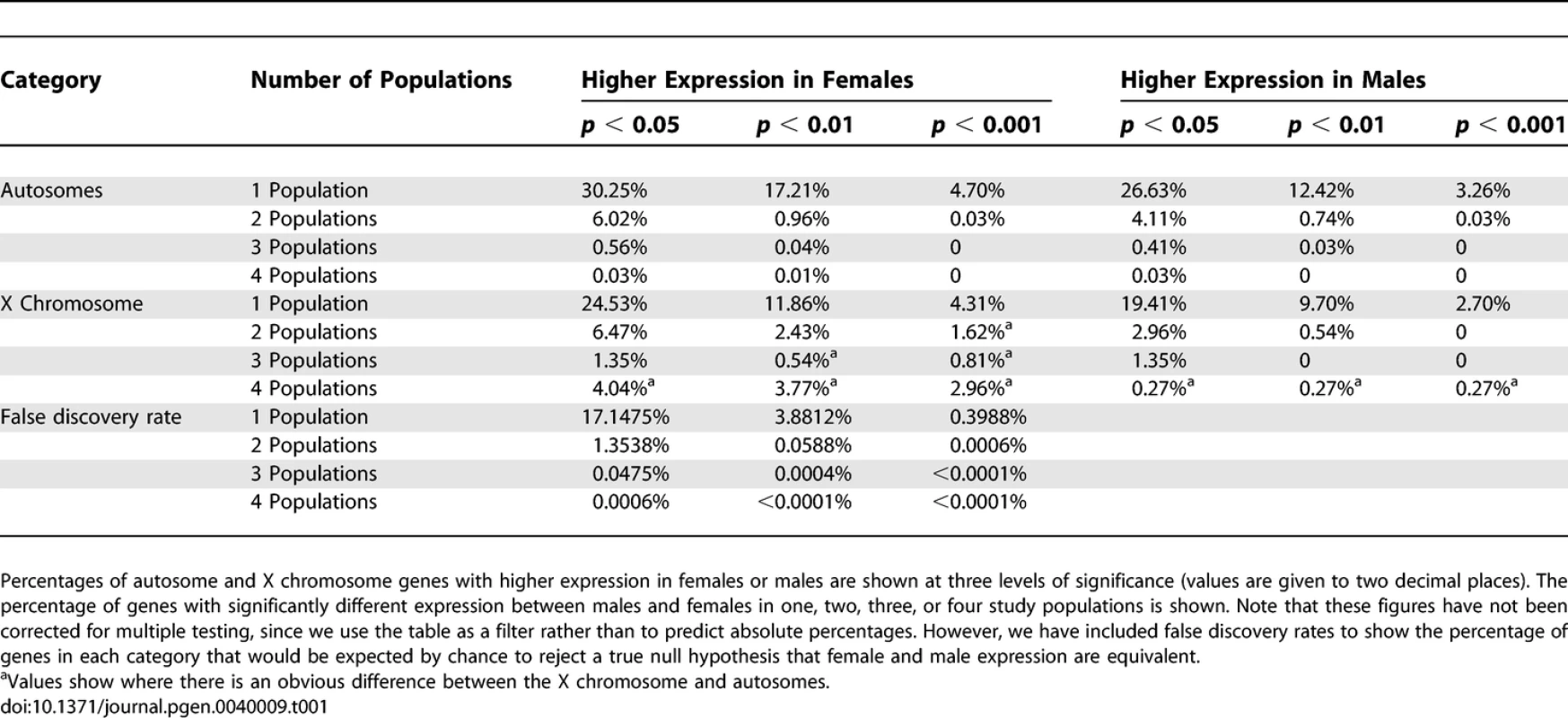
Observing a Clear Difference between X Chromosome and Autosome Genes in Respect of Higher Female Expression Using the combination of significance values and population commonality as a filter (Table 1), we identified a group of 20 X chromosome genes that are remarkable in the consistency of their elevation across females (Table 2): ALG13, CA5B, DDX3X, EIFIAX, EIF2S3, FUNDC1, HDHD1A, JARID1C, MSL3L1, PCTK1, PNPLA4, PRKX, RPS4X, SMC1L1, STS, UBE1, USP9X, UTX, ZFX and ZRSR2. Eleven of these were expressed more highly in females at p < 0.001 in all four populations (Table 2). This situation was not observed for any of 11,952 autosomal genes tested.
Tab. 2. 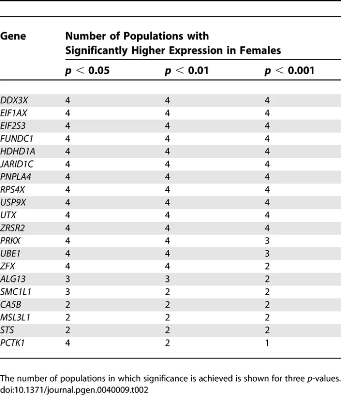
Genes Expressed More Highly in Females That Fit the Criteria for Escaping X Inactivation Figure 4A illustrates the female to male ratio of expression for each of the genes in the four populations. Taking the mean of the four populations, there is a subset of six genes (JARID1C, UTX, HDHD1A, PNPLA4, DDX3X and EIF1AX) for which expression in females is around 1.5-fold greater than in males. Most genes have a much smaller difference: EIF2S3, USP9X, CA5B and PCTK1, ZFX and SMC1L1 all have less than 1.2-fold higher expression in females compared to males. We interpret these ratios as expression from the active X that is equivalent to expression from the single X in males, combined with a lower level of expression from the inactive X that is more variable between genes. Some genes (e.g., DDX3X, STS) have a much higher ratio in some populations than others, which may represent a biological difference in the extent of escape from X inactivation in different human populations.
Fig. 4. Comparison of Female and Male Expression of Genes with Higher Female Expression 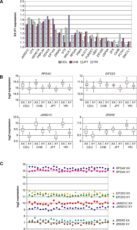
(A) Female to male expression ratio for 20 genes expressed more highly in females in four human populations. Expression in females is typically 10%–50% higher than in males. We observe higher female expression for 5.4% of the X chromosome genes expressed in the cell lines (20/371). The possibility remains that other X-linked genes may escape from XCI in lymphoblastoid cells. However, we have determined that these 20 genes account for almost all of the difference in gene expression between males and females seen in Figure 2B (data not shown). Therefore, any expression of additional genes from the inactive X chromosome must be very low and/or must occur in only a small fraction of female cell lines. In either case, the impact on dosage compensation at the population level would be minimal. Therefore, we conclude that 94.6% of X-linked genes are effectively dosage compensated in human lymphoblastoid cell lines.
Genes with Higher Female Expression Do Not Show Greater Variation in Expression among Females
We hypothesized that escape from XCI may be partly stochastic, and that this might lead to greater variation in expression levels among females than males for some genes. Figure 4B shows box and whisker plots for four of the genes that can be considered to escape from XCI on the basis of their higher female expression. The other 16 genes follow a similar pattern (data not shown). The size of the box (interquartile range) is a good indicator of the similarity of distribution between females and males. In the majority of cases there is little difference in the distribution or range of values between the sexes, although the female values have a higher median and therefore the entire plot is shifted upwards. There is no correlation between the size of the interquartile range and median expression level. We also calculated the variance for each gene and found no significant difference between males and females within each population (unpublished data). We conclude that genes escaping XCI in these cell lines are not expressed more variably in females than males, which suggests that escape may be a tightly regulated rather than a stochastic event.
While the medians and interquartile ranges are clearly different between males and females, there is considerable overlap between the distributions of the expression levels of the two datasets. Individual data points from males and females of the YRI population are shown for four genes escaping XCI as a scatter graph (Figure 4C). JARID1C is unique in that all data points for females are higher than all data points for males. For the other 19 genes, the female and male datasets overlap to varying extents (contrast EIF2S3 with RPS4X in Figure 4C). This can also be seen as overlap of whiskers in Figure 4B. This observation illustrates the extent of inter-individual variability of gene expression and highlights the importance of comparing large samples of males and females to identify differences in gene expression that are a consequence of escape from XCI.
PAR1 Genes Have Equal Expression from X and Y Chromosomes
The genes from the pseudoautosomal regions (PARs) are a special case as they lie within regions of XY recombination and are essentially equivalent on the X and Y chromosomes. For genes in PAR1, escape from XCI is generally believed to be a prerequisite for dosage compensation between the two female X chromosomes and the male X and Y. We found that twelve PAR1 genes show no significant difference between females and males across populations and are therefore dosage compensated (Table S1). The only exception is CD99, which is expressed significantly more highly in males in all four populations (p < 0.001).
We identified single nucleotide polymorphisms (SNPs) in PAR1 genes SLC25A6, CXYorf3, ZBED1 and CD99 and used a quantitative assay to measure relative expression from the X and Y alleles in heterozygous males. As shown in Table 3, the relative contribution from the X and Y chromosomes is very similar for each of the four genes. We conclude that the majority of PAR1 genes escape from XCI and are dosage compensated, consistent with the expectation above.
Tab. 3. 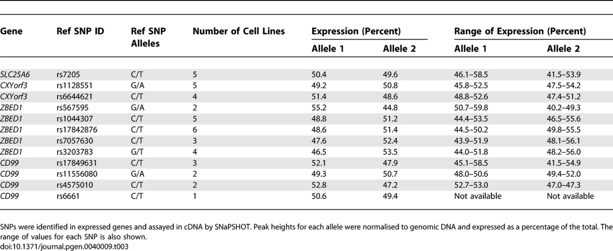
Genes in the Pseudoautosomal Region (PAR1) Are Expressed Equally from the X and Y Chromosomes in Male Cell Lines Evidence That Dosage Compensation of Some X-Linked Genes Is Maintained through Expression of a Y-Linked Copy
The majority of genes that have a functional Y chromosome homologue were found to be expressed in hybrid cells containing the Xi [16]. Therefore, we decided to test the possibility that genes with higher female expression might, like the genes in PAR1, be compensated by functionally equivalent Y-linked copies. Eight of the 20 genes with higher female expression have functional Y-linked gametologues. We excluded PRKY and EIF1AY from the analysis. The PRKY probe has 94% sequence identity to PRKX and gives a strong signal in females. Expression of EIF1AY is approximately 13-fold greater than EIF1AX in males, suggesting that EIF1AY is not involved in a compensation mechanism. Gene expression for the remaining six X-Y gene pairs is shown in Figure 5.
Fig. 5. Evidence That Expression of the Y-linked Copy of XY Gene Pairs Can Maintain Dosage Compensation 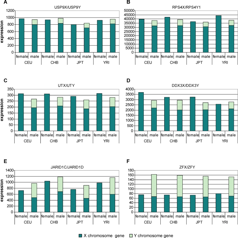
(A–F) Stacked bars show the cumulative expression of X and Y gametologues in males alongside expression from the X chromosomes in females. Note that the y-axis is a linear scale. USP9X expression is significantly higher in females at p < 0.001 in all populations. Interestingly, the sum of USP9X and USP9Y expression in males is not significantly different from the level of USP9X in females (p = 0.822 [CEU], p = 0.024 [CHB], p = 0.245 [JPT] and p = 0.610 [YRI]). USP9Y expression therefore completely restores the USP9X dosage imbalance between males and females. Figure 5B–5D shows similar results for gene pairs RPS4X/RPS4Y1, UTX/UTY and DDX3X/DDX3Y. In each case dosage from the X and Y copies in males is similar to that of the X copies in females. Expression of the Y copy is always much lower than expression of the X copy and appears to reflect the expression from the Xi. For these four genes, dosage compensation appears to be achieved by expression of the Y copy. In contrast, expression levels of JARID1C (X) and JARID1D (Y) (Figure 5E) are approximately equal and their combined expression in males is significantly greater than JARID1C expression in females. A similar picture is obtained with ZFX and ZFY in males (Figure 5F). We have identified an X-Y gene pair (TMSB4X/TMSB4Y) whose X copy is dosage compensated according to our data. TMSB4Y is expressed at less than 1% of the level of TMSB4X and therefore does not affect dosage compensation between males and females.
Genes with Higher Female Expression Are Not Distributed Evenly on the X Chromosome
Genes that escape from XCI in hybrid cell lines are non-randomly distributed on the X chromosome [16,17]. In light of identifying a smaller proportion of genes with higher female expression, we assessed the relationship between gene expression and chromosomal location. We observed that the distribution of genes with elevated female expression is also non-random, with most lying on the short arm (Figure 6). The chromosome can be divided into strata that ceased to recombine with the Y chromosome at different times in evolutionary history [17,26]. The most ancient parts of the chromosome (strata S1 and S2), covering the long arm and proximal short arm, contain 287 of the 371 expressed genes, but only six that have higher female expression. By contrast, a larger fraction of genes have elevated female expression levels in regions that stopped recombining with the Y chromosome more recently, either in early eutherian mammals (S3) or in primates (S4, S5). Ten out of 66 S3 genes fit this picture, while all three expressed genes in S4, together with the single example in S5, are more highly expressed in females. These findings support the model that X-linked genes are recruited into the XCI system following the Y chromosome degeneration that occurs when regions cease to recombine [27].
Fig. 6. Genes Expressed More Highly in Females Are Distributed Non-Randomly on the X Chromosome 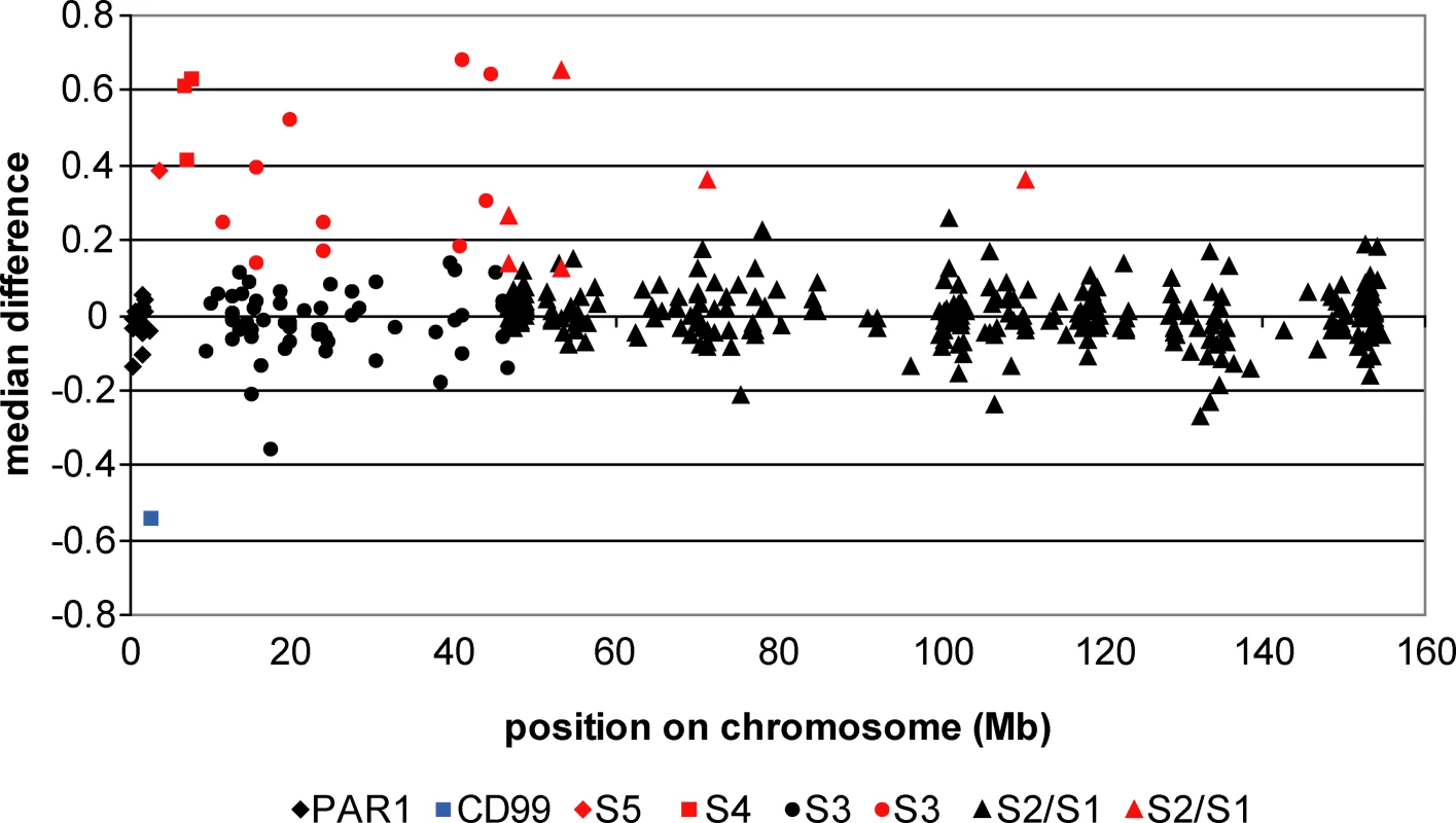
For each gene, the median expression value for males was subtracted from the median expression value for females to give a median difference. Illustrated is the median difference for each gene based on averaging the values for the four populations, plotted according to location on the X chromosome. A median difference of 1 represents a 2-fold difference in expression. Different symbols represent different evolutionary strata. Genes expressed significantly more highly in females are coloured red, and the single gene expressed significantly more highly in males (CD99) is coloured blue. Discussion
On the basis of these data, we suggest that dosage compensation in human lymphoblastoid cells is virtually complete. Gene expression from the single X chromosome in males is upregulated 2-fold compared to the autosomes. Expression from the female X chromosome pair is almost the same as from the male X, suggesting that few genes escape the silencing process to any great extent. Twenty genes in this study (5.4%) have significantly higher female expression, and four of these could have dosage balance maintained through expression of a Y-linked homologue.
Ohno predicted 40 years ago that the evolution of a dosage compensation mechanism in mammals must have involved a doubling of expression of each X-linked gene as the Y chromosome degenerated [4]. Two-fold upregulation of the X chromosome has now been demonstrated for both humans and mice [5,6] and in a range of tissues [5]. In D. melanogaster, a slight but significant overexpression of the X chromosome compared to the autosomes in all XX;AA samples has been reported [6] which may be due to inherent hypertranscription of the X chromosome. We have determined that human X chromosome expression is not significantly elevated above the autosome average, suggesting that the X chromosome is not hypertranscribed in the cell lines over and above the 2-fold upregulation. Our data show that upregulation occurs precisely and consistently in 105 individual male samples, also leading us to conclude that in lymphoblastoid cell lines gene expression is appropriately regulated.
Upregulation of the X is not seen in mouse germ cells suggesting that it takes place in the developing embryo [5]. Two-fold upregulation may be a general feature of X chromosomes, affecting genes on Xa and those on Xi that escape inactivation. Alternatively, upregulation and silencing could be mutually exclusive choices for X chromosomes in embryogenesis, simultaneously achieving correct X gene expression and dosage compensation. Previously, genes have been classified as partially escaping XCI if their female to male expression ratio is below two. However, under the second model described, genes fully expressed from both Xi and an upregulated Xa in females would have a theoretical maximum expression that is 1.5-fold greater in females than males. We favour this model as we have identified six genes expressed approximately 1.5-fold more highly in females and the greatest ratio we observe for any gene is 1.56, averaged across four populations.
Is 2-fold upregulation a chromosome-wide phenomenon? The PAR1 region is the only surviving remnant of a large autosomal addition to both sex chromosomes that still undergoes recombination in male meiosis. The X and Y chromosomes are equivalent in PAR1 and genes here are predicted to escape XCI. Accordingly, all PAR1 genes tested were expressed from Xi in hybrid cell lines [16]. The majority of PAR1 genes included in our study showed no significant difference in expression between females and males. This gives rise to two models for PAR1 gene expression. In the first, PAR1 genes have equal expression from Xa, Xi and Y copies and so no dosage compensation is necessary. Under this model, PAR1 is excluded from both the upregulation and the silencing components of dosage compensation. In the second model, the PAR1 is 2-fold upregulated on the Xa only and dosage compensation is achieved through equal expression from the Xi and Y copies. A corollary of this model is unequal expression of PAR1 alleles within a cell, but equal expression between males and females. We have tested three dosage compensated PAR1 genes in males and find that expression levels are similar from the X and Y alleles. Therefore, we favour the first model and propose that PAR1 is protected from both upregulation and silencing.
CD99 is the only PAR1 gene found to be more highly expressed in males, yet has equivalent expression from the X and Y copies. CD99 is the gene that lies closest to the boundary between the PAR1 and X-linked material, and we suggest that spreading of the XCI signal across the pseudoautosomal boundary results in partial silencing of CD99. Consistent with this hypothesis, the protein product of the CD99 gene was found to be present at lower levels in hybrids containing Xi than those containing Xa [28].
Outside the pseudoautosomal regions, twenty genes are expressed at significantly higher levels in female cell lines. We hypothesized that these genes would escape from XCI. Formally, the alternative explanation for the elevated female expression could be that these genes are hypertranscribed from the female active X chromosome. However, since all of these genes are included in previous reports of escape from XCI [12,15,16,29–33], we prefer the former explanation for higher female expression. An intriguing observation is that the dosage of some of these genes could be effectively compensated by expression of a Y copy. An underlying assumption of this analysis is that the X and Y gametologues are functionally equivalent, despite their evolutionary divergence. The DEAD box RNA helicase proteins DDX3X and DDX3Y appear to be interchangeable, as both rescue a temperature-sensitive mutant hamster cell line incapable of growth at a non-permissive temperature [34]. The ribosomal proteins RPS4Y1 and RPS4X show functional equivalence in a similar rescue assay and can function interchangeably in ribosomes [35]. RPS4Y1 and RPS4X are among a very small number of genes from the ancient sex chromosomes that have functional Y copies [17]. Despite their considerable divergence time and very high synonymous substitution rate, their protein products share 93% identity and are the same length, consistent with their being functionally equivalent.
Previous microarray studies have assessed escape from XCI by looking for increased expression in cell lines with supernumerary X chromosomes [18] or by comparing female and male expression [19,20]. More recently, a larger study assessed genome-wide sex differences in gene expression using lymphoblastoid cell lines from monozygotic twin pairs [21]. Each study reports some genes that are considered to be well established as escaping from XCI, but the four vary considerably in the number and identity of genes documented. These differences might be explained in part by variation in escape from X inactivation in different tissues, for which there is evidence [20]. However, a further possibility is that inter-individual variability in expression that is unrelated to XCI could increase the risk of false positives or negatives where the sample size is small. The scale of our study, which measured gene expression in 210 individuals for 81% of protein coding genes on the X chromosome, means that we can confidently detect significantly higher expression in females at the population level in spite of this factor. Notably the study by McRae et al. [21], which assayed 38 lymphoblastoid cell lines, agrees most closely with our study in the identity and proportion of genes with higher female expression.
We find that 5.4% of X-linked genes have increased female expression in the cell lines. Analysis of somatic-cell hybrids that retain Xi and of fibroblast cells with non-random XCI has put the proportion of genes escaping XCI at 15%–25% [16]. This difference could be explained by a lower proportion of genes escaping XCI in lymphoblastoid compared with fibroblast (or hybrid) cells. However, perhaps more important are the differences of approach between the two studies. Our study used a population based analysis of dosage compensation, whereas Carrel and Willard [16] detected expression in hybrid cell lines, or compared expression levels from Xa and Xi alleles in female cell lines, using methods capable of detecting very low levels of expression from Xi. Some genes, therefore, could be expressed from Xi but at a level that is insufficient to cause a dosage imbalance. Other genes could escape to a larger extent in a small number of females, as suggested by Carrel and Willard [16]. Neither of these, though, is substantial enough to generate a significant sex-difference at the population level.
We conclude that dosage compensation in human lymphoblastoid cell lines is highly effective and tightly controlled. It will be interesting to extend these studies to other tissues, but it seems unlikely that this level of regulation would be restricted to this single cell type. Therefore, we propose that the contribution of escape from XCI to male-female phenotypic differences may be small and furthermore, we suggest that the number of genes contributing to phenotype in X chromosome aneuploidies is lower than previously thought.
Materials and Methods
Generation of dataset.
Gene expression was assayed in lymphoblastoid cell lines of all 210 unrelated HapMap individuals [25] from four populations (CEU: 60 (30 Male/30 Female) Utah residents with ancestry from northern and western Europe; CHB: 45 (22M/23F) Han Chinese in Beijing; JPT: 45 (23M/22F) Japanese in Tokyo and YRI: 60 (30M/30F) Yoruba in Ibadan, Nigeria). RNA preparation, labeling, hybridization to Sentrix Human-6 BeadChip (Illumina), gene expression quantification and normalization of raw data were described previously [23]. Briefly, each RNA sample was labelled in duplicate and each labelled sample was hybridised to two separate arrays. Data were subjected to quantile normalization then were median normalized across all individuals. Final data points for each gene are the mean of the four normalized hybridisation values. Log2 transformed mRNA expression values were used throughout except where otherwise stated. Data can be downloaded from http://www.sanger.ac.uk/humgen/genevar/ and the Gene Expression Omnibus (http://www.ncbi.nlm.nih.gov/geo/) entry GSE6536.
Selection of expression data for analysis.
We established an appropriate cut-off point for evaluating gene expression by examination of four non-human probes (lysA, pheA, thrB and trpF) in 210 individuals: 836/840 data points had log2 expression values <6.4. We also evaluated signals from Y-linked genes in female samples: 593/620 data points were found to have values <6.4, excluding two probes that apparently cross-hybridised with X-linked genes. We conservatively chose to analyse genes with log2 median expression >6.4 in all four populations. We excluded redundant probes (n = 21) for X chromosome genes and any X probes that matched autosomal exons. Data for 11,952 expressed autosome sequences and 371 expressed X chromosome genes were used in all downstream analyses. The complete set of X chromosome and autosomal genes represented in this study and median expression values are shown in Table S2.
Statistical analysis.
Data from the four populations were considered separately throughout. We tested male and female datasets separately for skewness and kurtosis for 371 X chromosome probes and found no evidence for them, except for a very small number of genes in some populations where log2 expression was close to the expression cutoff of 6.4. We therefore concluded that the gene expression data generally follow a normal distribution. We used median values to illustrate chromosome or population averages except where we have shown the standard deviation (Figure 2B). To make comparisons between autosome genes and X genes, or between groups of individuals, we normalized all gene expression values to the median value of 11,952 autosomal probes for each individual. We compared the variances of male and female samples for X chromosome genes by placing the larger variance over the smaller to form an F statistic. We found that variances for females and males are not significantly different. We tested significance by calculating p-values associated with a Student's two sample homoscedastic t-test with a two-tailed distribution. The complete list of p-values is shown in Table S1.
Allele-specific gene expression analysis.
SNaPshot was carried out on cDNA and genomic DNA from heterozygous males using the SNaPshot Multiplex Kit (Applied Biosystems) according to the manufacturer's instructions with the following modifications. Initial template generation was carried out using Platinum Taq polymerase (Invitrogen) in a standard reaction using touchdown polymerase chain reaction (PCR): denaturation: 94°C 15 min; 20 cycles: 94°C 30 sec, 70°C, 30 sec reducing by 1°C per cycle, 72°C 45 sec; then 15 cycles: 94°C 30 sec, 50°C 30 sec, 72°C 45 sec; final extension 72°C for 7 min. PCR products were treated with 2 units of shrimp alkaline phosphatase (USB) and 1.5 units of Exonuclease I (USB) for 1 hour at 37°C to remove primers and nucleotides, then at 80°C for 15 mins. Primer extension products were analysed on an ABI 3730 DNA Analyzer with a POP-7 Polymer and a 36cm capillary array with the ABI standard run module. SNP data were analysed using ABI PRISM GeneMapper Software Version 3.0. Primers used to generate template DNA for analysis and SNaPshot extension primers are shown in Table S3. Peak heights from cDNA were normalized to genomic DNA values and expressed as an allelic ratio.
Supporting Information
Zdroje
1. BakerBSGormanMMarinI
1994
Dosage compensation in Drosophila.
Annu Rev Genet
28
491
521
2. MellerVHKurodaMI
2002
Sex and the single chromosome.
Adv Genet
46
1
24
3. LyonMF
1961
Gene action in the X-chromosome of the mouse (Mus musculus L.).
Nature
190
372
373
4. OhnoS
1967
Sex chromosomes and sex-linked genes
Berlin
Springer
5. NguyenDKDistecheCM
2006
Dosage compensation of the active X chromosome in mammals.
Nat Genet
38
47
53
6. GuptaVParisiMSturgillDNuttallRDoctoleroM
2006
Global analysis of X-chromosome dosage compensation.
J Biol
5
3
7. ChowJCYenZZiescheSMBrownCJ
2005
Silencing of the mammalian X chromosome.
Annu Rev Genomics Hum Genet
6
69
92
8. DavidsonRGNitowskyHMChildsB
1963
Demonstration of two populations of cells in the human female heterozygous for glucose-6-phosphate dehydrogenase variants.
Proc Natl Acad Sci U S A
50
481
485
9. MigeonBRDer KaloustianVMNyhanWLYoughWJChildsB
1968
X-linked hypoxanthine-guanine phosphoribosyl transferase deficiency: heterozygote has two clonal populations.
Science
160
425
427
10. DeysBFGrzeschickKHGrzeschickAJaffeERSiniscalcoM
1972
Human phosphoglycerate kinase and inactivation of the X Chromosome.
Science
175
1002
1003
11. GartlerSMChenSHFialkowPJGiblettERSinghS
1972
X Chromosome inactivation in cells from an individual heterozygous for two X-linked genes.
Nat New Biol
236
149
150
12. ShapiroLJMohandasTWeissRRomeoG
1979
Non-inactivation of an X-chromosome locus in man.
Science
204
1224
1226
13. MigeonBRShapiroLJNorumRAMohandasTAxelmanJ
1982
Differential expression of steroid sulfatase locus on active and inactive human X chromosome.
Nature
299
838
840
14. BrownCJCarrelLWillardHF
1997
Expression of genes from the human active and inactive X chromosomes.
Am J Hum Genet
60
1333
1343
15. CarrelLCottleAAGoglinKCWillardHF
1999
A first-generation X-inactivation profile of the human X chromosome.
Proc Natl Acad Sci U S A
96
14440
14444
16. CarrelLWillardHF
2005
X-inactivation profile reveals extensive variability in X-linked gene expression in females.
Nature
434
400
404
17. RossMTGrafhamDVCoffeyAJSchererSMcLayK
2005
The DNA sequence of the human X chromosome.
Nature
434
325
337
18. SudbrakRWieczorekGNuberUAMannWKirchnerR
2001
X chromosome-specific cDNA arrays: identification of genes that escape from X-inactivation and other applications.
Hum Mol Genet
10
77
83
19. CraigIWMillJCraigGMLoatCSchalkwykLC
2004
Application of microarrays to the analysis of the inactivation status of human X-linked genes expressed in lymphocytes.
Eur J Hum Genet
12
639
646
20. TalebizadehZSimonSDButlerMG
2006
X chromosome gene expression in human tissues: male and female comparisons.
Genomics
88
675
681
21. McRaeAFMatigianNAVadlamudiLMulleyJCMowryB
2007
Replicated effects of sex and genotype on gene expression in human lymphoblastoid cell lines.
Hum Mol Genet
16
364
373
22. StrangerBEForrestMSClarkAGMinichielloMJDeutschS
2005
Genome-wide associations of gene expression variation in humans.
PLoS Genet
1
e78
doi: 10.1371/journal.pgen.0010078
23. StrangerBEForrestMSDunningMIngleCEBeazleyC
2007
Relative impact of nucleotide and copy number variation on gene expression phenotypes.
Science
315
848
853
24. StrangerBENicaACForrestMSDimasABirdCP
2007
Population genomics of human gene expression.
Nat Genet
39
1217
1224
25. The International HapMap Consortium
2005
A haplotype map of the human genome.
Nature
437
1299
1320
26. LahnBTPageDC
1999
Four evolutionary strata on the human X chromosome.
Science
286
964
967
27. JegalianKPageDC
1998
A proposed path by which genes common to mammalian X and Y chromosomes evolve to become X inactivated.
Nature
394
776
780
28. GoodfellowPPymBMohandasTShapiroLJ
1984
The cell-surface antigen locus, MIC2X, escapes X-inactivation.
Am J Hum Genet
36
777
782
29. FisherEMBeer-RomeroPBrownLGRidleyAMcNeilJA
1990
Homologous ribosomal protein genes on the human X and Y chromosomes: escape from X inactivation and possible implications for Turner syndrome.
Cell
63
1205
1218
30. AgulnikAIMitchellMJMatteiMGBorsaniGAvnerPA
1994
A novel X gene with a widely transcribed Y-linked homologue escapes X-inactivation in mouse and human.
Hum Mol Genet
3
879
884
31. CarrelLClemsonCMDunnJMMillerAPHuntPA
1996
X inactivation analysis and DNA methylation studies of the ubiquitin activating enzyme E1 and PCTAIRE-1 genes in human and mouse.
Hum Mol Genet
5
391
401
32. EhrmannIEEllisPSMazeyratSDuthieSBrockdorffN
1998
Characterization of genes encoding translation initiation factor eIF-2 gamma in mouse and human: sex chromosome localization, escape from X-inactivation and evolution.
Hum Mol Genet
7
1725
1737
33. GreenfieldACarrelLPennisiDPhilippeCQuaderiN
1998
The UTX gene escapes X inactivation in mice and humans.
Hum Mol Genet
7
737
742
34. SekiguchiTIidaHFukumuraJNishimotoT
2004
Human DDX3Y, the Y-encoded isoform of RNA helicase DDX3, rescues a hamster temperature-sensitive ET24 mutant cell line with a DDX3X mutation.
Exp Cell Res
300
213
222
35. WatanabeMZinnARPageDCNishimotoT
1993
Functional equivalence of human X - and Y-encoded isoforms of ribosomal protein S4 consistent with a role in Turner syndrome.
Nat Genet
4
268
271
Štítky
Genetika Reprodukční medicína
Článek vyšel v časopisePLOS Genetics
Nejčtenější tento týden
2008 Číslo 1
Nejčtenější v tomto čísle- Application of Ancestry Informative Markers to Association Studies in European Americans
- Large-Scale Population Study of Human Cell Lines Indicates that Dosage Compensation Is Virtually Complete
- Haplotype Mapping of a Diploid Non-Meiotic Organism Using Existing and Induced Aneuploidies
Kurzy
Zvyšte si kvalifikaci online z pohodlí domova
Současné možnosti léčby obezity
nový kurzAutoři: MUDr. Martin Hrubý
Všechny kurzyPřihlášení#ADS_BOTTOM_SCRIPTS#Zapomenuté hesloZadejte e-mailovou adresu, se kterou jste vytvářel(a) účet, budou Vám na ni zaslány informace k nastavení nového hesla.
- Vzdělávání


